INTRODUCTION
Panniculitis is a diagnostically challenging arena for both dermatologists and pathologists. Terminology is difficult, partly because various names have been applied to the same disorder (e.g. nodular vasculitis and erythema induratum), and partly because new discoveries have resulted in the introduction of new terms and the abandonment of others. From a clinical standpoint, many forms of panniculitis with diverse etiologies closely resemble one another, presenting as tender, erythematous, subcutaneous nodules. Some panniculitides can be a manifestation of different disease processes (erythema nodosum is the classic example), and, even if the type of panniculitis is correctly identified, this is only the first step in a series of investigations required to determine the underlying cause. From a pathologic standpoint, the subcutaneous fat responds to a variety of different insults in a limited number of ways, and, therefore, histopathologic differences among the various forms of panniculitis may be subtle. Management can also be difficult, since there are often at least two therapeutic desiderata:
●specific treatment of the panniculitis
●treatment of the underlying illness. In this chapter, these issues will be addressed by introducing a schema for the classification of these disorders, recommending an approach to the histopathologic diagnosis, and providing information about the specific forms of panniculitis and their management. The categories are determined partly by clinical characteristics such as anatomic location (Fig. 100.1), partly by histopathology (Fig. 100.2), and partly by etiology. A word should be said about septal versus lobular panniculitis. These are largely artificial constructs, since there is no purely septal or purely lobular panniculitis.
Table 100.1 provides an approach to the histopathologic diagnosis of an unknown case of panniculitis. When performing a biopsy in a patient with panniculitis, it is absolutely critical that the specimen includes a generous portion of subcutaneous fat. Therefore, excisional biopsies extending through the subcutis or narrow incisional biopsies that incorporate a broad expanse of subcutaneous fat are preferable to punch biopsies.
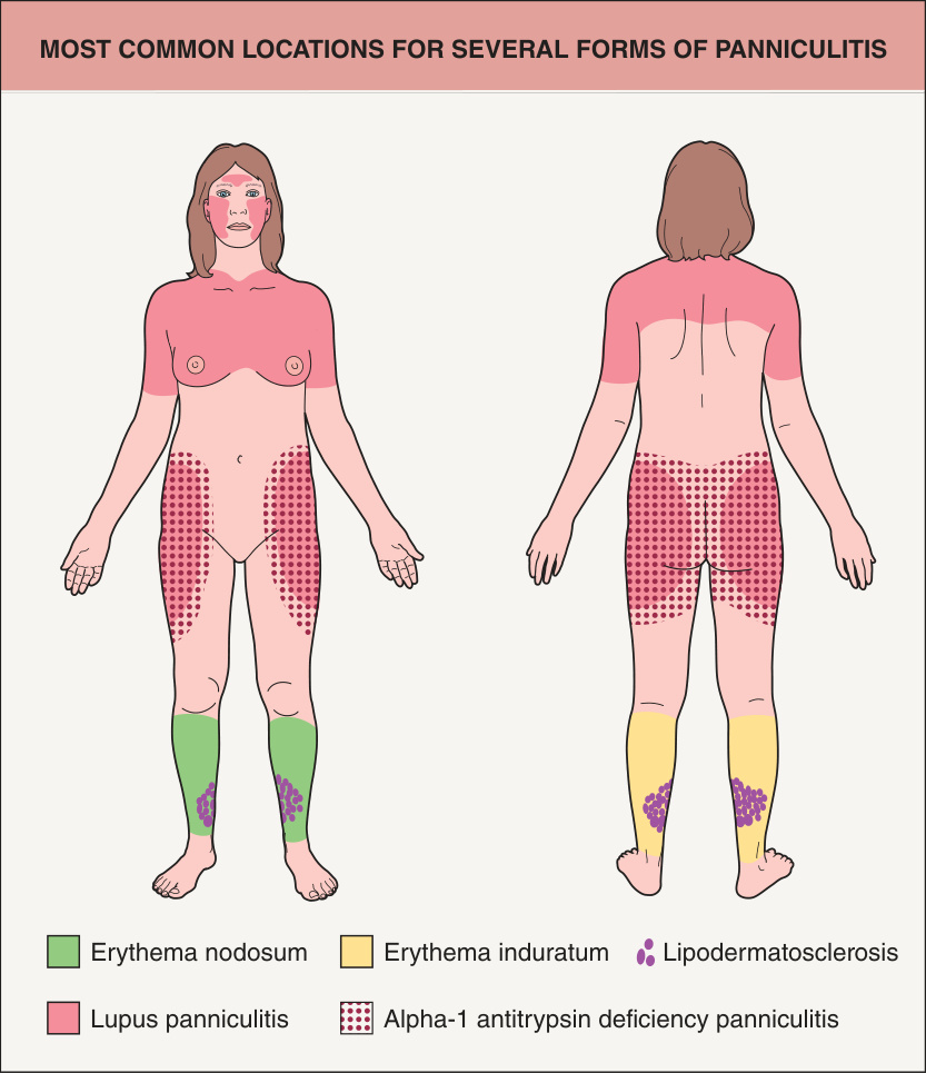
Fig. 100.1 Most common locations for several forms of panniculitis.
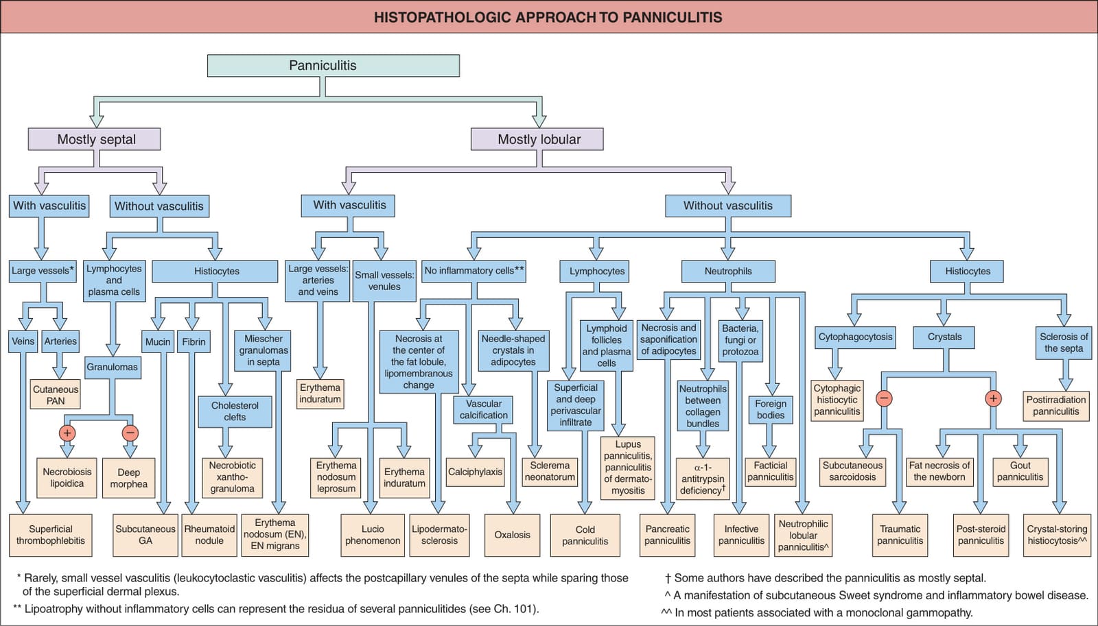
Fig. 100.2 Histopathologic approach to panniculitis. Drug-induced panniculitis can be primarily septal or lobular and is reviewed in Table 100.3. Leukemia cutis or lymphoma cutis may mimic panniculitis as in subcutaneous panniculitis-like T cell lymphoma, and the subcutaneous nodules observed in ANCA-associated vasculitides can contain extravascular necrotizing granulomas. GA, granuloma annulare; PAN, polyarteritis nodosa.
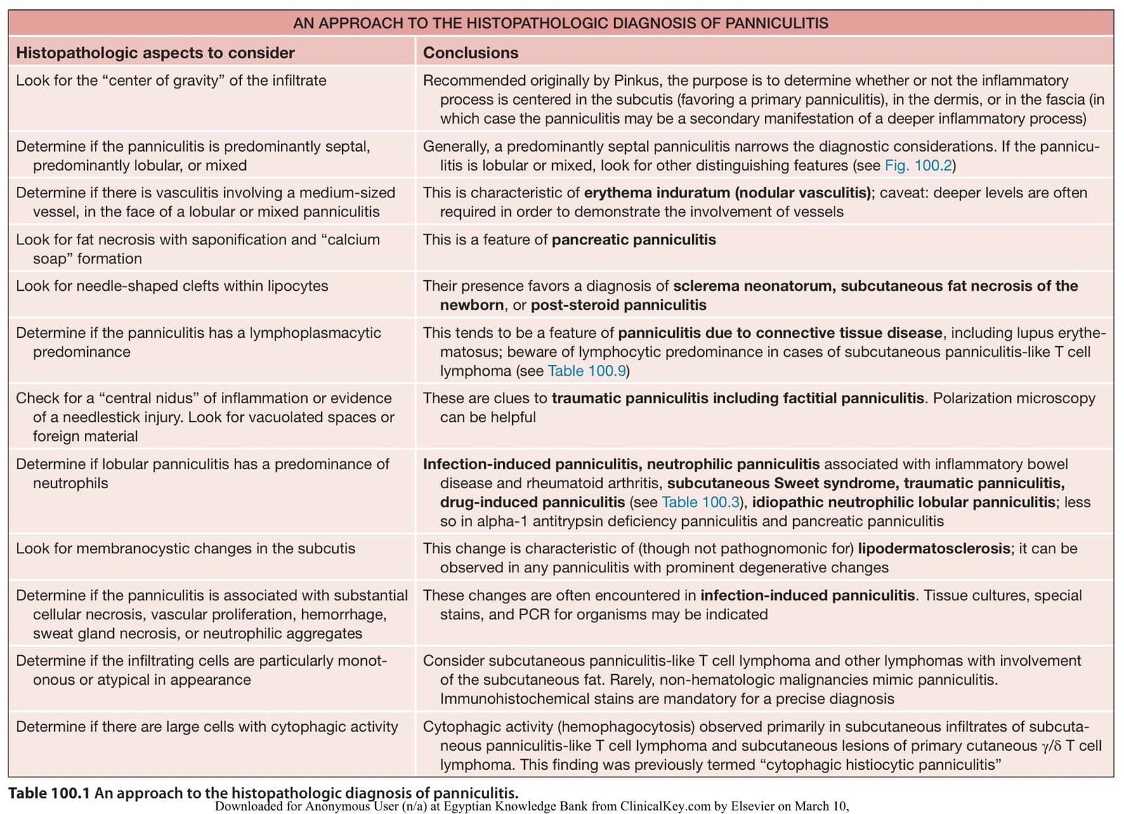
Table 100.1 An approach to the histopathologic diagnosis of panniculitis.