ERYTHEMA NODOSUM
Synonyms: Erythema contusiformis Erythema nodosum migrans (variant form)
Key features
Tender, erythematous, subcutaneous nodules
Usually distributed symmetrically and favor the pretibial areas;
occasionally occur elsewhere
In later stages, lesions acquire a bruise-like appearance
May be accompanied by fever, arthralgias, and malaise
Associated with a wide variety of systemic disorders
Introduction
Erythema nodosum is the best known of the various forms of panniculitis, as well as the most common. It typically presents as an acute eruption of tender, erythematous, subcutaneous nodules in the pretibial areas bilaterally. It is widely regarded as a delayed hypersensitivity response to a variety of antigenic challenges, although the mechanisms of its development are more complex than this statement would indicate. Histopathologically, it is the prototype of a “septal”
Classification of panniculitis depends upon multiple factors, including clinical characteristics such as location and ulceration as well as histopathology and etiology.
panniculitis. Identification and treatment of the underlying disorder, if found, is of primary importance, but therapy directed toward the lesions themselves is also an option, especially when idiopathic.
History
At the beginning of the eighteenth century, Robert Willan gave the first clear description of erythema nodosum, and he provided its name in his famous work, On Cutaneous Disease.
Epidemiology
Erythema nodosum can occur at any age, in both sexes, and in all racial groups. It is more common among women and is more frequently observed during the second through fourth decades of life. The relative ranking of underlying causes may vary according to geographic location; for example, in areas where Coccidioides immitis is endemic or regions where Behçet disease is more prevalent.
Pathogenesis
Erythema nodosum has been considered a delayed hypersensitivity response to a variety of antigenic stimuli, including bacteria, viruses, and chemical agents. Llorente et al. observed expression of mRNA for Th1 cytokines (interferon-γ, interleukin-2) in the skin lesions and peripheral blood of patients with erythema nodosum, and a Th1 pattern of cytokine synthesis is associated with delayed-type hypersensitivity reactions. However, a complex series of intermediate steps is involved in the development of these lesions. A variety of adhesion molecules and inflammatory mediators appear to be associated with erythema nodosum. For example, in erythema nodosum lesions, vascular cell adhesion molecule-1 (VCAM-1; CD106), platelet endothelial cell adhesion molecule-1 (PECAM-1; CD31), HLA-DR, and E-selectin are expressed on endothelial cells, while very late antigen-4 (VLA-4), L-selectin, and HLA-DR are expressed by inflammatory cells (see Ch. 102).
Neutrophils are often numerous in early lesions, and it has been shown that a higher percentage of circulating neutrophils in patients with erythema nodosum leads to the production of reactive oxygen intermediates; these intermediates, in turn, may provoke inflammation and tissue damage. Support for a pathogenic role for these cells and molecules is provided by studies on the effects of colchicine. This inhibitor of neutrophil chemotaxis has been shown to diminish L-selectin expression on the neutrophil surface, inhibit E-selectinmediated endothelial adhesiveness for neutrophils, and diminish stimulated expression of ICAM-1 on the endothelium.
Additional indirect evidence for the role of inflammatory cells and mediators includes reports of erythema nodosum following treatment with granulocyte colony-stimulating factor, and improvement (as well as flares) of erythema nodosum lesions with administration of tumor necrosis factor (TNF) inhibitors. Erythema nodosum, especially in its chronic phase, is characterized by granuloma formation and TNF is known to play a role in granuloma formation. A link between deregulation of TNF production and granuloma formation is further supported by the strong correlation of a polymorphism in the promoter region of the gene that encodes TNF and the development of sarcoidosis-associated erythema nodosum.
A wide range of precipitating factors has been linked with erythema nodosum. Infectious causes are common, particularly upper respiratory infections (both streptococcal and non-streptococcal). Other commonly reported causes are listed in Tables 100.2 and 100.3.
Clinical Features
Erythema nodosum presents with bilateral, tender, erythematous nodules. These arise in crops and clearly the most common site is the shins (Fig. 100.3). Other locations are occasionally involved, particularly the thighs and forearms. Nodules may also appear on the trunk, neck, and face, but this is sufficiently rare that development of lesions in these locations should prompt consideration of other diagnoses. Unlike other forms of panniculitis, ulceration is not a feature of erythema nodosum. Systemic symptoms may occur that are not necessarily related to a specific coexisting systemic disorder; these include arthritis, arthralgia, fever, and malaise.
Because of its close association with a variety of disorders and infections, erythema nodosum is an important skin sign of systemic disease.
For example, its development may precede or accompany a flare of inflammatory bowel disease. It may also have some value as a prognostic indicator in certain disorders. For example, erythema nodosum is associated with a protective effect against disseminated disease in patients with coccidioidomycosis, and it is closely aligned with a more benign and self-limited form of sarcoidosis. Nevertheless, a significant percentage of cases – more than one-third – have no known disease association, even when followed for a year or more. Clinical or laboratory data that tend to predict that the development of erythema nodosum may be secondary to a systemic disease are listed in Table 100.4.
Erythema nodosum lesions usually last a few days or weeks and then slowly involute, without scar formation. Discoloration suggestive of a bruise may be seen as the erythema subsides. More chronic forms do occur, some of which show a tendency toward migration or centrifugal spread; the latter have been termed subacute nodular migratory panniculitis or erythema nodosum migrans (see below). Up to one-third of cases of erythema nodosum recur. Annual recurrences are particularly common among idiopathic cases.
Pathology
Erythema nodosum is the prototypic septal panniculitis (Fig. 100.4), but this should not be taken to imply that histopathologic changes are entirely confined to subcutaneous septa. Biopsy specimens of early lesions tend to show edematous septa and mild lymphocytic infiltrates. Of note, neutrophils may predominate in early lesions, and a variant with a predominance of eosinophils has been reported. True vasculitis of the type seen in leukocytoclastic vasculitis is not observed, and erythema nodosum is not generally regarded as a vasculitic process. However, “secondary” vasculitis may be observed in lesions when they contain relatively heavy, mixed, or neutrophil-rich inflammatory infiltrates. Erythema nodosum-like lesions in Behçet disease may demonstrate leukocytoclastic or lymphocytic vasculitis involving subcutaneous venules or muscular veins; the latter changes are prone to occur in patients with more severe forms of Behçet disease.
In early lesions, one may also find Miescher microgranulomas, a characteristic if not pathognomonic feature of erythema nodosum. These are small collections of cells (macrophages or perhaps “histiocytoid cells” [neutrophil precursors]) that are found within septa or at a septal–lobular interface and tend to surround neutrophils or small cleft-like spaces. Reported variations in the frequency of these granulomas in erythema nodosum may result in part from differences in definition, in the acceptance of subtle changes, and in the rigor of the search. In older lesions, Miescher microgranulomas may feature epithelioid and multinucleated giant cells (Fig. 100.4, inset).
As lesions progress, the septa become widened and contain a mixed, partly granulomatous infiltrate. These cells infiltrate the periphery of fat lobules in a lace-like configuration. The extent of lobular involvement may vary, and in some cases can be prominent. Nevertheless, in the case of a lobular panniculitis without the characteristic septal changes, a diagnosis of erythema nodosum should be made with caution.
Additional evaluation can include viral hepatitis panel and, in women of child-bearing age, serum β-human chorionic gonadotropin.
Frequently, there is also a mild to moderate perivascular lymphocytic infiltrate in the overlying dermis. In later stages, the septa become fibrotic, partially replacing the fat lobules. Residual granulomas and lipophages can be observed, and a degree of vascular proliferation may be present. Over the long term, a remodeling process takes place that usually results in minimal residual scarring.
Differential Diagnosis
The clinical scenario of an acute eruption of tender subcutaneous nodules over both shins of a young person is highly characteristic of erythema nodosum. However, when lesions are few in number, are located in sites other than the lower legs, or are of longer duration (>6 weeks), erythema nodosum can be difficult to distinguish from other forms of panniculitis. Lesions of erythema induratum (nodular vasculitis) can resemble those of erythema nodosum, but they tend to occur on the posterior aspect of the lower legs and may ulcerate. Ulceration is also a feature of pancreatic panniculitis, which occurs more frequently in other locations (although still favoring the lower legs), is more likely to be accompanied by arthritis and serositis, and is associated with elevated serum amylase and lipase levels.
Histopathologically, the picture of a predominantly septal panniculitis usually limits the differential diagnosis and tends to exclude those conditions that are chiefly lobular or mixed (see Fig. 100.2). Pancreatic panniculitis may show predominantly septal changes in its earliest stages, but eventually these lesions exhibit the characteristic fat necrosis, with saponification and “ghost cell” formation. Infection-induced panniculitis can sometimes mimic erythema nodosum, but there are often more extensive neutrophilic infiltrates, cellular necrosis (including sweat gland necrosis), vascular proliferation, and hemorrhage.
Treatment
Treatments most often recommended for uncomplicated erythema nodosum include bed rest, salicylates, and nonsteroidal anti-inflammatory drugs (NSAIDs; Table 100.5). Potassium iodide has been used with success, with adult dosages ranging from 450 to 1500 mg/day (Table 100.6). Improvement can be seen within 2 weeks. Potassium iodide may work through inhibition of cell-mediated immunity, as well as via inhibition of neutrophil chemotaxis and suppression of neutrophil-generated oxygen intermediates. In light of this treatment response, reports of erythema nodosum triggered by potassium iodide seem contradictory.
Treatment of erythema nodosum is influenced by underlying conditions. Thus, colchicine is useful in management of erythema nodosum that accompanies Behçet disease. Various treatments for inflammatory bowel disease are also effective in managing coexistent erythema nodosum. Both etanercept and infliximab have been reported to be effective in treating erythema nodosum, but paradoxically both have been noted to produce erythema nodosum as a cutaneous side effect. In case reports, adalimumab has led to improvement of refractory chronic erythema nodosum. Additional systemic therapies are listed in Table 100.5.
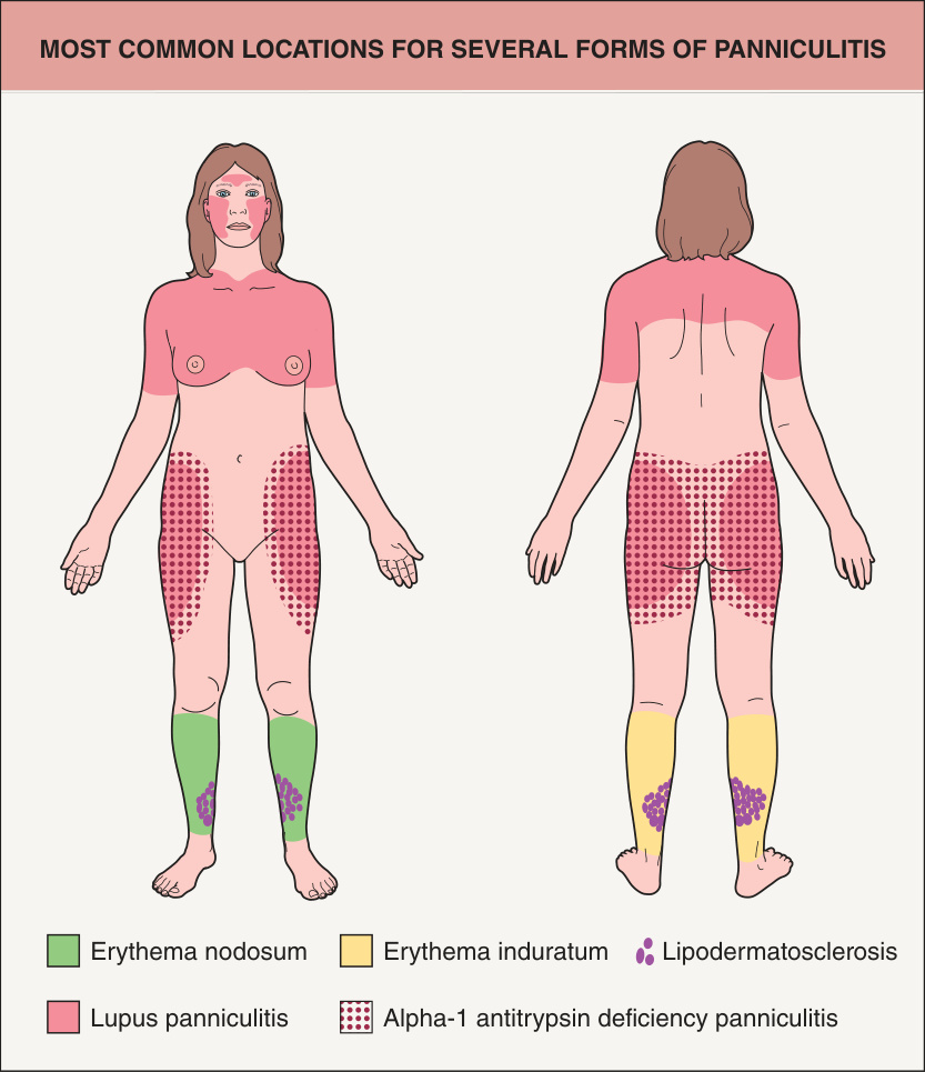
Fig. 100.1 Most common locations for several forms of panniculitis.
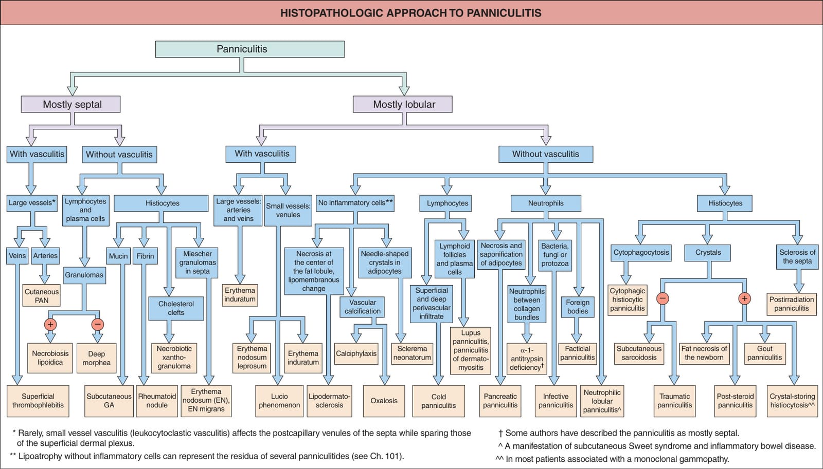
Fig. 100.2 Histopathologic approach to panniculitis. Drug-induced panniculitis can be primarily septal or lobular and is reviewed in Table 100.3. Leukemia cutis or lymphoma cutis may mimic panniculitis as in subcutaneous panniculitis-like T cell lymphoma, and the subcutaneous nodules observed in ANCA-associated vasculitides can contain extravascular necrotizing granulomas. GA, granuloma annulare; PAN, polyarteritis nodosa.
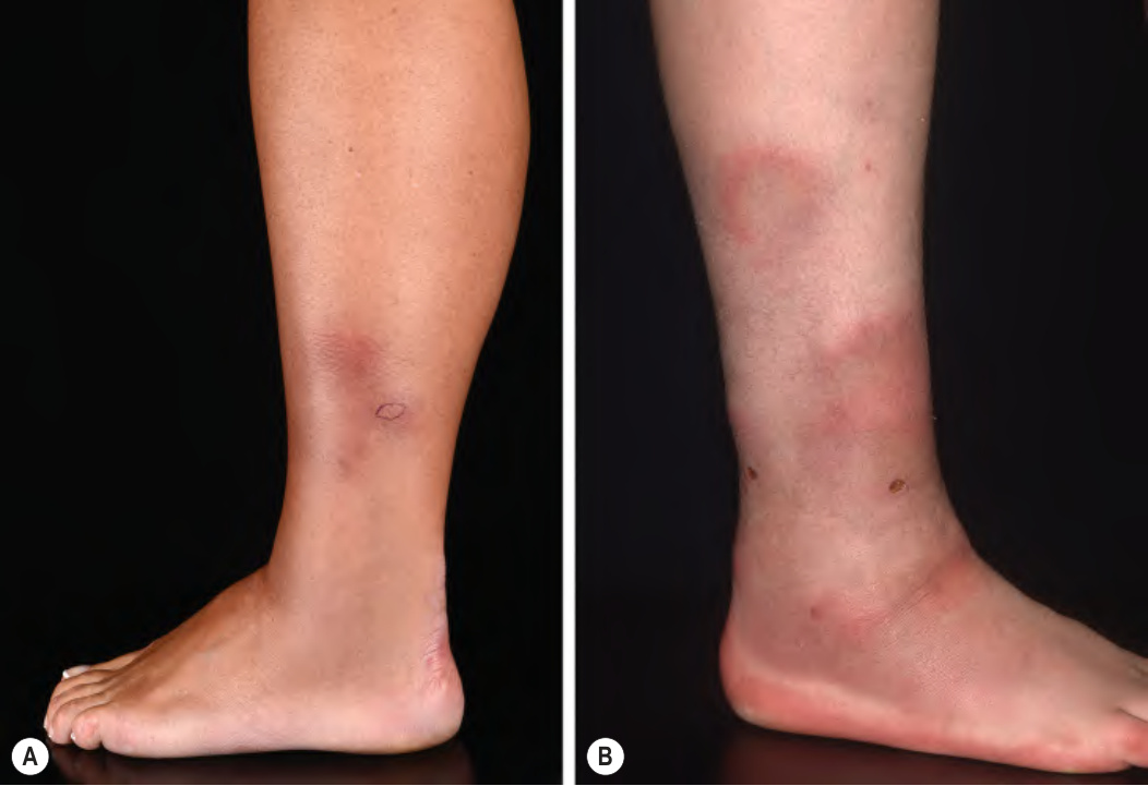
Fig. 100.3 Erythema nodosum – clinical appearance.A Cluster of dull red–brown, tender nodules on the lower leg. B The plaques may develop an arciform configuration and bruise-like appearance. Courtesy Lorenzo Cerrroni, MD.
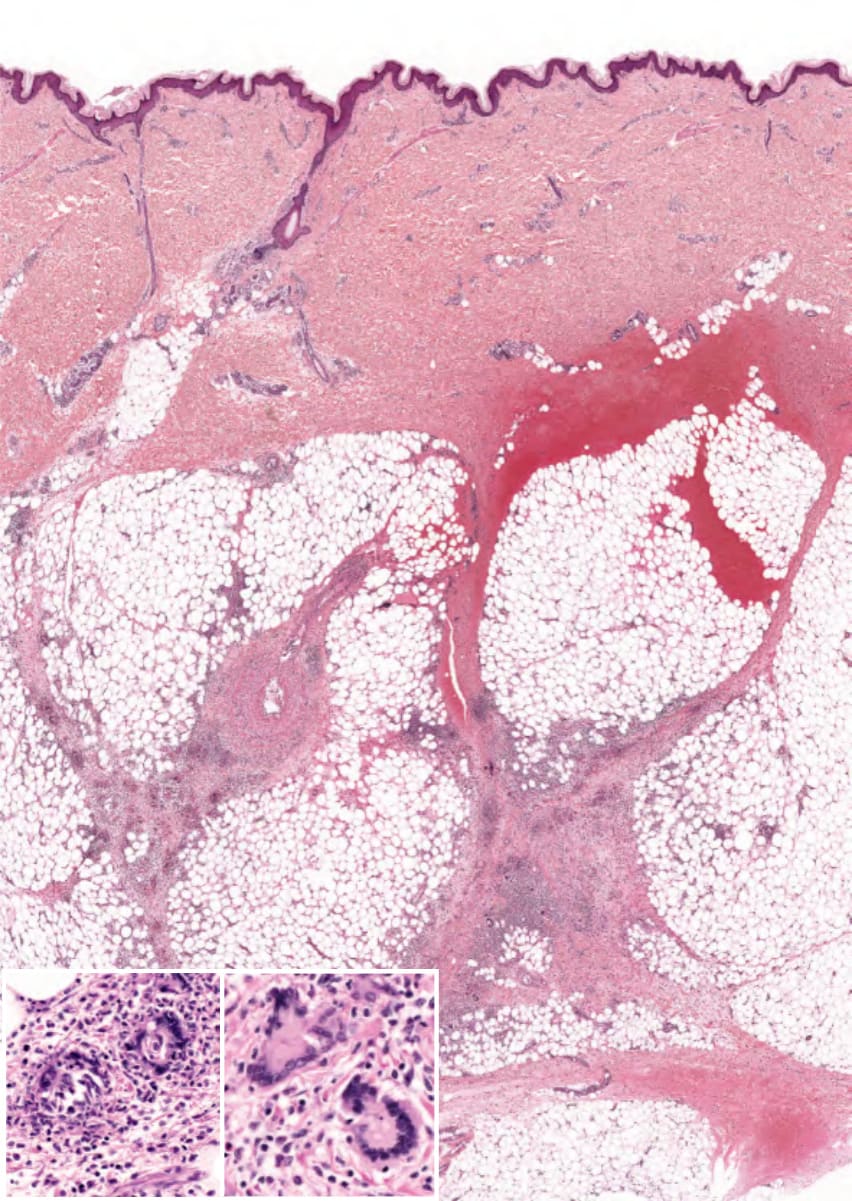
Fig. 100.4 Erythema nodosum – histopathologic features. A predominantly septal panniculitis with Miescher microgranulomas within the septa (left inset). Note the multinucleated giant cells (right inset).
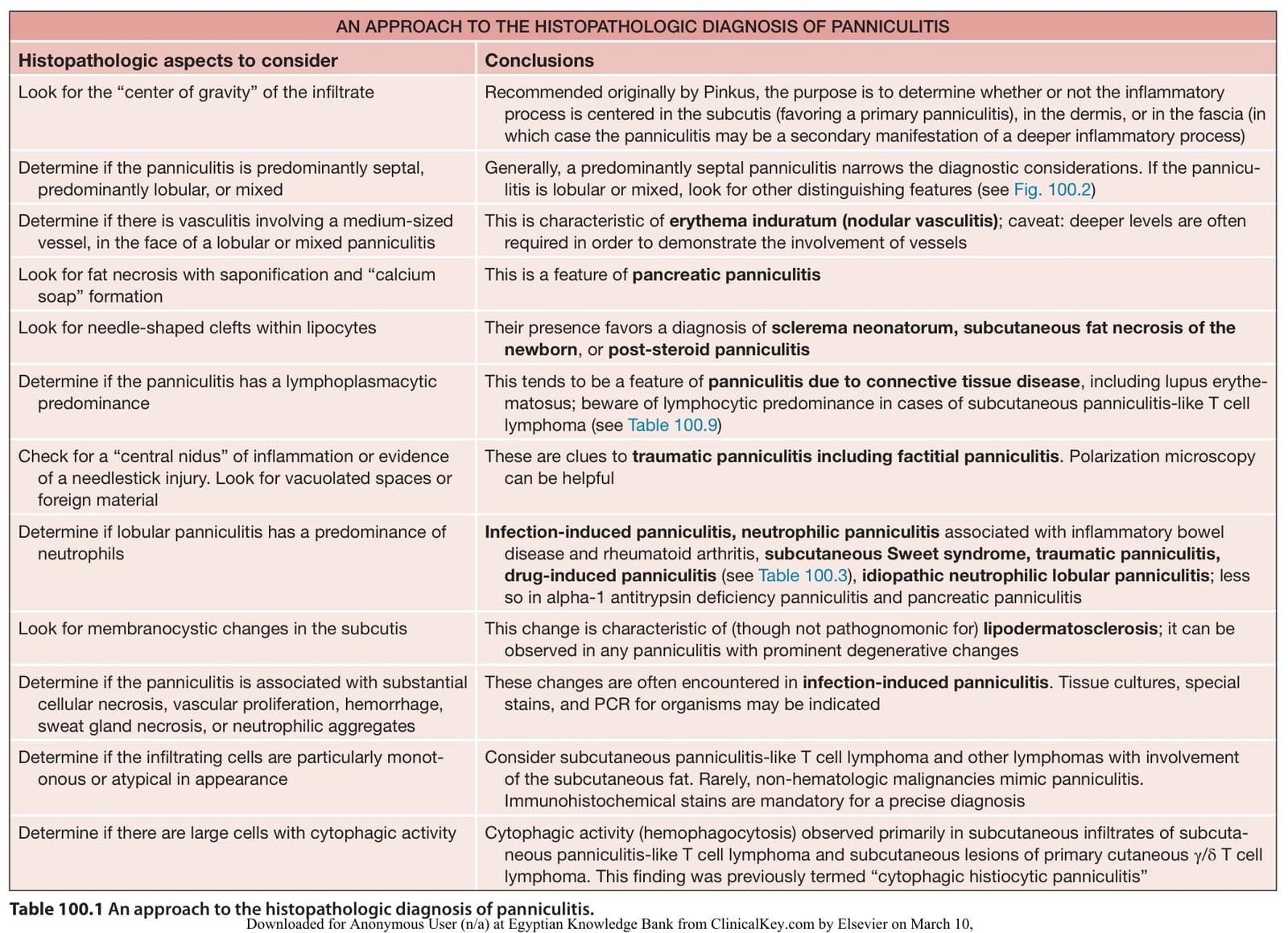
Table 100.1 An approach to the histopathologic diagnosis of panniculitis.
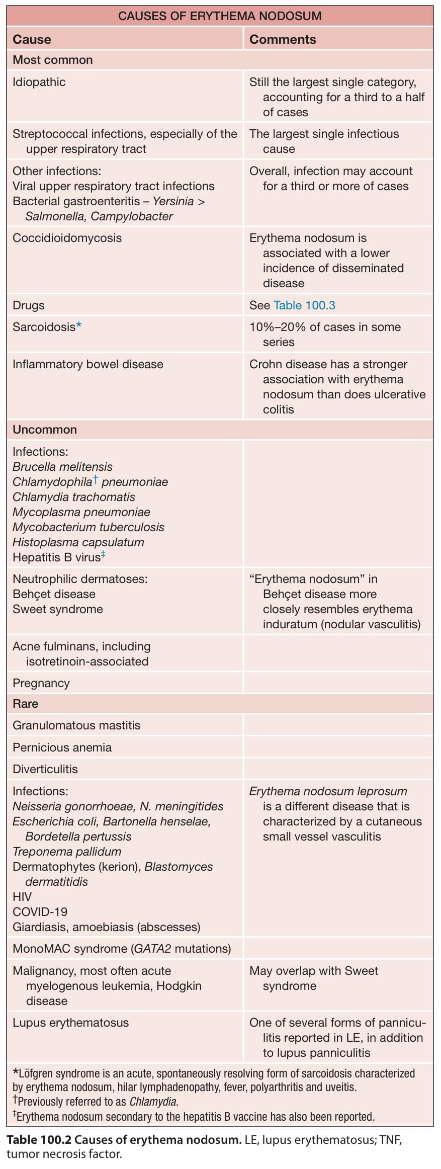
Table 100.2 Causes of erythema nodosum. LE, lupus erythematosus; TNF, tumor necrosis factor.
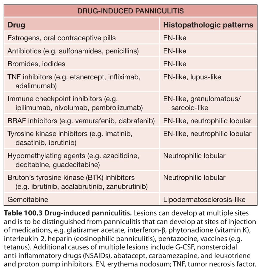
Table 100.3 Drug-induced panniculitis. Lesions can develop at multiple sites and is to be distinguished from panniculitis that can develop at sites of injection of medications, e. g. glatiramer acetate, interferon-β, phytonadione (vitamin K), interleukin-2, heparin (eosinophilic panniculitis), pentazocine, vaccines (e. g. tetanus). Additional causes of multiple lesions include G-CSF, nonsteroidal anti-inflammatory drugs (NSAIDs), abatacept, carbamezapine, and leukotriene and proton pump inhibitors. EN, erythema nodosum; TNF, tumor necrosis factor.
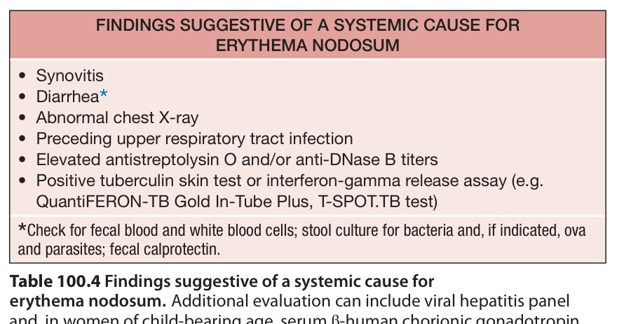
Table 100.4 Findings suggestive of a systemic cause for erythema nodosum.
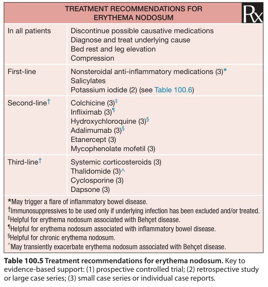
Table 100.5 Treatment recommendations for erythema nodosum. Key to evidence-based support: (1) prospective controlled trial; (2) retrospective study or large case series; (3) small case series or individual case reports.
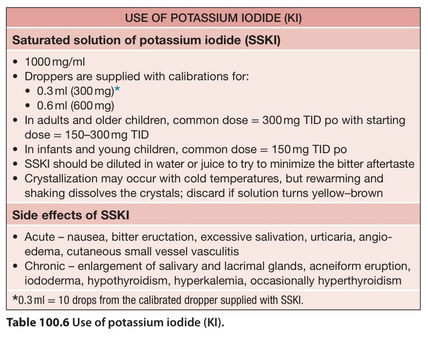
Table 100.6 Use of potassium iodide (KI).