MORPHEA/SCLERODERMA PANNICULITIS
(See also Chs. 43 & 44.)
Clinical Features
Both morphea and systemic sclerosis (scleroderma) can affect the subcutaneous fat (Table 100.7), which is the primary site of involvement in deep morphea (morphea profunda). Subcutaneous extension also occurs in variants such as disabling pansclerotic morphea of childhood, generalized morphea, and linear morphea. In addition, eosinophilic fasciitis can involve the subcutaneous fat as well as the fascia. These entities may be associated with peripheral eosinophilia, polyclonal gammopathy, and serologic abnormalities. It has been proposed that the sclerosing process in systemic sclerosis is initiated by the changes that take place in the subcutis.
Pathology
Table 100.7 outlines the microscopic changes in morphea- and systemic sclerosis-associated septal panniculitis (Fig. 100.5). Lymphocytes and plasma cells predominate, although macrophages and eosinophils may be present. In some cases, the number of plasma cells is striking. In late stages of morphea, the subcutis is largely replaced by hyalinized connective tissue, very often accompanied by changes of lipoatrophy.
Differential Diagnosis
The combination of septal panniculitis with lymphoplasmacytic predominance and dermal and subcutaneous sclerosis is unique, and this helps to separate morphea/systemic sclerosis panniculitis from other septal forms of panniculitis. Differentiation among the several variants outlined above is more difficult, requiring clinical data for accurate classification. Sclerosis with lesser degrees of subcutaneous inflammation characterizes systemic sclerosis, while a predominance of fascial involvement with extension into the subcutis is more typical of eosinophilic fasciitis.
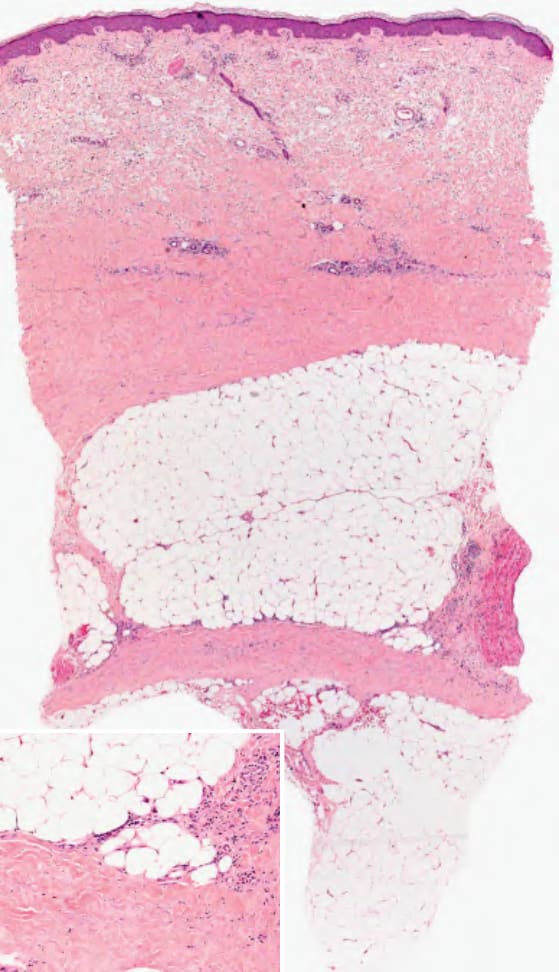
Fig. 100.5 Morphea panniculitis. Septal thickening, mucin deposition and mild lymphocytic infiltrate. Lymphoplasmacytic infiltrates concentrate at the dermal–subcutaneous interface (inset). Courtesy Lorenzo Cerrroni, MD.
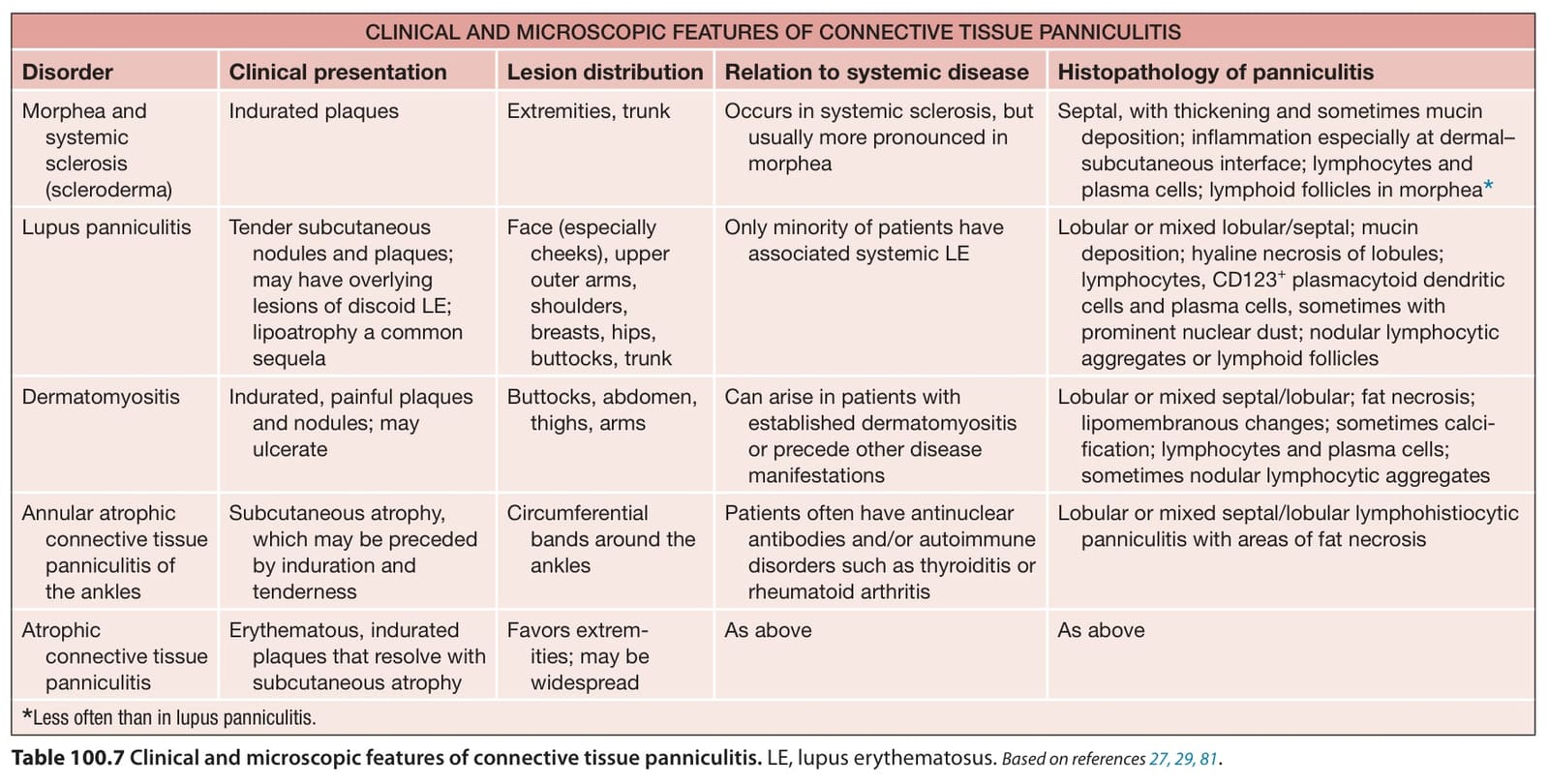
Table 100.7 Clinical and microscopic features of connective tissue panniculitis. LE, lupus erythematosus. Based on references