ALPHA-1 ANTITRYPSIN DEFICIENCY PANNICULITIS
Synonym: Alpha-1 protease (proteinase) deficiency panniculitis
Key features
Erythematous, painful, subcutaneous nodules or plaques that often ulcerate and drain
Associated with alpha-1 antitrypsin deficiency; patients with the most severe disease are homozygotes for the Z allele of SERPINA1 (PiZZ)
A characteristic histopathologic finding is liquefactive necrosis of the dermis and subcutaneous septa, but lobular or mixed septal– lobular changes with neutrophils may occur
Introduction
Alpha-1 antitrypsin deficiency is a well-established, but uncommon, cause of panniculitis. The most severely affected individuals, with markedly decreased levels of the protease inhibitor, are most prone to the development of ulcerating neutrophilic panniculitis. Recognition of the disorder is important not only in the selection of appropriate therapy, but also in addressing other systemic manifestations of the disease and in dealing with its genetic aspects.
History
Alpha-1 antitrypsin deficiency is an inborn error of metabolism that was first delineated by Eriksson and others in the early 1960s. In 1972, Warter and colleagues identified members of a family with alpha-1 antitrypsin deficiency and “Weber–Christian syndrome”. Subsequent investigations linked the clinical and microscopic findings to known effects of proteinase inhibitor deficiency.
Epidemiology
In the US, alpha-1 antitrypsin deficiency occurs most commonly in White, followed by Hispanic and Black, individuals. The incidence of panniculitis is approximately equal in men and women. Age of onset ranges from infancy to the eighth decade of life.
Pathogenesis
Alpha-1 antitrypsin, a glycoprotein produced in the liver, is the most abundant circulating serine protease inhibitor (serpin). The more than 120 different alleles of the gene that encodes this protein (SERPINA1; formerly known as PI) are divided into categories based upon the electrophoretic mobility of their protein products (M = medium, S = slow, Z = very slow). The most common protease inhibitor (Pi) phenotype is MM (homozygous for M alleles), which is associated with normal serum levels of alpha-1 antitrypsin (100–200 mg/dl). Heterozygotes with one copy of the S or Z allele have mild to moderate deficiencies of the inhibitor (PiMS and PiMZ; prevalences of 1%–3% in White populations). Patients who are homozygous for the Z allele (PiZZ; prevalence of 1 : 150–1 : 5000 in White populations) or whose genotype is the rare PiZnull have severe alpha-1 antitrypsin deficiency, with serum levels in the range of 20–45 mg/dl. In these individuals, most of the aberrant alpha-1 antitrypsin protein accumulates within the endoplasmic reticulum of hepatocytes, and the small amounts that enter the circulation have decreased function and a tendency to form inactive polymers that may stimulate neutrophil chemotaxis. Of note, Z-type polymers have been detected in the skin of a patient with alpha-1 antitrypsin deficiency panniculitis, further suggesting a possible proinflammatory role.
Alpha-1 antitrypsin acts upon a wide range of proteolytic enzymes that play a direct role in degradation of tissues, including trypsin, collagenase, and elastase. It also has important effects on immune function, e.g. inhibition of membrane-bound serine proteases involved in the activation of lymphocytes and macrophages. It may also inhibit complement activation, both through a direct effect on complementrelated proteases and by inhibiting the neutrophil proteases that activate enzymes of the complement system.
In addition to panniculitis, the consequences of alpha-1 antitrypsin deficiency include chronic liver disease with cirrhosis (resulting from retention of the aberrant protein within the liver), emphysema, pancreatitis, membranoproliferative glomerulonephritis, rheumatoid arthritis, c-ANCA (cytoplasmic antineutrophil cytoplasmic antibody)-positive vasculitis, other cutaneous vasculitides such as Henoch–Schönlein purpura, and angioedema (resulting from deficiency of the protease inhibitor). The initiating event in individuals who develop panniculitis is not always clear; trauma appears to play a role in some patients. Postpartum flares of the disease have been reported in genetically susceptible individuals. This is attributed to the estrogen-promoted increase in proteinase inhibitor levels during pregnancy, followed by a precipitous decline to subnormal levels postpartum.
Absence of the alpha-1 antitrypsin protease inhibitor results in activation of lymphocytes and macrophages, lack of restraint upon the complement cascade, release of chemotactic factors, accumulation of neutrophils with release of their proteolytic enzymes, and consequent attack upon fat and nearby connective tissues. The subcutis may be particularly vulnerable to this process, since fatty acids make nearby elastin more susceptible to proteolytic degradation.
Clinical Features
Large, erythematous to purpuric, tender nodules or plaques appear in a variety of sites (Fig. 100.6), especially the lower trunk and proximal extremities (flanks, buttocks, and thighs). Ulcers develop that may be deep and necrotic, accompanied by an oily discharge. A history of antecedent trauma can be elicited in approximately one-third of patients. Panniculitis may be accompanied by fever, pleural effusions, and pulmonary emboli. The clinical course of the panniculitis is often prolonged, and lesions are resistant to immunosuppressive therapy. Healing is accompanied by scarring and subcutaneous atrophy. The most severe manifestations arise in those with profound proteinase inhibitor deficiency (PiZZ), although the panniculitis can also occur in heterozygotes.
Pathology
Descriptions of the pathology of alpha-1 antitrypsin deficiency panniculitis have varied. Early, there is a neutrophilic panniculitis, followed rapidly by necrosis and destruction of fat lobules. Splaying of neutrophils
between collagen bundles in the reticular dermis has been described as an early clue to the diagnosis. Dissolution of dermal collagen, with resultant liquefactive necrosis and separation of fat lobules from adjacent septa, is a principal change in most cases. Another characteristic feature is the presence of “skip areas” of normal fat adjacent to foci of severe necrotizing panniculitis. Chronic inflammation and hemorrhage may be present at the periphery of areas of involvement. Most authors have not found evidence for primary leukocytoclastic vasculitis, although there may be evidence for lymphocytic vasculitis, secondary vasculitis in areas of heavy neutrophilic infiltration, or thrombosis. In individuals with intermediate levels of protease inhibitor deficiency, lipophage and giant cell accumulation may be prominent. Lesions heal with scarring and obliteration of fat lobules.
Views differ on whether alpha-1 antitrypsin deficiency panniculitis should be regarded as a primarily septal or lobular panniculitis. Clearly, involvement of fat lobules can be significant, and, as a result, there are authors who have labeled the process a lobular panniculitis. On the other hand, some descriptions have emphasized early septal inflammation, collagenolysis of the fibrous septa (Fig. 100.7), and the prominent septal fibrosis in late-stage lesions.
Differential Diagnosis
Clinically, the degrees of inflammation, ulceration, and drainage associated with alpha-1 antitrypsin deficiency panniculitis may actually elicit a differential diagnosis more focused upon ulcerative skin disorders (see Fig. 105.1). Ulcers associated with alpha-1 antitrypsin deficiency generally lack the necrotic, “undermined” borders associated with pyoderma gangrenosum. Tissue culture to exclude infectioninduced panniculitis may be required.
Entities in the microscopic differential diagnosis include traumatic (factitial) panniculitis, infection-induced panniculitis, pancreatic panniculitis, and erythema induratum (nodular vasculitis). Each of these can be associated with infiltrates that include neutrophils and varying degrees of necrosis, yet each has other distinct findings (see below). In the appropriate clinical setting, subcutaneous Sweet syndrome, druginduced panniculitis, and neutrophilic panniculitis in patients with rheumatoid arthritis or inflammatory bowel disease (IBD) may represent additional diagnostic considerations. Of note, the underlying IBD can be clinically silent. When no underlying disorder is detected, the term idiopathic neutrophilic lobular panniculitis may be applied.
Treatment
Treatments that are usually ineffective include corticosteroids and other immunosuppressants, cytotoxic agents, colchicine, danazol, and hydroxychloroquine. Doxycycline is sometimes effective, particularly in mild cases; one suggested dosage schedule is 200 mg twice daily for 3 months. Dapsone may also be beneficial in mild cases, by suppressing neutrophil migration and inhibiting oxidation reactions
induced by myeloperoxidase. Reduction of alcohol intake has also been recommended, since ethanol (as a hepatotoxin) may precipitate alpha-1 antitrypsin-associated hepatitis.
The most effective therapeutic intervention is replacement of alpha-1 antitrypsin via intravenous infusions. Dosages are generally 60 mg/kg per week, administered over a period of 3 to 7 weeks. Improvement is relatively rapid, in that clearing of the panniculitis can occur after three weekly doses. Recurrences are possible when alpha-1 antitrypsin levels fall below 50 mg/dl, but these typically respond to further replacement therapy. Other successful therapies include plasma exchange and liver transplantation (in appropriate clinical circumstances). There is evidence that autophagy-enhancing drugs, such as carbamazepine, may reduce systemic manifestations, including hepatic fibrosis. Most recently, fazirsiran, an RNA interference therapy, has led to hepatic improvement42a.
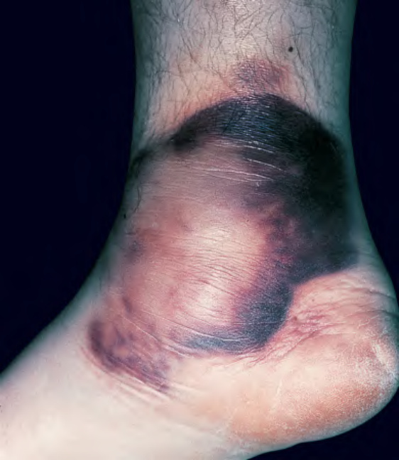
Fig. 100.6 Alpha-1 antitrypsin deficiency panniculitis – clinical appearance. Purpuric nodules on the ankle. Courtesy Kenneth E. Greer, MD.
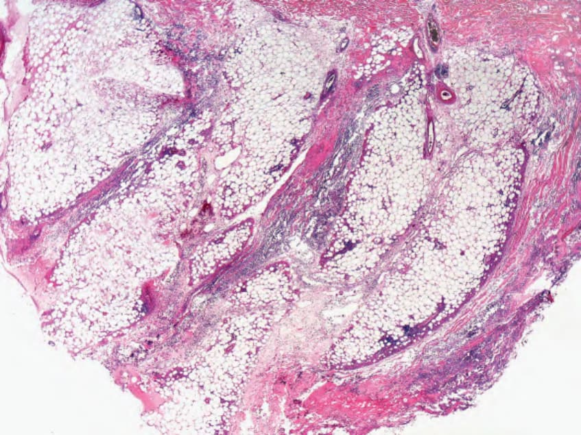
Fig. 100.7 Alpha-1 antitrypsin deficiency panniculitis – histopathologic features. Predominantly septal involvement is seen, with inflammation and collagenolysis of fibrous septa.