ERYTHEMA INDURATUM
Synonyms: Nodular vasculitis Erythema induratum of Bazin or Bazin disease (tuberculous etiology) Erythema induratum of Whitfield (non-tuberculous etiology)
Key features
Erythematous nodules or plaques, usually on the posterior lower legs of young to middle-aged women
Ulceration and drainage may occur
Microscopic features of lobular or mixed panniculitis with evidence of vasculitis involving arteries or veins
Classically associated with tuberculosis, but similar lesions can be idiopathic or induced by other infectious agents or drugs
Introduction
Erythema induratum is a condition characterized by nodules on the lower extremities, which may ulcerate and drain. Originally regarded as a tuberculid, detection of mycobacterial DNA within cutaneous lesions in a subset of patients supports its relationship to tuberculosis, at least in a proportion of cases.
History
Erythema induratum was first described by Ernest Bazin in 1861. It was generally regarded as a tuberculid because of a strong association with tuberculosis, although Koch’s postulates could not be fulfilled. In
1945, Montgomery and colleagues proposed the term nodular vasculitis for cases with similar clinical and pathologic features that were not of tuberculous origin. Since the early 1970s, erythema induratum and nodular vasculitis have generally been considered as synonyms referring to a clinicopathologic entity with several possible causes, one of which is tuberculosis. The detection of mycobacterial DNA in cutaneous lesions has confirmed a tuberculous origin in some patients. There are certain clinicians, however, who prefer to use the term nodular vasculitis when referring to individuals with a non-tuberculous etiology.
Epidemiology
In erythema induratum, an overwhelming female predominance is observed, but men can also develop the disease. There is no apparent racial predilection, and although there is a wide age range among affected patients, the mean is between 30 and 40 years. Erythema induratum of tuberculous etiology occurs more frequently in populations with a high prevalence of tuberculosis.
Pathogenesis
As mentioned previously, some cases have been strongly associated with Mycobacterium tuberculosis infection. This can be substantiated by the detection of mycobacterial DNA in skin lesions by PCR, with specific primers used to distinguish M. tuberculosis complex DNA from that of other mycobacterial pathogens. Non-tuberculous cases have been related to other infectious agents (e.g. Nocardia, hepatitis C virus) or to drugs (e.g. propylthiouracil). There is a report of erythema induratum being induced by a tuberculin skin test.
Although it has been suggested that erythema induratum results from an immune complex-mediated vasculitis, most investigators believe that the process represents a type IV, cell-mediated response to an antigenic stimulus. Biopsy specimens show a predominance of T lymphocytes, macrophages and dendritic cells, including Langerhans cells. One study of peripheral blood mononuclear cells from a patient with erythema induratum and active tuberculosis showed a high proliferative response to purified protein derivative (PPD) and marked production of interferon-γ, suggesting a pathogenic role for these PPD-specific T cells in a delayed hypersensitivity response to mycobacterial antigens.
Clinical Features
Erythema induratum is characterized by tender, erythematous to violaceous nodules and plaques that most often develop on the lower legs, especially the calves. Lesions have also been reported on the feet, thighs, buttocks, and arms. An annular arrangement of nodules has been described in M. tuberculosis-related cases. Ulceration can occur (Fig. 100.8). Lesions are persistent, tend to heal with scarring, and are prone to recurrence. In erythema induratum associated with M. tuberculosis, there may be clinical and radiographic evidence for active
tuberculosis, positive skin tests to PPD, or a positive interferon-gamma release assay such as the QuantiFERON®-TB Gold Plus test. In addition, other tuberculids, e.g. papulonecrotic, may be present. Clinical differences between tuberculous and non-tuberculous cases are minor.
Pathology
Erythema induratum is generally described as a lobular or mixed septal/ lobular panniculitis. Inflammation is mixed, and can include neutrophils, lymphocytes, macrophages, and multinucleated giant cells (Fig. 100.9A). Vasculitis is identifiable in the vast majority of cases, and most frequently involves veins or arteries of connective tissue septa and small venules of the fat lobules (Fig. 100.9B). It may be predominantly neutrophilic, lymphocytic, or granulomatous. Necrosis with a coagulative or caseous appearance may be present, sometimes with palisading granulomas. Necrosis has been described in both tuberculous and non-tuberculous cases, and the incidence and degree of necrosis are greater in those cases that are positive for M. tuberculosis DNA by PCR methods. However, this finding is absent in over half of cases.
Differential Diagnosis
Infection-induced panniculitis tends to show a more prominent neutrophilic component, granular basophilic necrosis, sweat gland necrosis and proliferation of small vessels, and organisms may be identified on
special staining. Lupus panniculitis tends to be less granulomatous, has a prominent lymphoplasmacellular infiltrate, may show mucin deposits, and frequently has overlying epidermal and dermal changes typical for lupus erythematosus. Both polyarteritis nodosa and thrombophlebitis tend to show inflammation limited to the immediate perivascular zone, in contrast to the extensive lobular panniculitis often encountered in erythema induratum. Histologically, perniosis can be difficult to distinguish from erythema induratum, but there is typically a history of cold exposure, and on microscopic examination prominent involvement of dermal vessels is often observed, with “fluffy edema” of their walls.
Treatment
Treatment should be directed at the underlying cause, if found. This includes multi-drug antituberculous therapy (see Ch. 75) and discontinuing possible inciting medications. Therapeutic options for nontuberculous erythema induratum include NSAIDs, corticosteroids, tetracyclines, potassium iodide, and mycophenolate mofetil. Supportive care is similar to that for erythema nodosum (see Table 100.5).
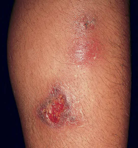
Fig. 100.8 Erythema induratum – clinical appearance. Inflamed nodular lesions on the lower leg, with evidence of ulceration. Courtesy Kenneth E. Greer, MD.
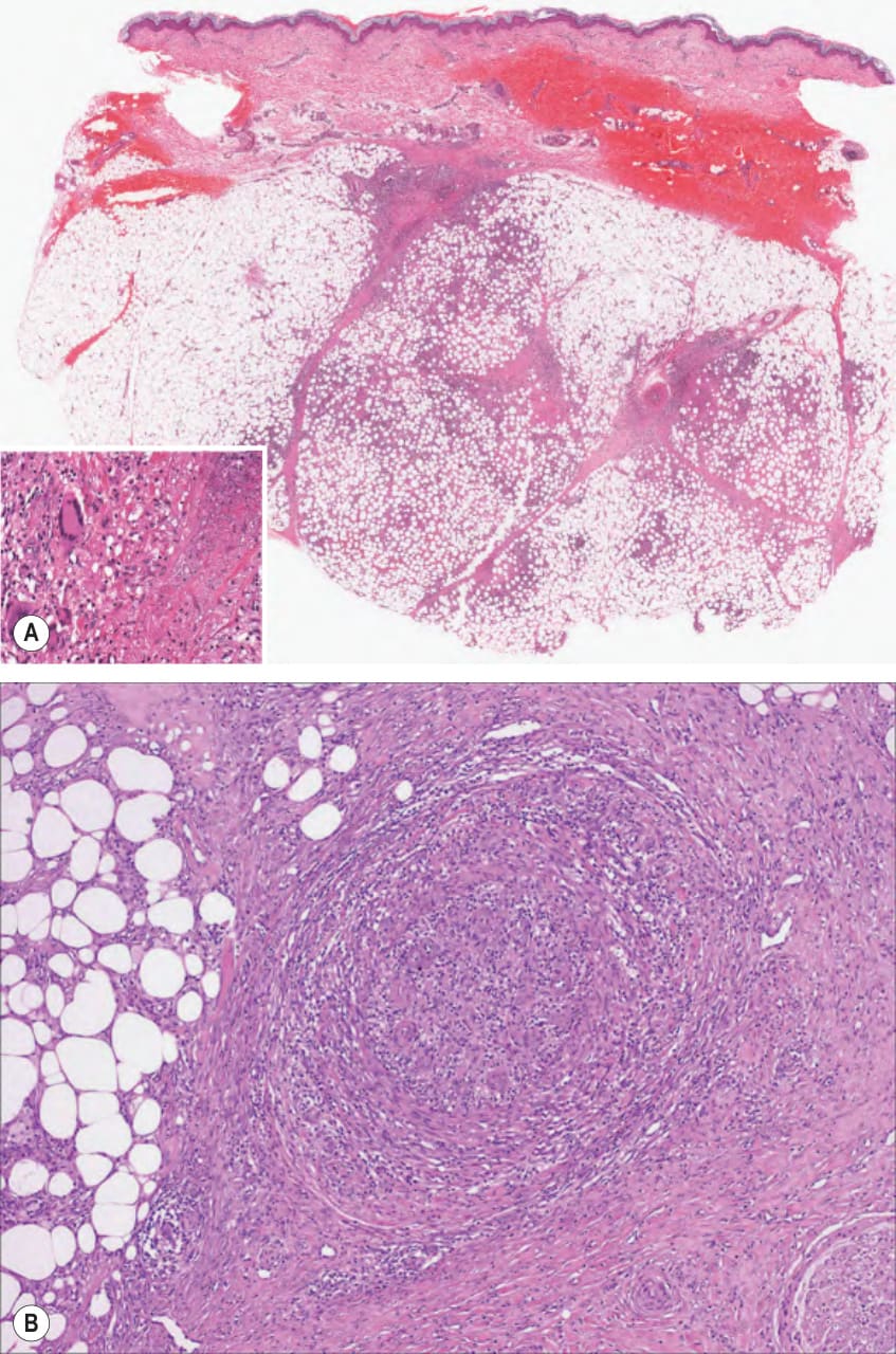
Fig. 100.9 Erythema induratum – histopathologic features.A A predominantly lobular panniculitis is seen with an infiltrate that is lymphocytic and granulomatous; note the multinucleated giant cells (inset). B Vasculitis involving a medium-sized vessel in the subcutis.
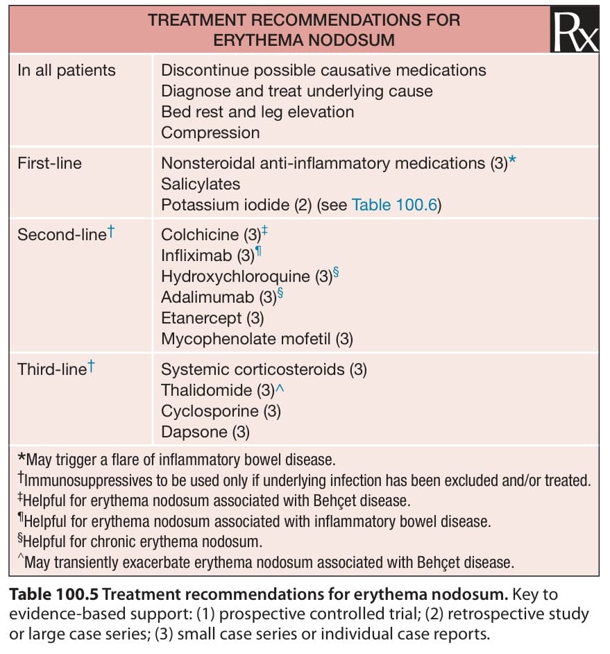
Table 100.5 Treatment recommendations for erythema nodosum. Key to evidence-based support: (1) prospective controlled trial; (2) retrospective study or large case series; (3) small case series or individual case reports.