PANCREATIC PANNICULITIS
Synonyms: Pancreatic fat necrosis Enzymatic panniculitis
Key features
Subcutaneous nodules, sometimes accompanied by fever, arthritis, or abdominal pain
Associated with pancreatic disorders, including acute and chronic pancreatitis and pancreatic carcinoma
Mixed septal/lobular panniculitis featuring “ghost cell” formation and the deposition of basophilic material due to saponification of fat by calcium salts
Treatment primarily directed towards the underlying pancreatic disorder
Introduction
Pancreatic panniculitis is an unusual complication of pancreatic disease. In addition to the symptoms associated with fat necrosis, its chief importance is as a sign of a significant systemic disorder, particularly because the panniculitis may be recognized prior to detection of the underlying pancreatic disease. The association of pancreatitis, panniculitis, and polyarthritis is sometimes referred to as the PPP syndrome.
History
Chiari first described pancreatic panniculitis in 1883. By 1999, fewer than 100 cases had been reported. Since then, ~100 additional patients with pancreatic panniculitis have been described, primarily as case reports.
Epidemiology
Panniculitis develops in up to 2% of patients with pancreatic disorders. No geographic, racial or sex predilections have been reported.
Pathogenesis
There is considerable evidence that the enzymes lipase, amylase and trypsin are involved in producing the lesions of pancreatic panniculitis. Elevated enzyme levels have been detected in the blood, urine, and skin lesions, even in the absence of detectable pancreatic disease. Lipase has the clearest relationship with the panniculitis, with a number of patients having elevated serum lipase levels but normal amylase levels. Utilizing an anti-pancreatic lipase monoclonal antibody, positive intracellular adipocyte staining was observed in a biopsy of lesional skin.
Amylase levels, when elevated, tend to peak 2–3 days after eruption of the skin lesions and return to normal 2–3 days after regression of the lesions. Trypsin and perhaps amylase may act by promoting increased permeability of vessel walls, thereby permitting lipase to hydrolyze neutral fat to form glycerol and free fatty acids, with resulting fat necrosis and inflammation. Venous stasis may promote this process, possibly explaining the predilection for the lower extremities. Elevated enzyme levels may not be the complete explanation for the changes of pancreatic panniculitis; immunologic factors probably also play a role.
Clinical Features
Subcutaneous nodules develop in association with acute or chronic pancreatitis, pancreatic carcinoma (acinar cell > other types [e.g. neuroendocrine carcinomas]), pancreatic pseudocysts, pancreas divisum, traumatic pancreatitis, or drug-induced panniculitis (e.g. azathioprine). The possibility of a pancreaticoportal fistula should be considered when panniculitis develops in individuals with chronic pancreatitis, and in some of these patients, an acinar cell carcinoma can also be found. Panniculitis may precede detection of pancreatic disease by 1–7 months, and in the case of pancreatic carcinoma, its onset may signal the presence of metastatic disease.
In pancreatic panniculitis, subcutaneous nodules develop, frequently on the legs (Fig. 100.10) but also on the anterior trunk, arms, and scalp. Erythematous, edematous, sometimes painful lesions arise singly or in crops. They can become fluctuant and ulcerate, discharging an oily material. Panniculitis may also involve visceral fat, including the omentum and preperitoneal fat. Associated findings include fever, abdominal pain, inflammatory polyarthritis, ascites, and pleural effusions.
The association of subcutaneous nodules, polyarthritis, and eosinophilia is known as Schmid’s triad, and it is associated with a poor prognosis. Some patients have radiographic evidence of multiple lytic areas involving the cortical bone near large joints. Cutaneous lesions may involute within a period of weeks, leaving hyperpigmented scars. In acute pancreatitis, the panniculitis resolves as the acute inflammatory phase passes. However, lesions may also persist and expand until the underlying pancreatic abnormality has been treated.
Pathology
Pancreatic panniculitis may begin as a septal panniculitis, but with progression, the lesion takes on the appearance of a lobular or mixed septal/lobular process. Even in early stages, fat necrosis with liquefaction and microcyst formation is observed. Lipocytes lose their nuclei and develop thick, shadowy walls, forming the characteristic “ghost cells”. Saponification of fat by calcium salts results in deposition of granular or homogeneous basophilic material (Fig. 100.11). Neutrophils, occasional eosinophils, macrophages, and multinucleated giant cells are sometimes present and may encroach upon the septa. Fibrosis and lipoatrophy are seen in late stages as the process resolves.
Differential Diagnosis
Clinically, the nodules of pancreatic panniculitis can resemble those of a number of other forms of panniculitis. Ulceration and discharge would argue against erythema nodosum, and an association with fever, polyarthritis, and/or abdominal pain should raise suspicion of associated pancreatic disease. Serum amylase and lipase levels, if elevated, may also be helpful. Histologic evidence of “ghost cell” formation and saponification of fat distinguishes pancreatic panniculitis from other panniculitides, with the exception of ghost cell formation in mucormycosis panniculitis. Eosinophilic “hyaline necrosis” is seen in lupus panniculitis, rather than the granular or homogeneous basophilic necrosis typical of pancreatic panniculitis.
Treatment
Although supportive measures such as compression and elevation can be helpful, effective management of pancreatic panniculitis is dependent upon treatment of the underlying pancreatic disease. In chronic pancreatitis, pancreatectomy is curative but is rarely performed because of potential complications. If a fistula or cyst is involved, minimally invasive drainage techniques are initially employed. Somatostatin analogues (e.g. octreotide) can be used to inhibit pancreatic enzyme production. Resection of pancreatic cancer may be followed by regression of the panniculitis.
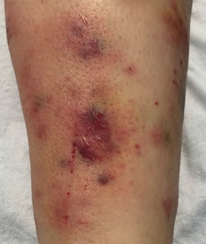
Fig. 100.10 Pancreatic panniculitis – clinical appearance. Multiple painful pink–violet to dark purple nodules and plaques on the shins. There is also evidence of hemorrhage. Courtesy Sa Rang Kim, MD, and Jean L. Bolognia, MD.
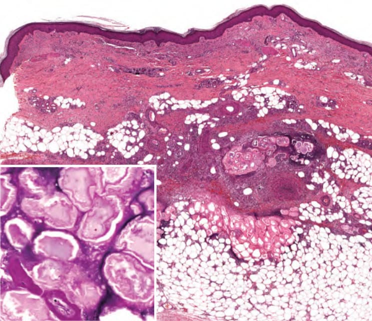
Fig. 100.11 Pancreatic panniculitis – histopathologic features. Lobular and septal panniculitis with neutrophilic inflammation, cellular necrosis, ghost cells (inset), and deposition of homogeneous basophilic material due to saponification of fat by calcium salts (seen here near ghost cells).