LUPUS ERYTHEMATOSUS PANNICULITIS (LUPUS PANNICULITIS)
Synonyms: Lupus profundus Subcutaneous lupus erythematosus
Key features
Tender subcutaneous nodules and plaques arising on the face, proximal extremities, hips, and trunk
Associated with discoid lupus erythematosus in at least one-third of patients; less often associated with systemic lupus erythematosus (10%–15% of patients)
Often precedes onset of other manifestations of lupus erythematosus
Characteristic microscopic changes include lobular panniculitis with hyaline necrosis and a predominantly lymphoplasmacytic infiltrate; nodular aggregates of lymphocytes are common
Overlying epidermal or dermal changes of lupus erythematosus are frequently present
History
Kaposi first described the clinical characteristics of lupus panniculitis in 1869. In 1940, it was recognized as a manifestation of lupus erythematosus (LE) by Irgang. Lupus panniculitis was firmly established as a specific subtype of LE in 1956 by Arnold.
Epidemiology
Lupus panniculitis constitutes a small subset of all cases of cutaneous LE, representing 2%–3% of this group. It usually occurs in adults, with a median age of onset of 30–40 years. Children can also develop lupus panniculitis, and an association with neonatal lupus has been described. There is a predominance among women, with a female : male ratio ranging from 2 : 1 to 4 : 1. Sometimes there is a family history of either LE or another autoimmune connective tissue disease.
Pathogenesis
The autoimmune basis of lupus panniculitis is thought to be similar to that of other types of LE (see Ch. 41). The cells comprising the infiltrates of lupus panniculitis are T lymphocytes and macrophages. Lupus panniculitis, often with a childhood onset and widespread distribution, has been described in patients with partial C4 deficiency. This complement deficiency causes defective opsonization of immune complexes, which may play a role in the pathogenesis of the disease. Immunohistochemical analysis has shown an interferon-driven Th1-biased immune response in active lesions of lupus panniculitis; this may result in recruitment of cytotoxic CXCR3-positive lymphocytes.
Clinical Features
Lupus panniculitis is characterized by tender subcutaneous nodules and plaques that may arise in crops (see Table 100.7). A history of trauma can sometimes be elicited. Lesions tend to develop on the face, upper arms, hips, and trunk (Fig. 100.15). Linear scalp lesions are more common among, but not restricted to, East Asian populations. The lack of involvement of the distal extremities is noteworthy. Changes in the overlying skin range from a light pink color to those of discoid LE, i.e. scaling, follicular plugging, dyspigmentation, telangiectasias, atrophy, and scarring. At times, findings of discoid LE are too subtle to be recognized clinically but can be seen on microscopic examination of biopsy specimens. The overlying skin may also be “tethered” to the subcutaneous nodule or plaque, creating a surface depression, and ulceration occasionally occurs. Lupus panniculitis has a chronic, relapsing clinical course. It usually eventuates in subcutaneous atrophy, which can be disfiguring (see Chs. 41 & 101).
Lupus panniculitis often occurs prior to other manifestations of LE and in the absence of other autoimmune connective tissue diseases. For example, lesions of discoid LE have developed up to 10 years after the appearance of the panniculitis. However, manifestations of systemic LE or discoid LE can also occur long before or simultaneously with the panniculitis. There is a closer relationship of lupus panniculitis to other forms of chronic cutaneous LE (e.g. discoid LE) than to systemic LE. Coexistent discoid lesions are observed in at least one-third of patients, whereas only 10%–15% meet the diagnostic criteria for systemic LE. Most individuals in the latter group have relatively mild systemic manifestations, usually arthralgias or Raynaud phenomenon. It is common for patients to have low-titer antinuclear antibodies; they can also have other circulating autoantibodies (e.g. anti-dsDNA, anti-RNP), leukopenia, hypocomplementemia, and an elevated ESR.
Pathology
Lupus panniculitis is a predominantly lobular process with a variable degree of inflammation (Fig. 100.16A). Nodular aggregates of lymphocytes, usually B cells, are often present (Fig. 100.16B), as are variably
large clusters of CD123+ plasmacytoid dendritic cells. The characteristic microscopic changes, e.g. hyaline necrosis of the fat lobules (Fig. 100.16C), are outlined in Table 100.7. Granulomas can occur and tend to encroach upon the septa, but they are usually not prominent. Other features include lymphocytic vasculitis and mucin or calcium deposition.
The subcutaneous findings alone are considered sufficiently characteristic to permit a diagnosis of lupus panniculitis. However, overlying epidermal or dermal changes of discoid LE occur in one-half to two-thirds of cases and are also helpful diagnostically. With direct immunofluorescence, a positive lupus band can be identified in the overlying skin in a high percentage of cases, even in those where the histopathologic changes are nonspecific.
Differential Diagnosis
Clinically, lesions of lupus panniculitis can resemble other forms of panniculitis. The rarity of involvement of the distal lower extremities helps to distinguish it from conditions such as erythema nodosum and erythema induratum. It should also be remembered that other forms of subcutaneous inflammation may occur in patients with LE, including erythema nodosum, thrombophlebitis, pancreatic panniculitis, and juxta-articular, rheumatoid nodule-like lesions.
Microscopically, lupus panniculitis can resemble the panniculitis associated with morphea and dermatomyositis (see Table 100.7), traumatic panniculitis (which often has evidence of foreign material), and, in its later stages, localized lipoatrophy due to other etiologies. The most important entity in the differential diagnosis is subcutaneous panniculitis-like T cell lymphoma, as there can be considerable histopathologic overlap between the two disorders. The distinguishing features are outlined in Table 100.9. It should be noted, however, that changes such as vacuolar alteration of the basilar layer and dermal mucin deposition have been reported in panniculitis-like T cell lymphoma. In some cases, overlapping histopathological features of lupus panniculitis and subcutaneous panniculitis-like T cell lymphoma have been reported, and these cases probably represent examples of “atypical” lupus panniculitis.
A, B A primarily lobular panniculitis with nodular aggregates of lymphocytes. C Hyaline necrosis of fat lobules is also a characteristic feature. A focus of lipomembranous changes, a nonspecific finding observed in the late stages of several types of panniculitis, is present. Courtesy Lorenzo Cerroni, MD.
Treatment
Antimalarials are frequently used to treat lupus panniculitis, and they produce improvement in most patients. Addition of a second antimalarial may prove useful in patients who do not respond to a single agent. In view of the chronic, relapsing nature of the disease, treatment may be required for several years.
Systemic corticosteroids, when used, are often restricted to initial phases of the disease. Other systemic therapies include dapsone, immunosuppressive agents (e.g. mycophenolate mofetil), thalidomide, IVIg, and rituxumab. Overlying discoid lesions, if present, can be treated with potent topical or intralesional corticosteroids.
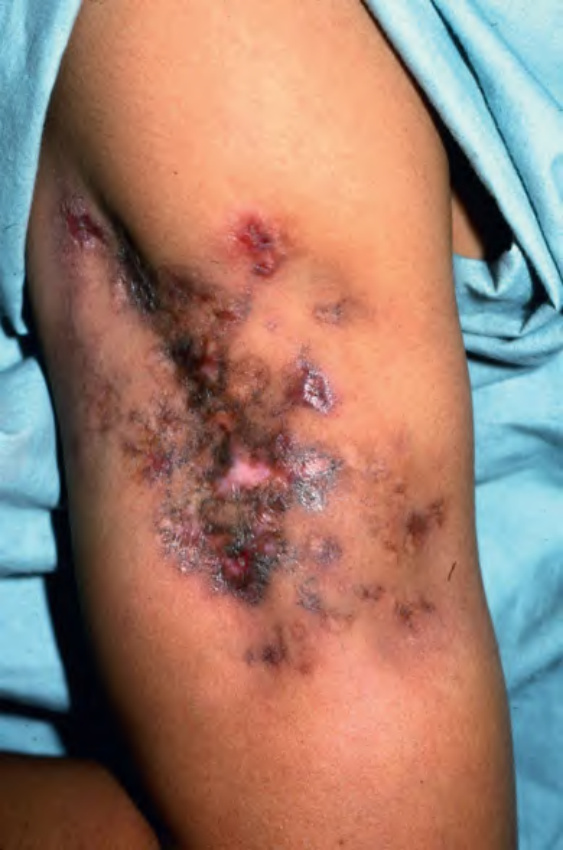
Fig. 100.15 Lupus panniculitis – clinical appearance. Red–brown plaques on the upper outer arm. Note the significant subcutaneous atrophy and overlying lesions of discoid lupus erythematosus. Courtesy Kenneth E. Greer, MD.
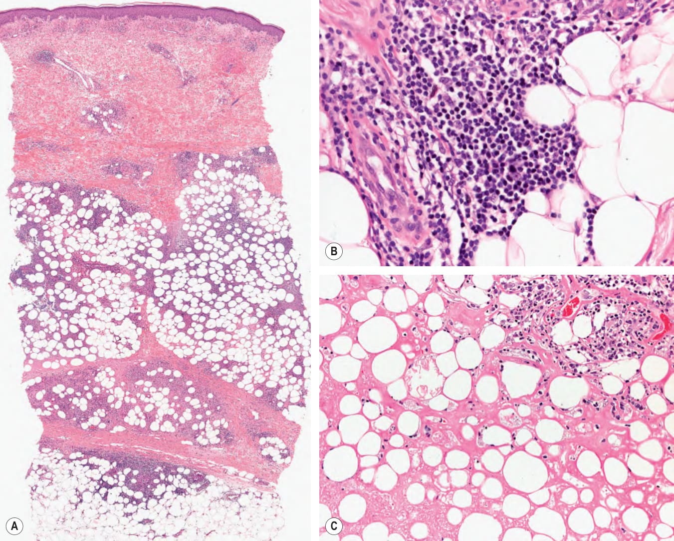
Fig. 100.16 Lupus panniculitis – histopathologic features.
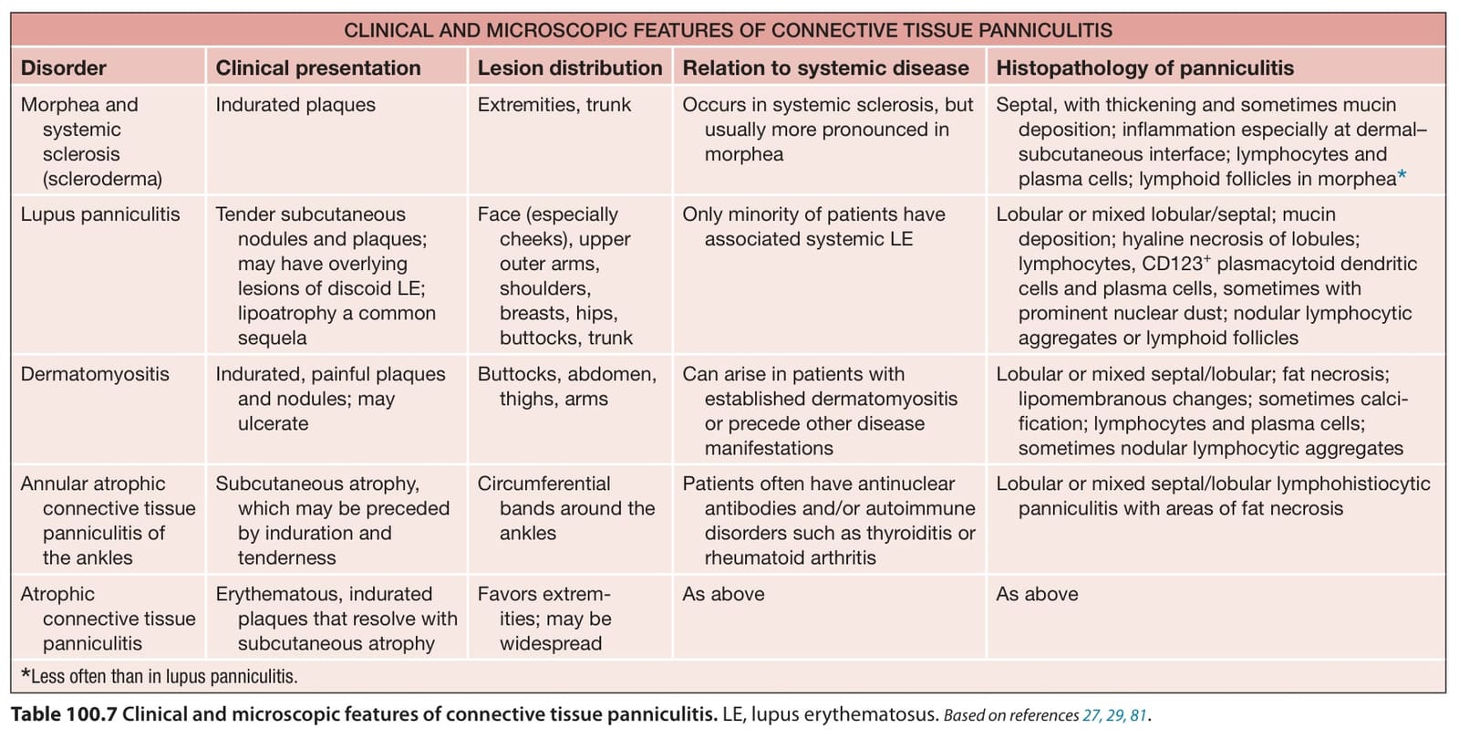
Table 100.7 Clinical and microscopic features of connective tissue panniculitis. LE, lupus erythematosus. Based on references
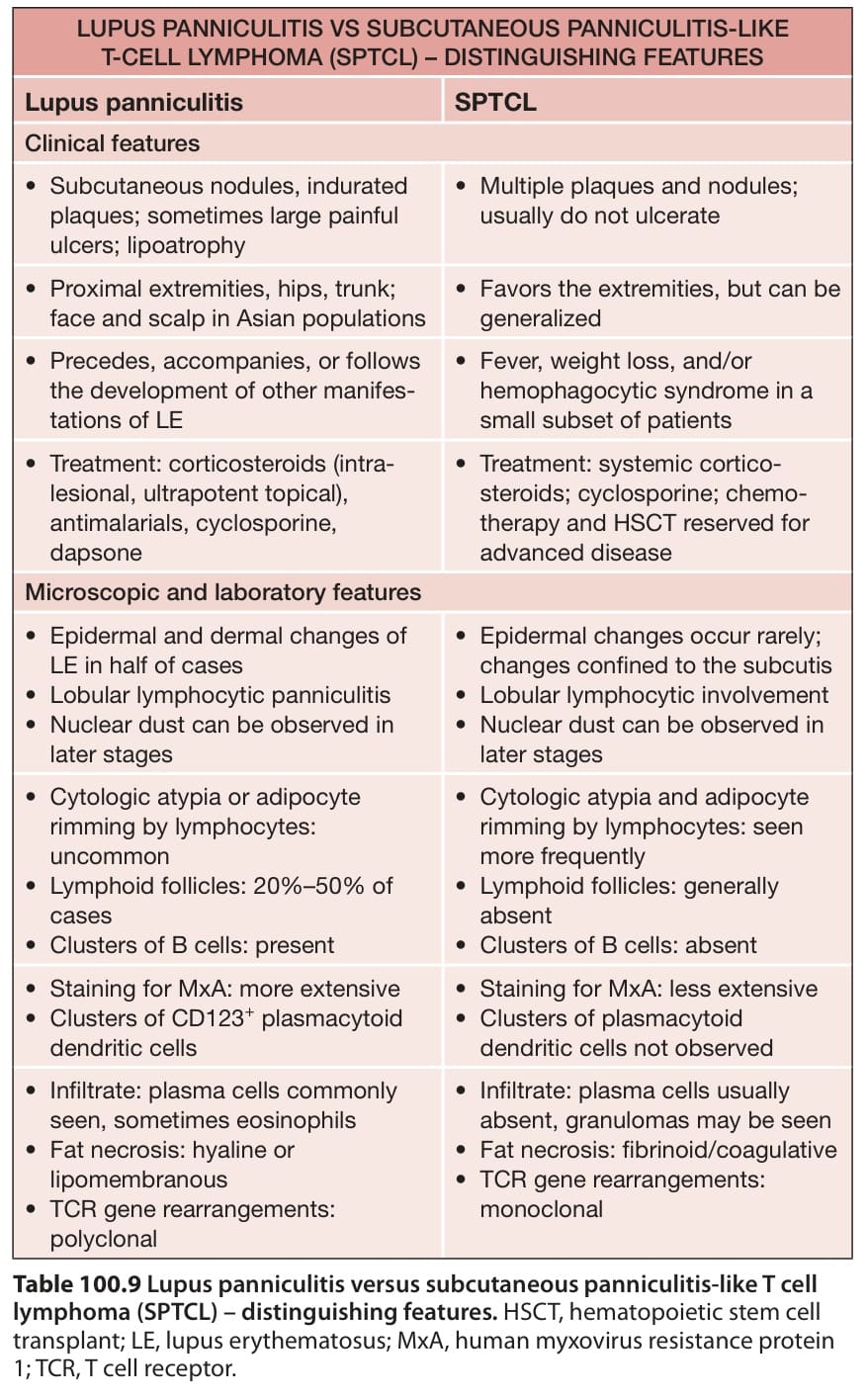
Table 100.9 Lupus panniculitis versus subcutaneous panniculitis-like T cell lymphoma (SPTCL) – distinguishing features. HSCT, hematopoietic stem cell transplant; LE, lupus erythematosus; MxA, human myxovirus resistance protein 1; TCR, T cell receptor.