PANNICULITIS OF DERMATOMYOSITIS
Key features
An uncommon manifestation of dermatomyositis
More frequently, incidental microscopic changes of panniculitis (in the absence of clinical lesions) are observed
Can precede or follow other manifestations of dermatomyositis
Microscopically, it is a lobular or mixed panniculitis with lympho- plasmacytic predominance
Usually responds to therapy
Introduction
Panniculitis is an uncommon but established clinical manifestation of dermatomyositis. However, there is some evidence that microscopic involvement of subcutaneous fat without overt clinical features of panniculitis is more frequently observed.
Clinical Features
The characteristic lesions are persistent, indurated, painful plaques and nodules that may ulcerate and lead to lipoatrophy (see Table 100.7). They can arise in the setting of established dermatomyositis or represent the initial manifestation of the disease. In affected individuals, the other clinical and laboratory features as well as the incidence of malignancy appear to be similar to those of dermatomyositis patients without panniculitis. Partial or generalized lipoatrophy without preceding clinical lesions of panniculitis (occasionally accompanied by increased abdominal fat) occurs in up to one-quarter of patients with juvenile dermatomyositis, and it is often associated with metabolic abnormalities such as hypertriglyceridemia and insulin resistance (see Ch. 101).
Pathology
The microscopic features are outlined in Table 100.7. Lymphocytic vasculitis has been described, and fat necrosis has been noted in several reports. Membranocystic changes have been observed in some cases (see section on lipodermatosclerosis), and lesions with these changes may be particularly resistant to therapy. Calcification is variable and is an expected finding in cases of dermatomyositis associated with calcinosis of deep soft tissues and skeletal muscle. Vacuolar alteration of the basal layer of the overlying epidermis has been described, and the dermis may be edematous or mucinous with perivascular lymphocytic inflammation. Direct immunofluorescence is reported to be negative for deposits along the dermal–epidermal junction, although immunoreactants have been detected in vessel walls.
Differential Diagnosis
Lupus panniculitis is more likely to show hyaline necrosis and lymphoid nodules, but clinical and laboratory findings may be needed to make a distinction. Overlying poikilodermatous changes can occur in both diseases, but lupus is more likely to show appendageal involvement and is associated with positive basement membrane zone fluorescence.
Treatment
Good responses to therapy have generally been reported, and treatments include prednisone, methotrexate, azathioprine, JAK inhibitors, cyclosporine, and IVIg. Panniculitis lesions have shown variable responses to hydroxychloroquine. Residual facial lipoatrophy has been successfully treated with hyaluronic acid and poly-L-lactic acid dermal fillers.
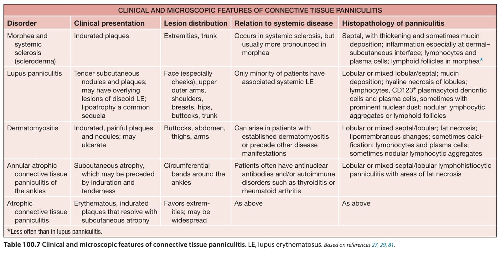
Table 100.7 Clinical and microscopic features of connective tissue panniculitis. LE, lupus erythematosus. Based on references
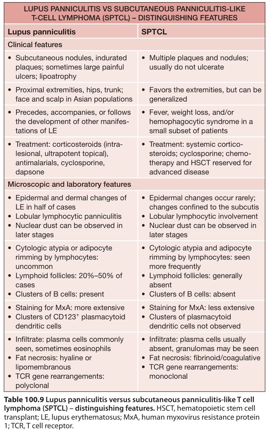
Table 100.9 Lupus panniculitis versus subcutaneous panniculitis-like T cell lymphoma (SPTCL) – distinguishing features. HSCT, hematopoietic stem cell transplant; LE, lupus erythematosus; MxA, human myxovirus resistance protein 1; TCR, T cell receptor.