TRAUMATIC PANNICULITIS
Key features
Inflammation in the subcutis resulting from external injury
The injurious event may be accidental, purposeful, or iatrogenic, and it may be a manifestation of an underlying psychiatric disturbance
A variety of histopathologic changes are observed, depending upon the inciting agent
Identification of foreign material is of greatest help in diagnosis
Introduction
Extrinsic injury of varying types can produce panniculitis. There are four broad categories: cold panniculitis (Haxthausen disease), sclerosing lipogranuloma, panniculitis due to other injectable substances or therapies (e.g. megavoltage radiation), and panniculitis due to blunt trauma.
History
Lemez described cold panniculitis in 1928, when he noted that newborns and infants up to 6 months of age were particularly susceptible to cold injury, as demonstrated by the production of a subcutaneous nodule following application of an ice cube. In 1941, Haxthausen described a condition occurring in small children a few days after exposure to cold, consisting of firm infiltrated nodules of the cheeks and chin. A similar type of cold injury due to popsicles was reported by Epstein and Oren in 1970.
Injection of foreign lipid material into the skin for cosmetic or other purposes has been performed for centuries. In 1950, Smetana and Bernhard reported 14 cases of what they termed sclerosing lipogranuloma of the male genitalia. They believed it was an endogenous process, but subsequent studies demonstrated the presence of mineral oil in similar cases.
Epidemiology
Infants and small children are most at risk for cold panniculitis. A form of the disease also occurs on the thighs of young women who are equestrians. Sclerosing lipogranuloma of the male genitalia is seen mostly in young adults. A sclerosing, pseudosclerodermatous panniculitis has been reported following megavoltage radiation for metastatic carcinoma and as a radiation recall reaction after cyclophosphamide therapy.
Pathogenesis
Cold injury to fat favors small children, due in part to the previously discussed characteristics of their fat. Cold injury is also related to fluctuations in blood flow that occur with declining temperatures (the “hunting phenomenon”), ice crystal formation, and the changes that occur with thawing. Injections of oils (and associated impurities) are known for producing subcutaneous inflammation. Substances include mineral oil (paraffin) as well as camphor, cottonseed, and sesame oils. Even medical grade silicone may contain impurities, and since encapsulation of this material is desirable when used for cosmetic purposes, fibrosis-inducing substances such as olive oil or castor oil are often added. Injected substances responsible for factitial panniculitis have included milk, feces, and a host of other substances.
Panniculitis has also been produced at the sites of injection by numerous therapeutic agents, such as pentazocine, phytonadione (vitamin K), glatiramer acetate, interferon-β, interleukin-2, heparin, and vaccines (e.g. tetanus), as well as substances that have been used for tissue augmentation such as bovine collagen or poly-L-lactic acid. More recently, panniculitis due to “mesotherapy” (injection of substances such as phosphatidylcholine and deoxycholate to treat localized fat accumulations; see Ch. 156) and electroacupuncture have been described. In addition to the foreign body response elicited by many of these agents, other immune mechanisms may also be involved. With blunt trauma, granulomas contain material that may be derived from the breakdown of erythrocyte membranes.
Clinical Features
In cold panniculitis (including popsicle panniculitis), erythematous, firm nodules develop, particularly on the cheeks and chin. In equestrian cold panniculitis, erythematous to violaceous, tender plaques appear on the thighs following exposure to cold while wearing tightfitting clothing (Fig. 100.17). While equestrian cold panniculitis is a commonly used term in the literature, it has been suggested that equestrian perniosis would be a more apt term given that the inflammatory infiltrate is primarily in the dermis.
In lipogranuloma, nodules are sometimes migratory and can be accompanied by varying degrees of swelling, erythema, abscess formation, lymphangitis, and fibrosis. The term sclerosing lipogranuloma often refers to lesions arising on the male genitalia due to self-injection of oily materials (see Ch. 94). There has also been a report of a sclerosing lipogranuloma that apparently resulted from topical application of vitamin E cream. In several Japanese patients, Y-shaped induration of the scrotum (in which exogenous lipids could not be detected) was described as eosinophilic sclerosing lipogranuloma. Patients with sclerosing lipogranuloma frequently deny self-injection, making diagnosis difficult. Another variant of lipogranuloma is the grease gun granuloma, which results from accidental firing of the grease gun used by mechanics. This results in formation of a verrucous nodule, often on the dorsum of the hand.
Inflamed nodules with varying degrees of pain and fibrosis have been observed in other forms of panniculitis due to injection, with the distribution of lesions sometimes providing a clue to their cause. A dramatic example of this is Texier disease, a panniculitis due to phytonadione (vitamin K) injections. In this disorder, sclerotic lesions with lilac-colored borders form on the buttocks and thighs, in a configuration resembling a “cowboy gunbelt and holster”. Lesions due to blunt trauma often have an ecchymotic character and involve locations such as the shin, arm, or hand. Hypertrichosis may also be present, possibly the consequence of local hyperemia or angiogenesis.
Pathology
Table 100.10 outlines the microscopic changes in various forms of traumatic panniculitis. In addition, sclerosing lipogranuloma is discussed in Chapter 94.
Differential Diagnosis
In cold panniculitis, the absence of needle-shaped clefts in lipocytes and location of the most intense inflammation near the dermal–subcutaneous interface help to distinguish this condition from subcutaneous fat necrosis of the newborn. In sclerosing lipogranuloma and related lipogranulomas, distinctive large vacuoles are found in the dermis and subcutis (Fig. 100.18). Mineral oil in non-processed tissue can be identified by infrared spectroscopy or mass spectrometry.
Panniculitis due to injectable substances can be diagnosed when foreign material (often identified by polarization microscopy) is present. Cosmetic fillers have distinctive histologic features (see Fig. 94.11), and silicone can be distinguished from other cosmetic fillers by cutaneous ultrasonography. Cases with acute inflammation and necrosis may resemble infection-induced panniculitis, and, in fact, infection may accompany injection panniculitis; special stains and cultures for organisms (including atypical mycobacteria) are useful in this regard. Sclerosing traumatic panniculitis (e.g. due to phytonadione or pentazocine injections) may resemble morphea clinically, but would not present as a septal panniculitis histologically.
Treatment
Treatment of these disorders should focus on removal of the inciting stimulus and eradication of any associated infection. Intralesional or systemic corticosteroids can be helpful in controlling the inflammation, and they have been used in the management of sclerosing lipogranuloma and granulomatous panniculitis due to other injected substances. Surgical excision may also be an option for sclerosing lipogranuloma.
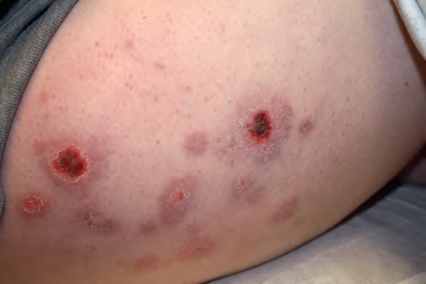
Fig. 100.17 Cold panniculitis. Violaceous nodules and plaques, some with central crusting, on the thigh of a young woman. Collarettes of scale developed as the lesions healed. While equestrian cold panniculitis is a commonly used term in the literature, it has been suggested that equestrian perniosis would be a more apt term given that the inflammatory infiltrate is primarily in the dermis. Courtesy Kendra Lesiak, MD.
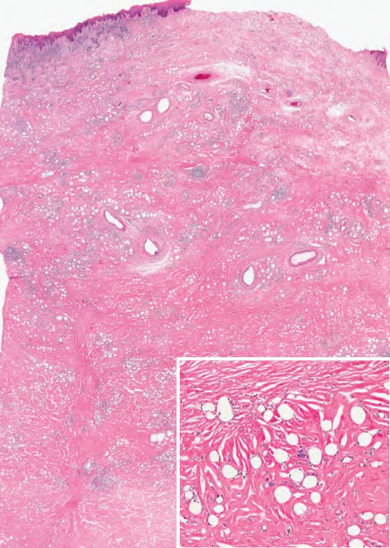
Fig. 100.18 Mineral oil granuloma
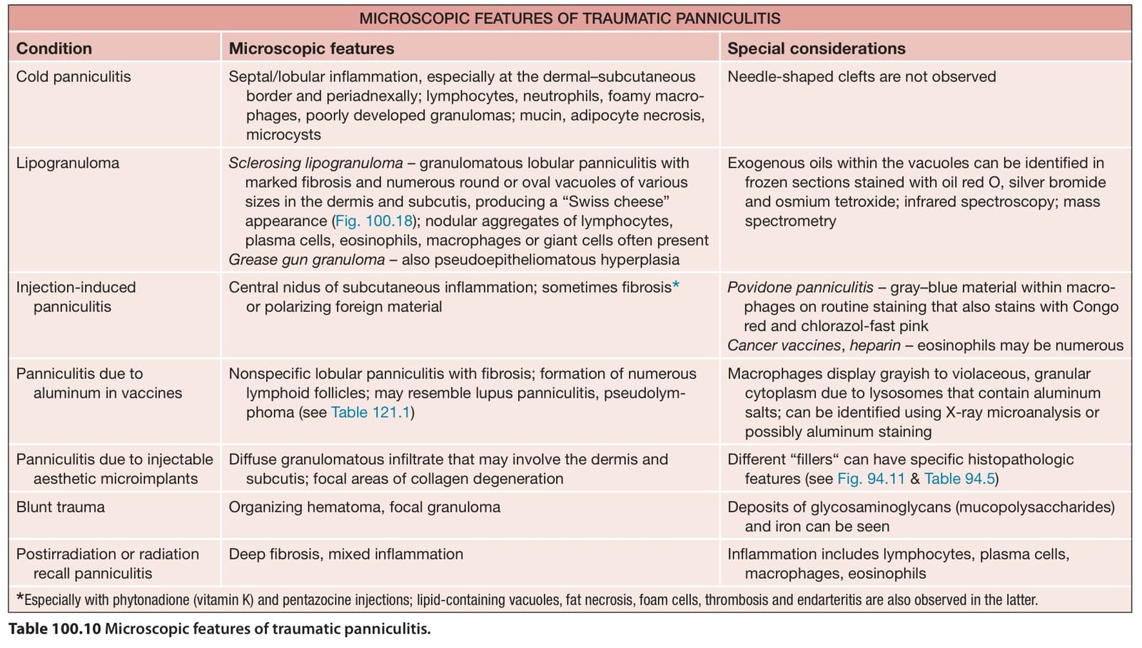
Table 100.10 Microscopic features of traumatic panniculitis.