LIPODERMATOSCLEROSIS
Synonyms: Sclerosing panniculitis Hypodermitis sclerodermiformis Chronic panniculitis with lipomembranous changes
Key features
Favors the medial aspect of the lower extremities above the malleolus
Acute phase with erythema, warmth, and tenderness, which may be misdiagnosed as infectious cellulitis
Chronic phase with induration and red–brown to violet–brown discoloration
Usually develops in the setting of chronic venous insufficiency
Both septal and lobular panniculitis; lipomembranous changes are common, particularly in chronic lesions
Introduction
Erythema, induration, and pigmentary changes have long been known to be associated with venous insufficiency. Because of the variety of clinical appearances and histopathologic findings that can occur at different stages of the disease, a number of diagnostic terms have been
(paraffinoma). There is sclerosis throughout the dermis and subcutis as well as numerous vacuolated spaces (inset). Scattered histiocytes and lymphocytes are also seen. Courtesy Lorenzo Cerroni, MD.
employed to explain the changes. Recently, the various manifestations of this panniculitis have been consolidated under the heading of lipodermatosclerosis or sclerosing panniculitis.
History
In 1955, Huriez drew attention to a related indurated lesion, which he termed hypodermitis sclerodermiformis.
Epidemiology
Most patients are women over the age of 40 years, but appearance after the age of 75 years has also been reported.
Pathogenesis
There is considerable evidence for venous insufficiency in patients with lipodermatosclerosis, and fibrinolytic abnormalities are also present in these individuals. Venous hypertension leads to a compromised ability to reduce foot vein pressure during exercise. This results in increased capillary permeability, which causes leakage of fibrinogen, its polymerization to form fibrin cuffs around vessels, impedance of oxygen exchange, and tissue anoxia (see Ch. 105). Pericapillary fibrin deposits can be seen in uninvolved, clinically normal extremities of patients with healed venous ulcers of the opposite extremity, suggesting that this abnormality precedes the clinical changes of lipodermatosclerosis.
There may also be an abnormal regulation of angiogenesis within lesions of lipodermatosclerosis. In particular, increased expression of vascular endothelial growth factor receptor 1 (VEGFR-1), which can act as a negative regulator of VEGF-mediated angiogenesis, has been observed, along with increased expression of angiopoietin-2 (Ang-2). Additional factors contributing to the pathogenesis of lipodermatosclerosis may include protein C and S deficiencies, local stimulation of collagen synthesis, including an increased number of cells expressing procollagen type 1 mRNA, and obesity.
Clinical Features
The acute phase of lipodermatosclerosis presents with pain, warmth, erythema, and some induration (Fig. 100.19), most often initially on the medial lower leg above the malleolus. Other dependent sites such as the lower aspect of the abdominal pannus can also develop lipodermatosclerosis. At this point, the changes are relatively diffuse. In the chronic phase, there is marked sclerosis of the dermis and subcutis, resulting in induration that is more sharply demarcated from adjacent normal skin. Brown discoloration due to hemosiderin deposition may also be present. These features give the affected leg the appearance of an inverted wine bottle.
Pathology
Early lesions show mid-lobular ischemic necrosis, a lymphocytic infiltrate in the septa that rims the fat lobules, variable degrees of capillary congestion and thrombosis, and hemorrhage with
Lipomembranous change, consisting of cystic formation with elaborate papillary configurations.
hemosiderin deposition. With progression, septal thickening, hyaline sclerosis involving lipocytes, lipophage formation, and mixed inflammatory cell infiltrates appear. Advanced lesions show marked septal sclerosis and membranocystic change in the face of a marked reduction in inflammation.
Membranocystic change is a key feature in lipodermatosclerosis. This consists of thickened, undulating membranes that form cysts and papillary configurations (Fig. 100.20). The membranes are believed to result from degenerated cell membranes of lipocytes and/or macrophages. The material comprising the membranes is ceroid, an oxidation product of unsaturated fatty acids.
Short, frayed elastic fibers can be present within the subcutaneous septa, and these may be calcified, features that resemble pseudoxanthoma elasticum. Myospherulosis, a foreign-body type of granulomatous reaction to lipid-containing material and blood, has been described in lesions of lipodermatosclerosis. By phosphotungstic acid–hematoxylin stain or by immunofluorescent methods, pericapillary fibrin can also be demonstrated in lesions of lipodermatosclerosis. Dermal changes include fibrosis, tortuous thick-walled veins, and superficial and deep perivascular inflammation. Biopsy should
be avoided if the diagnosis is obvious, since poor wound healing and ulceration frequently result. When necessary, a thin elliptical excision should be obtained from the margin of a lesion.
Differential Diagnosis
Difficulties in clinical diagnosis arise most often in early lesions, when the process is more diffuse and erythematous. At this stage, consideration is often given to infectious cellulitis, erythema nodosum, or erythema induratum. Persistence of a lesion, association with stasis changes, and lack of response to antimicrobials suggest the correct diagnosis, perhaps aided by studies of venous function.
As induration develops and progresses, differentiation from morphea and scleromyxedema may be necessary. In morphea, subcutaneous involvement is predominantly septal, and lipophagic and lipodystrophic changes are not as prominent as they are in lipodermatosclerosis. Membranocystic changes, when present, can be of great diagnostic help; however, these findings can occur in a variety of other conditions, including lupus and dermatomyositis panniculitis, liposarcoma, erythema nodosum, and diabetic dermopathy.
Treatment
Leg elevation and consistent compression therapy are the mainstays of treatment for lipodermatosclerosis. Traditional anti-inflammatory therapies are usually ineffective in this condition, although intra lesional corticosteroids (e.g. triamcinolone 5–10 mg/cc) may be of benefit when used in conjunction with compression therapy. Good results were reported with the anabolic steroid stanozolol especially in the earlier phases of the disease, but this medication is no longer commercially available. As a result, danazol has been used (successfully) as a substitute. Anabolic steroids enhance fibrinolysis, and they can reduce pain, extent of involvement, and induration of the skin. However, side effects of sodium retention, lipid profile abnormalities, hepatotoxicity, and virilization in women do limit their use. Oxandrolone, an anabolic steroid with less hepatotoxicity and fewer androgenic effects, represents another therapeutic option. Other reported treatments include ultrasound, pentoxifylline, fasciotomy, and phlebectomy.
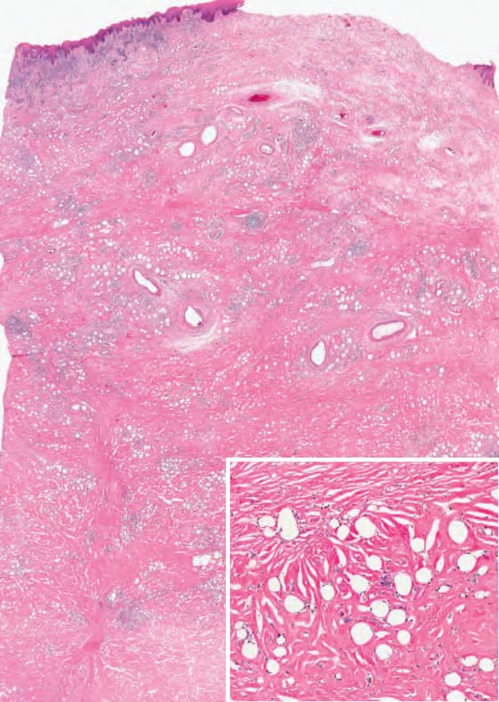
Fig. 100.18 Mineral oil granuloma
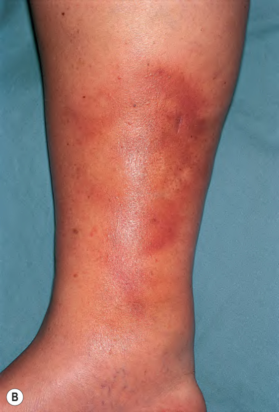
Fig. 100.19 Lipodermatosclerosis – clinical appearance.A Acute phase with tender erythematous plaques on both lower extremities. B Chronic phase with sclerotic red–brown plaque on the lower medial leg. B, Courtesy Kenneth E. Greer, MD.
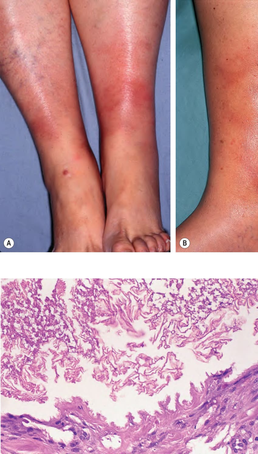
Fig. 100.20 Lipodermatosclerosis – histopathologic features.
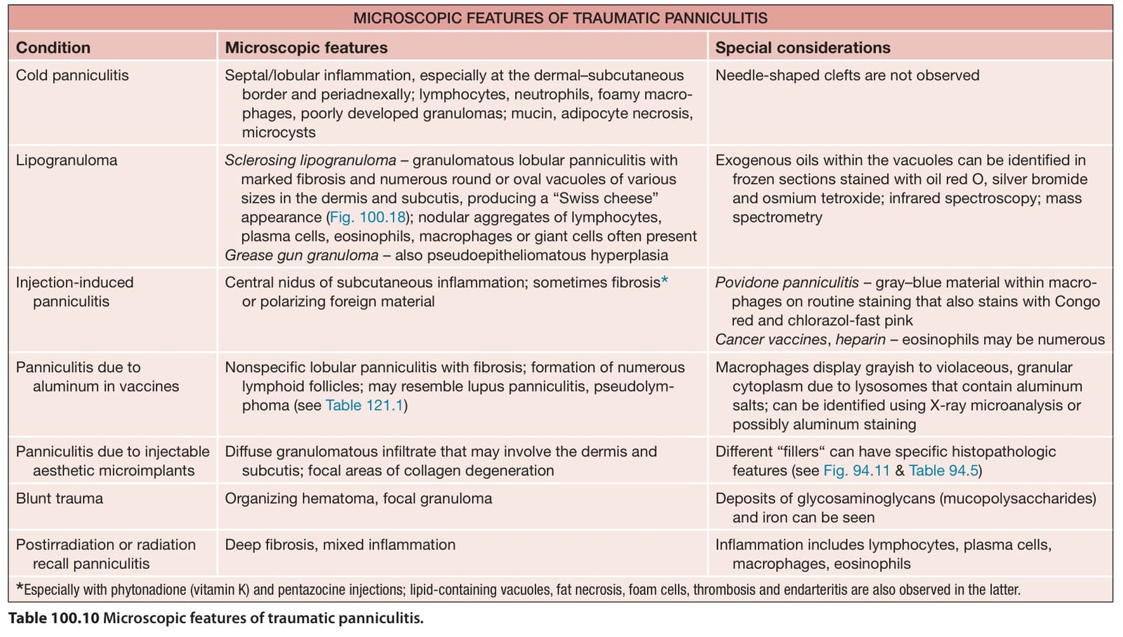
Table 100.10 Microscopic features of traumatic panniculitis.