INFECTION-INDUCED PANNICULITIS
Synonym: Infective panniculitis
Key features
A wide variety of infectious agents has been reported to produce panniculitis
Some degree of immunosuppression is common, but not invariable
Histopathologic findings vary, but often include mixed septal/ lobular panniculitis, neutrophilic infiltration, hemorrhage, and necrosis
Special staining and culture of drainage and tissue can provide a definitive diagnosis
Introduction
Panniculitis can result from a distant focus of infection (a classic example being erythema nodosum) or panniculitis can be directly induced by an infectious agent. The latter can produce a variety of clinical and microscopic appearances, although there are some features that infectioninduced lesions have in common, regardless of the etiologic agent.
History
While multiple reports of panniculitis directly caused by infectious agents have been published, most early studies of deep skin infections did not focus upon the changes in the subcutaneous fat. In 1989, Patterson et al. studied 15 cases of infection-induced panniculitis, with an emphasis upon the histopathologic features in the subcutis. Since that time, additional reports of panniculitis due to infection have continued to appear.
Epidemiology
There appears to be no age, sex, or racial predilection among cases of infection-induced panniculitis. Many of these patients are immunosuppressed or have predisposing medical conditions such as diabetes mellitus.
Pathogenesis
In this group of disorders, infectious agents are considered to be directly responsible for the panniculitis. Examples of reported microorganisms to date are listed in Table 100.11.
Involvement of the subcutis can result from direct inoculation or septicemia. Other potential modes of spread include transfascial from an enteric source, in the case of abdominal panniculitis or via “persorption”, a proposed mechanism by which Candida migrates across intact endothelium from the gut to a subcutaneous site. Immunosuppression is common, but not invariable, among individuals with infection-induced panniculitis.
Clinical Features
Patients develop local swelling and erythema. There may be one or more fluctuant nodules that ulcerate and drain. Lesions on the legs and feet are common, but other sites of involvement include the gluteal region, abdomen, axillae, arm, or hand. In addition to sepsis, underlying conditions include diabetes mellitus, leukemias or solid tumors, autoimmune connective tissue disease, AIDS, and organ transplantation.
Pathology
Individual cases can mimic other primary forms of panniculitis. Common changes (regardless of the infectious agent) include a mixed septal/lobular panniculitis, neutrophilic infiltration, vascular proliferation, hemorrhage, and necrosis that involves lipocytes, inflammatory cells, and eccrine sweat coils (Fig. 100.21). In Q fever due to Coxiella burnetii, there may be a “doughnut-like” granulomatous lobular panniculitis, in which fibrin and inflammatory cells form a ring around a central clear space; similar changes have been found in the liver and bone marrow of patients with Q fever. In lobular panniculitis due to Stenotrophomonas maltophilia (previously Pseudomonas maltophilia), phagocytosis of bacteria leads to foamy histiocytes whose cytoplasm is filled with bluish, granular material.
Differential Diagnosis
Fluctuant, ulcerating nodules also occur in pancreatic panniculitis, traumatic panniculitis, and alpha-1 antitrypsin deficiency panniculitis. Clinical and laboratory data can usually permit distinction. As noted previously, traumatic panniculitis may be accompanied by infection. Examples of infection-induced panniculitis with predominantly septal involvement or vasculitis could be confused with acute erythema nodosum or erythema induratum, respectively. Panniculitis due to mucormycosis may feature “ghost cells” and granular calcium deposits and thus resemble pancreatic panniculitis; it can also mimic gouty panniculitis due to the presence of intracellular crystalline deposits. Special stains for organisms and cultures of drainage and tissue are keys to the diagnosis. In one study, special stains were positive for organisms in 14 of 15 cases.
Subcutaneous nodules with purpura. Courtesy Kenneth E. Greer, MD.
Treatment
Treatment consists of appropriate antimicrobial therapy. Surgery may be indicated for isolated lesions caused by grain-forming fungi or bacteria, such as mycetoma or botryomycosis. A more radical surgical approach has been used successfully in treating abdominal panniculitis due to enteric bacteria.
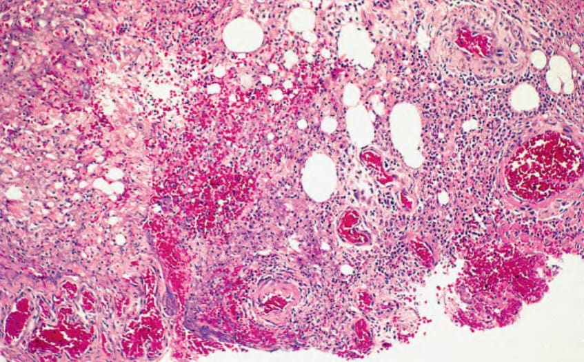
Fig. 100.21 Infection-induced panniculitis. Bacterial panniculitis, showing heavy neutrophilic infiltration, basophilic necrosis, vascular proliferation, and hemorrhage.
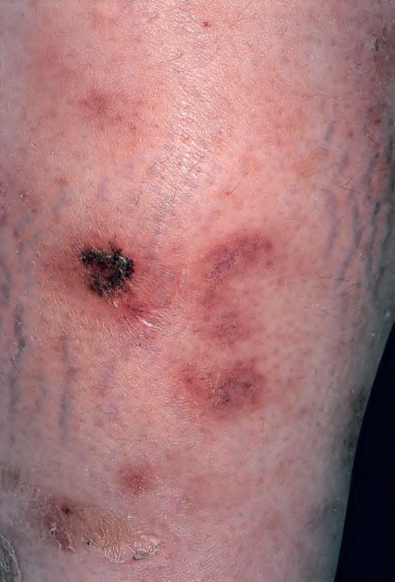
Fig. 100.22 Cytophagic histiocytic panniculitis – clinical appearance.
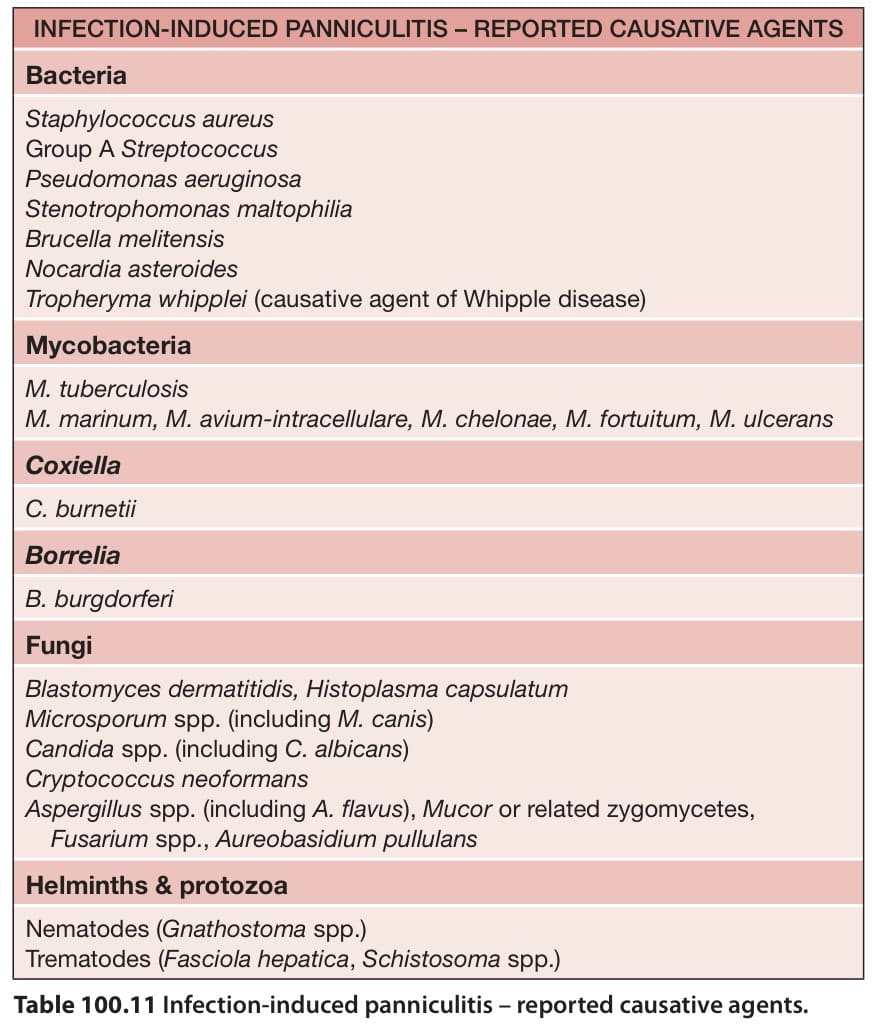
Table 100.11 Infection-induced panniculitis – reported causative agents.