CYTOPHAGIC HISTIOCYTIC PANNICULITIS
The term “cytophagic histiocytic panniculitis” (CHP) has been used to describe subcutaneous nodules or plaques that on histologic examination show infiltrates of macrophages containing erythrocytes, lymphocytes, and/or karyorrhectic debris, i.e. exhibiting hemophagocytosis (Figs. 100.22 & 100.23). These macrophages with cytophagic activity have been referred to as “bean bag” cells. With the advent of immunophenotyping and genetic techniques, it has become apparent that the vast majority of patients have lymphoma, with atypical lymphoid cells also present within the panniculus. In general, the lymphomas represent subcutaneous involvement of primary cutaneous γ/δ T cell lymphoma or EBV-associated extranodal NK/T cell lymphoma, nasal type (see Ch. 120). Occasionally, patients have subcutaneous panniculitislike T cell lymphoma, and, rarely, no lymphoma. Individuals in the latter group may still have a fatal course due to a hemophagocytic syndrome that involves the liver, spleen, and bone marrow (leading to pancytopenia). Mutations in a number of genes can predispose individuals to the development of hemophagocytic syndrome, including that which encodes perforin (see Table 91.1). Of note, a child with microscopic findings of CHP but no evidence of lymphoma was found to have a heterozygous nonsense mutation in the gene that encodes perforin.
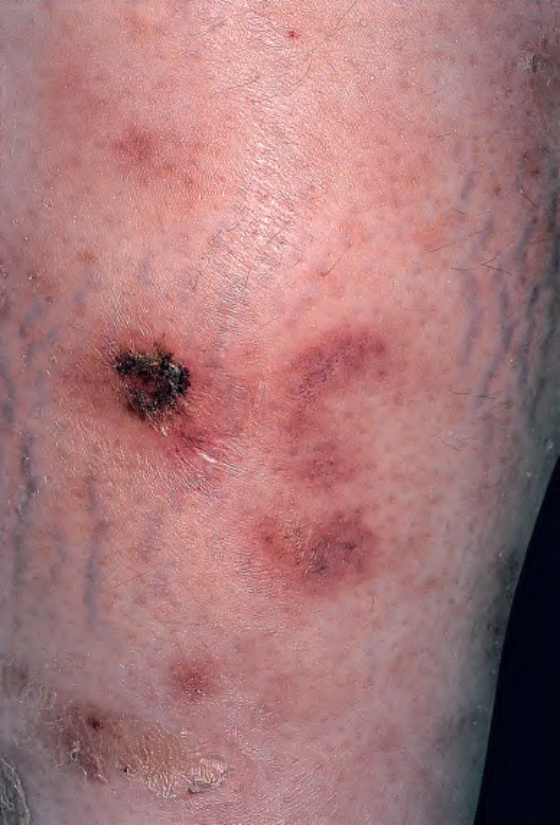
Fig. 100.22 Cytophagic histiocytic panniculitis – clinical appearance.