MALIGNANT SUBCUTANEOUS INFILTRATES
Malignant infiltrates may involve the subcutis, mimicking the clinical and microscopic appearance of panniculitis. The best known of these is subcutaneous panniculitis-like T cell lymphoma (see Ch. 120). While cytophagia can be seen in the latter disorder, it is particularly characteristic of subcutaneous involvement of primary cutaneous γ/δ T cell lymphoma and EBV-associated extranodal NK/T cell lymphoma, nasal type (see above). Other lymphomas (e.g. B cell), leukemias, and
Macrophages engaged in cytophagic activity. Some of them have the appearance of “bean bag” cells.
metastatic solid tumors may infiltrate the subcutis. Obviously, clinical information is crucial in such cases.
Histopathologic clues include the recognition of significant pleomorphism or monotony among infiltrating cells in the subcutis, infiltration between collagen bundles of the dermis or around adnexal structures, and supporting immunohistochemical studies. Of note, the diagnosis of subcutaneous panniculitis-like T cell lymphoma can sometimes be elusive as the initial impression may be lupus panniculitis (see Table 100.9) or even a nonspecific panniculitis with lipomembranous changes.
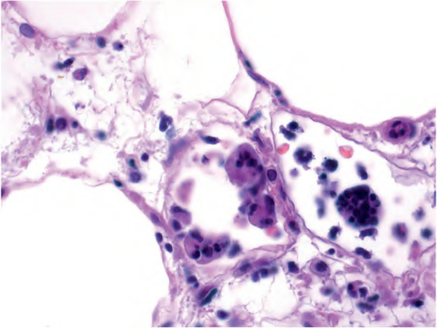
Fig. 100.23 Cytophagic histiocytic panniculitis – histopathologic features.
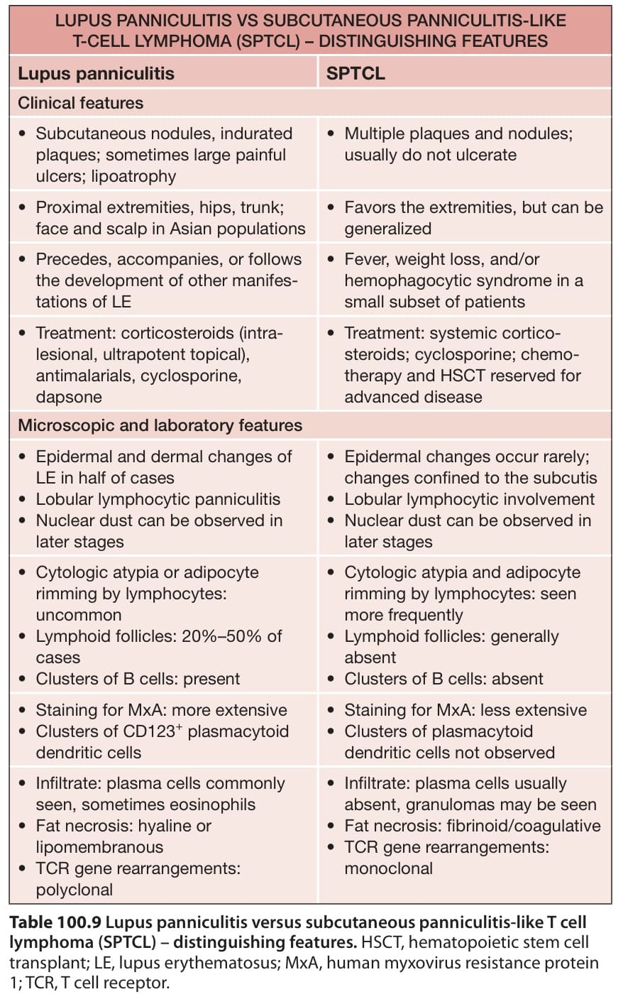
Table 100.9 Lupus panniculitis versus subcutaneous panniculitis-like T cell lymphoma (SPTCL) – distinguishing features. HSCT, hematopoietic stem cell transplant; LE, lupus erythematosus; MxA, human myxovirus resistance protein 1; TCR, T cell receptor.