INTRODUCTION
Lipodystrophy describes a heterogeneous group of diseases characterized by a selective fat loss in a characteristic body distribution pattern, often accompanied by secondary fat accumulation. Adipose tissue has crucial metabolic and endocrine functions, in addition to its role in mechanical protection. The loss of subcutaneous fat and a compensatory accumulation of visceral fat is closely associated with insulin resistance, diabetes mellitus, dyslipidemia, hypertension, and coronary artery disease. These metabolic abnormalities and their sequelae constitute the metabolic syndrome and underscore the important functions of fat, in its role as a diverse and crucial body organ.
More than a century after the first description of lipodystrophy and then delineation of generalized lipodystrophy into congenital and acquired forms, the genetic bases of several of the inherited lipodystrophies have now been elucidated. Mutations have been found in genes ranging from those encoding proteins essential for adipocyte differentiation to those encoding nuclear lamins (see below).
Clinically, lipodystrophy can be broadly classified into inherited or acquired forms and then further subclassified based on the extent of fat loss, in conjunction with genetic mutations, age of onset, and systemic manifestations (Fig. 101.1 & Table 101.1). Based upon the distribution pattern, lipodystrophy may be subdivided into three major groups: (1) generalized; (2) partial (extensive, but not generalized); and (3) localized (involving an isolated area).
Localized lipodystrophy, which may be due to injections of medications or vaccines, pressure, previous surgery, trauma or panniculitis, is the most commonly encountered form. It typically presents as lipoatrophy but occasionally lipohypertrophy develops, e.g. at sites of insulin injection (see Table 21.22). Less often, lipodystrophy associated with antiretroviral therapy (ART) for human immunodeficiency virus (HIV) infection and with administration of immune checkpoint inhibitors are seen. Dermatologists may also play a key role in diagnosing acquired generalized lipodystrophy and its preceding panniculitis. Finally, there are several rare genetic syndromes that will be discussed (see Table 101.1).

Fig. 101.1 Lipodystrophy syndromes and localized forms. Schematic representation of the predominant sites of lipoatrophy and lipohypertrophy. Additional syndromes, including familial partial lipodystrophy with mandibuloacral dysplasia, are outlined in Table 101.1. Tables 63.9 and 63.10 review progeroid syndromes in which lipoatrophy can be seen. AI-CTD, autoimmune connective tissue disease; ART, antiretroviral therapy; IR, insulin resistance; PD, programmed cell death. AGPAT2, encodes 1-acylglycerol-3-phosphate O-acyltransferase 2 (triglyceride and phospholipid synthesis); BSCL2, encodes seipin (lipid droplet formation); CAV1, encodes caveolin 1 (binds fatty acids and translocates them to lipid droplets); CAVIN1/PTRF, encodes caveolae associated protein 1 (biogenesis of caveolae and expression of caveolins 1 and 3); CIDEC, encodes cell death-inducing DNA fragmentation factor-like effector C (adipocyte apoptosis); LIPE, encodes lipase E, hormone sensitive type (hydrolysis of triglycerides to free fatty acids); LMNA, encodes lamins A/C (structural integrity of the nuclear lamina); LMNB2, encodes lamin B2 (structural integrity of the nuclear lamina); PLIN1, encodes perilipin 1 (formation, maturation, and function of lipid droplets within adipocytes); POMP, encodes proteasome maturation protein; PPARG, encodes peroxisome proliferator-activated receptor-gamma (essential role in lipogenesis); PSMA3, encodes proteasome subunit alpha 3; PSMB4, -B8, -B9, -B10, encode proteasome subunit beta 4, 8, 9, 10; PSMG2, encodes proteasome subunit gamma 2.
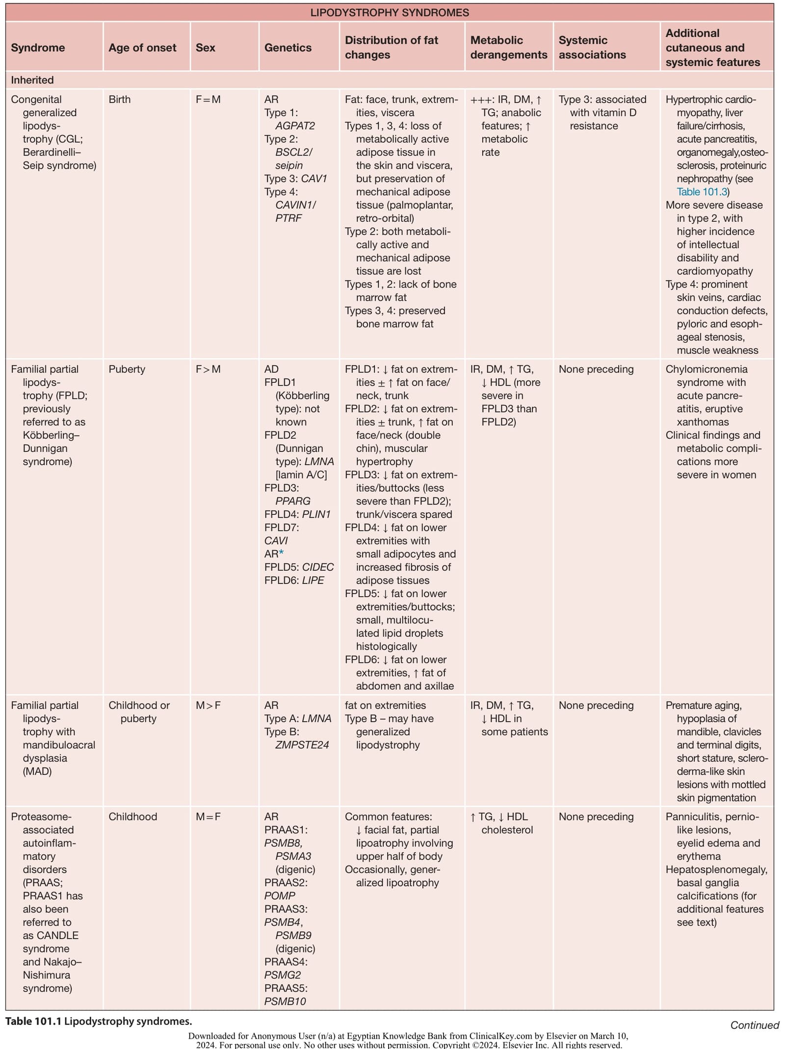
Table 101.1 Lipodystrophy syndromes. Continued