PATHOGENESIS
Adipose tissue functions as an endocrine organ via its secretion of hormones and adipocytokines, such as leptin, TNF, interleukin (IL)-6, and adiponectin. Altered expression and activity of these factors play a role in the development of insulin resistance and other metabolic sequelae seen in lipodystrophic syndromes, which are similar to those observed in the metabolic syndrome and obese individuals.
There appears to be a final common pathway of defective adipocyte triglyceride storage in conditions characterized by fat loss as well as fat excess. This defect can result from impaired triglyceride synthesis due to an enzyme deficiency (e.g. 1-acylglycerol-3-phosphate O-acyltransferase 2), defective adipocyte development caused by mutations in genes critical to adipocyte differentiation (e.g. BSCL2/seipin, PPARG, LMNA), adipocyte apoptosis, or autoimmune- or drug-mediated adipocyte destruction. These various processes impair normal adipocyte differentiation, development, lifespan, and/or function.
The destruction or impaired differentiation of adipocytes leads to a cascade of hormonal and metabolic consequences. Adiponectin, the protein product of ADIPOQ, is expressed in and secreted exclusively by differentiated adipocytes. It plays a positive role in regulating insulin sensitivity and glucose and lipid homeostasis. Plasma adiponectin levels are inversely correlated with fasting insulin levels and insulin resistance. Serum adiponectin and leptin levels are reduced both in murine models of lipoatrophy with insulin resistance and in humans with congenital and acquired lipodystrophies, including HIV/ ART-related lipodystrophy. Leptin replacement has been shown to reverse insulin resistance in mouse models with a deficiency of mRNAs that encode leptin. This replacement also results in significant and sustained improvements in hyperglycemia, dyslipidemia, and hepatic steatosis in patients with different forms of lipodystrophy.
Congenital Generalized Lipodystrophy (Berardinelli–Seip Congenital Lipodystrophy, Berardinelli–Seip Syndrome)
Congenital generalized lipodystrophy (CGL) is an autosomal recessive disorder with four known genetic subtypes. The two major forms, types 1 and 2, are due to mutations in AGPAT2, which encodes 1-acylglycerol- 3-phosphate O-acyltransferase 2, and BSCL2/seipin, respectively (see Table 101.1). It has been postulated that these genetic mutations cause lipodystrophy primarily by affecting adipocyte differentiation or lipid droplet formation in adipose tissue. In type 1, aberrant AGPAT2
enzyme activity causes a marked reduction in triglyceride and phospholipid synthesis, resulting in abnormal adipocyte function. In type 2, BSCL2 mutations affect the endoplasmic reticulum membrane protein seipin which is critical for lipid droplet morphology.
In types 3 and 4 CGL, genetic mutations alter the function of caveolae, which are invaginations of the plasma membrane involved in signal transduction and endocytosis, including internalization of the insulin receptor. Patients with type 3 CGL have mutations in CAV1 which encodes caveolin 1; caveolins are essential components of caveolae and caveolin 1 binds fatty acids and translocates them to lipid droplets. Type 4 CGL is due to mutations in CAVIN1/PTRF, whose protein product is involved in the biogenesis of caveolae and the expression of caveolins 1 and 3.
Differences in the molecular basis of CGL may account for the phenotypic heterogeneity. AGPAT2 has been found to be highly expressed in human omental adipose tissue, which may explain the preferential loss
of metabolically active intra-abdominal adipose tissue but preservation of mechanical palmoplantar adipose tissue in patients with type 1 CGL. BSCL2 has been found to also be highly expressed in the brain, which may account for the higher prevalence of intellectual disability in type 2 patients.
Familial Partial Lipodystrophy
Familial partial lipodystrophy (FPLD) is a heterogeneous group of autosomal dominantly, and rarely autosomal recessively, inherited disorders (see Table 101.1). The most prevalent subtype is FPLD2, which is due to mutations in LMNA. LMNA encodes lamins A/C, with lamins belonging to the intermediate filament family of structural proteins that compose the nuclear lamina. LMNA mutations lead to a disruption in nuclear function, resulting in apoptosis and premature cell death of adipocytes, and they lead to alterations in the extracellular matrix of adipose tissue. There is also aberrant TGF-β signaling and this is associated with abnormal responses to metabolic requirements. Decreased plasma leptin and increased fasting plasma insulin and C-peptide levels are additional findings, as is premature senescence of vascular cells; the latter contributes to the observed cardiovascular complications. Of note, LMNA mutations are responsible for a group of disorders known as laminopathies that includes premature aging syndromes (e.g. progeria), muscular dystrophies, cardiomyopathies, and one form of Charcot–Marie–Tooth neuropathy (see Tables 63.9 & 63.10). The clinical phenotype/syndrome is determined by the site and type of LMNA mutation(s).
While the genetic basis of FPLD1 is unknown, FPLD3 results from heterozygous missense mutations of the gene that encodes peroxisome proliferator-activated receptor-γ (PPAR-γ). PPAR-γ plays an essential role in adipogenesis (see Fig. 101.14), but the entire pathogenesis remains unclear. Although this subtype has a milder phenotype than FPLD2, with a later age of onset and involvement confined to the distal extremities, metabolic abnormalities are more severe, suggesting that PPARG mutations may have additional effects on metabolism.
In patients with FPLD4, heterozygous loss-of-function mutations have been described in the gene that encodes perilipin-1 (PLIN1). Perilipin is responsible for the formation, maturation, and function of lipid droplets within adipocytes. Lipoatrophy of the lower extremities is accompanied by marked hypertriglyceridemia and severe insulin resistance with type 2 diabetes.
To date, FPLD5 and FPLD6 are based on a single patient and three sets of siblings, respectively, and because of homozygous mutations (missense, nonsense) or deletions (FPLD5, FPLD6) and consanguinity (FPLD6), an autosomal recessive pattern is favored. The associated genes encode a member of the cell death-inducing DNA fragmentation factor-like effector family (CIDEC) that is thought to play a role in adipocyte apoptosis and lipase E, hormone sensitive type (LIPE). While homozygous mutations in CAV1 lead to congenital generalized lipodystrophy (see above), patients with FPLD7 are heterozygotic for CAV1 mutations.
Partial lipodystrophy may also be seen in association with mandibulo acral dysplasia (MAD), an autosomal recessive syndrome associated with mutations in LMNA (type A), or with mutations in ZMPSTE24, which encodes a zinc metalloproteinase involved in post-translational proteolytic processing of prelamin A (type B). The latter has been associated with severe mandibuloacral dysplasia, premature aging, and generalized lipodystrophy.
Mesangiocapillary glomerulonephritis type 2 (MCGN II) has been reported in a case of partial lipodystrophy due to a mutation in LMNA, suggesting that partial lipodystrophy of both the sporadic and familial subtypes may predispose to this condition and the observed renal abnormalities and hypocomplementemia may be secondary to other factors associated with lipodystrophy.
Acquired Generalized Lipodystrophy (Lawrence Syndrome)
There is no known genetic defect. A third of patients have an antecedent autoimmune disease or viral or bacterial infection, but a causal relationship with the latter has not been established. The preceding panniculitis observed in 25% of patients (see Table 101.1) and the frequent association of autoimmune disease imply immunologically mediated lipolysis. Autoantibodies against the adipocyte membrane have been reported in one patient with this condition, and anti-perilipin 1 antibodies in a second. This autoimmune diathesis could also explain the high incidence of type 1 diabetes mellitus and reports of co-existing common variable immunodeficiency (CVID).
Acquired Partial Lipodystrophy Syndrome (Barraquer–Simons Syndrome)
Acquired partial lipodystrophy syndrome occurs sporadically or may be autosomal dominant, with mutations in LMNB2 detected in some patients. Subcutaneous fat is often lost acutely after a viral illness. The exact pathogenesis is not known, but it may be related to adipsin, a protein produced by adipocytes which is identical to factor D (a component of the alternative complement pathway; see Ch. 60), as well as C3 nephritic factor (C3NeF), an IgG autoantibody against an alternative pathway enzyme. There is dysregulated activation of the alternative pathway, associated with C3NeF binding to the rate-limiting C3 convertase enzyme (C3bBb). This results in unopposed activation of the alternative complement pathway, excessive consumption of C3, and complement-dependent lysis of adipocytes.
Regional differences in factor D expression parallel the regional distribution of adipocyte loss in partial lipodystrophy, which may explain the cephalocaudal distribution of fat. Renal cells also express complement components, and a similar mechanism of complement-mediated injury may be responsible for the MCGN II seen in these patients.
Localized Lipoatrophy
The pathogenesis of localized lipoatrophy is heterogeneous. Circumscribed areas of lipoatrophy may follow inflammation from pyogenic abscesses, lobular panniculitides due to autoimmune connective tissue diseases (e.g. lupus erythematosus, dermatomyositis), or subcutaneous panniculitis-like T cell lymphoma.
Iatrogenic causes include traumatic and inflammatory responses to injected medications (Table 101.2). Anti-sense oligonucleotides (AONs), which modulate protein expression via hybridization with cellular RNA, represent the most recent addition to this list. One example of AONs is the experimental drug drisapersen.
In the case of insulin lipoatrophy, it may be induced by impurities and is significantly associated with the presence of anti-insulin antibodies; mononuclear infiltrates near insulin injections suggest a localized immune response. Because repeated use of the same injection site increases the risk of lipoatrophy, the latter can be largely prevented by regular rotation of injection sites. Of note, lipoatrophy is fairly rare with the use of human insulin and insulin pump therapy. Lipoatrophy may also occur at injection sites of growth hormone and glatiramer acetate due to a direct lipolytic effect and panniculitis, respectively.
Lipoatrophia semicircularis may represent repetitive trauma or pressure-induced changes (Fig. 101.2), due to constant or intermittent pressure from leaning against the edges of furniture, basins and counters, or from tight-fitting clothing and elastic undergarments. Both resolution of the lesions when trauma is avoided and the occurrence of similar lesions in multiple employees in the same workplace provide support for microtrauma as the etiology. Local hyperproduction of
Lipoatrophy can also be seen at sites of acupuncture.
TNF by macrophages has been implicated. Localized lipoatrophy of the upper and lateral calf due to pressure is commonly observed in women who cross their legs when seated (Fig. 101.3).
Up to 60% of involutional lipoatrophy (see below) may be associated with prior local injections, suggesting a trauma-related phenomenon. Although lipophagocytizing macrophages seen by electron microscopy suggest an initial stimulation by injectable material, an active foreign body reaction is typically absent.
Lipodystrophia centrifugalis abdominalis infantilis is usually idiopathic, but has been reported to be associated with mechanical trauma or focal infection. Predominance of this condition in East Asia is notable and reports in twins and siblings point to the possibility of an HLA predisposition. In one patient with lipodystrophia centrifugalis abdominalis infantilis, positive immunohistochemical staining for Fas, Bcl-2 and p53, as well as terminal transferase-mediated dUTP nick end-labeling (TUNEL) in degenerating fatty tissue, suggested apoptosis as a possible factor.
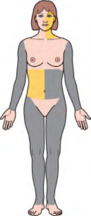
Fig. 101.1 Lipodystrophy syndromes and localized forms. Schematic representation of the predominant sites of lipoatrophy and lipohypertrophy. Additional syndromes, including familial partial lipodystrophy with mandibuloacral dysplasia, are outlined in Table 101.1. Tables 63.9 and 63.10 review progeroid syndromes in which lipoatrophy can be seen. AI-CTD, autoimmune connective tissue disease; ART, antiretroviral therapy; IR, insulin resistance; PD, programmed cell death. AGPAT2, encodes 1-acylglycerol-3-phosphate O-acyltransferase 2 (triglyceride and phospholipid synthesis); BSCL2, encodes seipin (lipid droplet formation); CAV1, encodes caveolin 1 (binds fatty acids and translocates them to lipid droplets); CAVIN1/PTRF, encodes caveolae associated protein 1 (biogenesis of caveolae and expression of caveolins 1 and 3); CIDEC, encodes cell death-inducing DNA fragmentation factor-like effector C (adipocyte apoptosis); LIPE, encodes lipase E, hormone sensitive type (hydrolysis of triglycerides to free fatty acids); LMNA, encodes lamins A/C (structural integrity of the nuclear lamina); LMNB2, encodes lamin B2 (structural integrity of the nuclear lamina); PLIN1, encodes perilipin 1 (formation, maturation, and function of lipid droplets within adipocytes); POMP, encodes proteasome maturation protein; PPARG, encodes peroxisome proliferator-activated receptor-gamma (essential role in lipogenesis); PSMA3, encodes proteasome subunit alpha 3; PSMB4, -B8, -B9, -B10, encode proteasome subunit beta 4, 8, 9, 10; PSMG2, encodes proteasome subunit gamma 2.

Fig. 101.2 Lipoatrophia semicircularis (semicircular lipoatrophy). Bilateral slightly curved depressions on the anterolateral thighs. Courtesy Diane Thaler, MD.
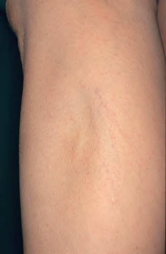
Fig. 101.3 Localized lipoatrophy of the upper lateral calf due to pressure. A fairly common finding in women who cross their legs while seated. Courtesy Jean L. Bolognia, MD.
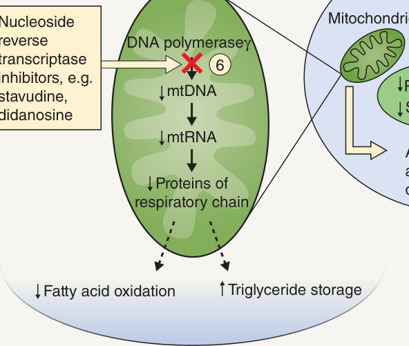
Fig. 101.14 Proposed mechanisms of HIV/ART-associated lipodystrophy. The combination of decreased PPAR-γ expression and decreased fatty acid oxidation capacity resulting from drug exposure could explain how these drugs cause adipocyte toxicity and lipoatrophy. See Table 101.6 for additional details. The numbers represent sites of drug effects. ART, antiretroviral therapy; at-RA, all-trans-retinoic acid; CRABP-1, cytoplasmic retinoic acid binding protein type 1; LPL, lipoprotein lipase; LRP, low-density lipoprotein receptor-related protein; mtRNA, mitochondrial RNA; mtDNA, mitochondrial DNA; PIs, protease inhibitors; PPAR-γ, peroxisome proliferator-activated receptor-γ; RXR, retinoid X receptor; SREBP-1c, sterol regulatory element-binding protein-1c. Adapted from Carr A. HIV protease inhibitor-related lipodystrophy syndrome. Clin Infect Dis 2000;30:S135–42.
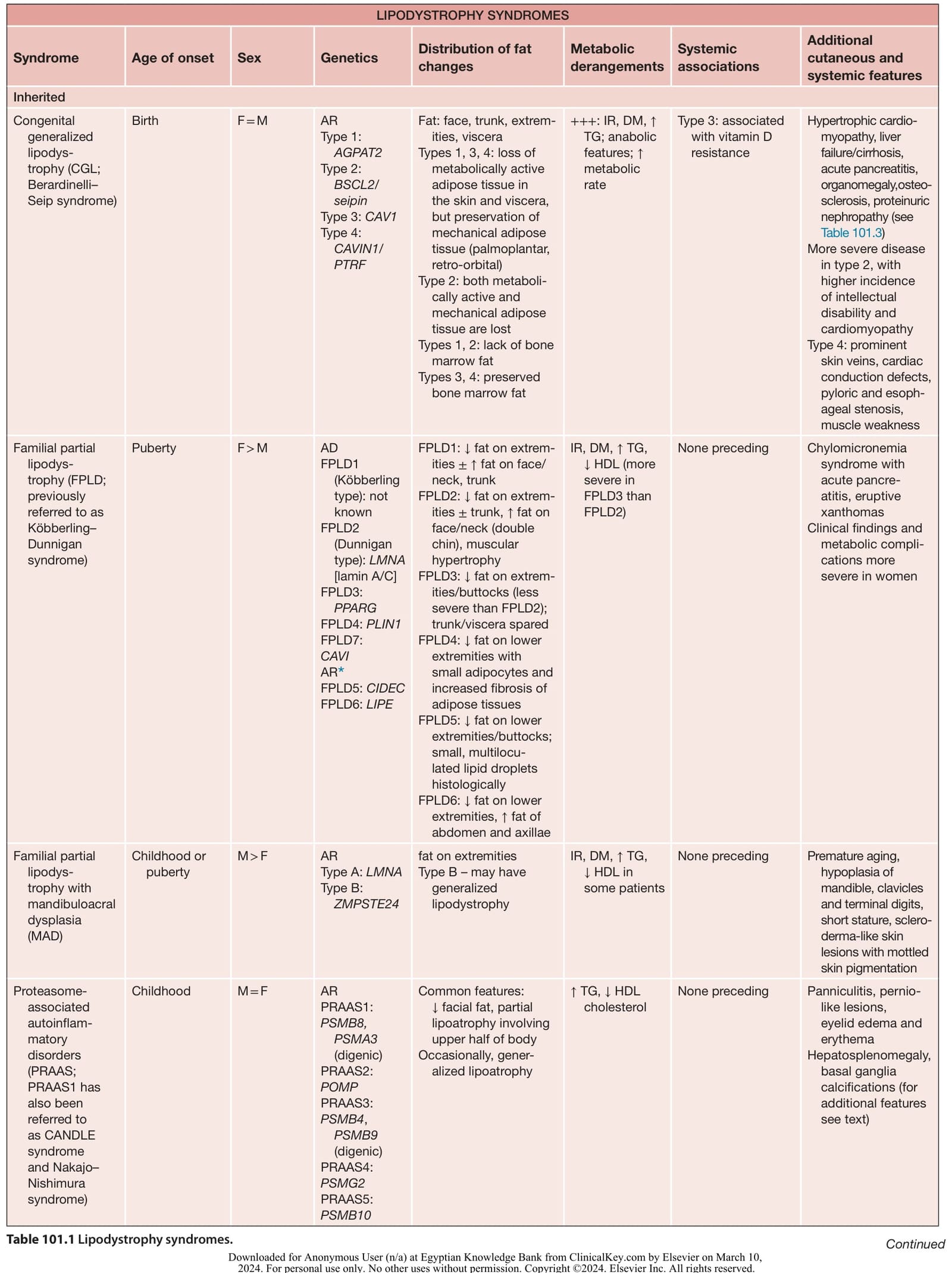
Table 101.1 Lipodystrophy syndromes. Continued
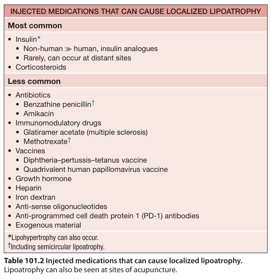
Table 101.2 Injected medications that can cause localized lipoatrophy.