CLINICAL FEATURES
Congenital Generalized Lipodystrophy (CGL)
CGL is rare, with an estimated prevalence of <1 case in 10 million. As an autosomal recessive disorder, there is often consanguinity. All subtypes are characterized by generalized loss or absence of metabolically active subcutaneous fat from birth, resulting in a cadaveric facies and distinctive muscular-appearing body habitus (Fig. 101.4A). Anabolic features are evident in early childhood (Table 101.3). There is also a deficiency of bone marrow fat (types 1, 2) and visceral fat (types 1, 3, 4). Type 2 CGL also lacks mechanical fat, in addition to metabolically active fat.
The inability to store sufficient fat in the adipocytes results in the metabolic syndrome (see Table 53.5), which becomes more profound during puberty. Diabetes mellitus is evident by adolescence, but hyperinsulinemia can be detected as early as infancy. Clitoromegaly is seen in women and may be associated with polycystic ovarian syndrome and infertility. Hepatosplenomegaly is typical and may be associated with umbilical herniation. Cutaneous findings (see Table 101.3) include acanthosis nigricans, which is noted by adolescence and may be extensive (Fig. 101.4B). Serious medical complications and metabolic derangements include cirrhosis from fatty liver, premature atherosclerosis, sequelae of diabetes, pancreatitis from hypertriglyceridemia, osteosclerosis, and a high mortality rate from hypertrophic cardiomyopathy. Type 2 appears to be a more severe disorder that is associated with intellectual disability, hypertrophic cardiomyopathy,
and a higher incidence of premature death while type 4 patients have arrhythmias from myocardial fibrofatty infiltration, esophageal dysmotility, and pyloric stenosis. In one series of CGL, there was an ~35-year reduction in lifespan, with liver disease and infection as the most common causes of death.
Familial Partial Lipodystrophy
Familial partial lipodystrophy (FPLD) was first described by Dunnigan and Köbberling, who outlined the clinical characteristics of large Scottish and German pedigrees with familial syndromes of partial lipodystrophy. FPLD is classified based upon genetic mutations and clinical phenotype (see Table 101.1). This group of disorders differs from other forms of inherited lipodystrophy by a later onset (after puberty) and fat loss predominantly affecting the extremities, with sparing of the face. FPLD is a rare disorder, with a prevalence of <1 in 15 million.
In FPLD2, a normal childhood is followed by a progressive, symmetric loss of subcutaneous fat; the latter uniformly involves the extremities and variably extends to the trunk (anterior > posterior). Compensatory accumulation of excess fat occurs and results in a fat head and neck, with a round face, as well as increased supraclavicular fat (see Fig. 101.1). Acromegalic facies with submental fullness (“double chin”) is characteristic. As a result of the loss of fat in the limbs, there is an accentuation of subcutaneous veins and muscular prominence (Fig. 101.5). Although FPLD3 has milder clinical features and an onset after the second decade of life, the metabolic disturbances may be more severe.
Metabolic disturbances in FPLD are similar to those in the generalized lipodystrophy syndromes. Glucose intolerance, ranging from mild to severe, develops during young adulthood. Acute pancreatitis and hepatic steatosis and cirrhosis may occur, with complications from diabetes mellitus, atherosclerotic cardiovascular disease, or hypertrophic cardiomyopathy leading to premature death. When compared to men, affected women have been reported to have more severe triglyceride elevations (chylomicronemia syndrome). Dermatologic manifestations include tuberous xanthomas, acanthosis nigricans and hirsutism, while gynecologic findings include menstrual irregularities, polycystic ovaries, and fat hypertrophy within the labia majora.
Familial partial lipodystrophy with mandibuloacral dysplasia is a rare variant of partial lipodystrophy characterized by mandibular and clavicular hypoplasia, short stature, a high-pitched voice, and ectodermal abnormalities of the skin, teeth, nails, and hair. There are multiple craniofacial defects, including dental overcrowding and bird-like facies. Skeletal abnormalities include osteolysis of the clavicles, acroosteolysis, delayed closure of cranial sutures, and joint contractures. Mottled hyperpigmentation, alopecia, atrophy of extremity skin, and nail dysplasia are the characteristic cutaneous findings. Less common clinical features include sensorineural hearing loss, delayed puberty, a high-arched palate, and cutaneous calcinosis. Features of the metabolic syndrome may be present (see Table 53.5).
Proteasome-Associated Autoinflammatory Syndromes
To date, five types of proteasome-associated autoinflammatory syndrome (PRAAS) have been identified and are due to mutations in seven genes that encode proteasome-associated proteins (see Table 45.7). PRAAS1 has also been referred to as CANDLE (chronic atypical neutrophilic dermatosis with lipodystrophy and elevated temperature) syndrome and Nakajo–Nishimura syndrome. In addition to recurrent fevers, annular violaceous plaques, eyelid erythema, and pernio-like lesions that begin during infancy or early childhood, patients can develop panniculitis-induced lipodystrophy. Partial (e.g.
PRAAS1) or generalized (e.g. PRAAS4) lipoatrophy has been described. In biopsy specimens of nodules, a dense dermal infiltrate of atypical myeloid cells that resembles leukemia cutis or “histiocytoid” Sweet syndrome is seen, along with mature neutrophils. Treatment options include JAK inhibitors, corticosteroids, and methotrexate.
Recurrent lipoatrophic panniculitis of children
In this syndrome, painful subcutaneous nodules are accompanied by fever, malaise, abdominal pain, and arthralgias. Localized lipoatrophy then develops at the sites of nodules. Histopathologically, there is a lobular panniculitis with a mixed infiltrate composed of neutrophils, lymphocytes, macrophages, and mononuclear myeloid cells. Methotrexate and/or corticosteroids led to clinical improvement. The authors proposed that this constellation may represent an autoinflammatory disease. Of note, whole genome sequencing did not detect a form of PRAAS (see previous section).
Acquired Generalized Lipodystrophy (Lawrence Syndrome)
To date, ~80 cases of acquired generalized lipodystrophy (AGL) have been reported in the English literature. New diagnostic criteria for AGL have been proposed, as well as a sub-classification into three subtypes (Table 101.4).
In approximately one-third to one-half of patients, there is a preceding systemic illness, in particular, a viral or bacterial infection or an autoimmune disorder (e.g. a connective tissue disease, thyroiditis). The rare patients who also have CVID can have recurrent infections (see Ch. 60). Drug-induced lipodystrophy, e.g. due to immune checkpoint inhibitors, needs to be excluded (see below).
Clinical features of AGL are similar to those of the congenital variant, but are of later onset and milder. The lipoatrophy typically does not become apparent until childhood or adolescence and rarely develops during adulthood. This disorder is three times as common in women. Large areas of the face, trunk, and extremities have fat loss (Fig. 101.6), and the palms and soles may even be involved. Loss of acral subcutaneous fat as well as perinephric and intra-abdominal fat can be documented by MRI scans. However, bone marrow fat is preserved, and this feature can be helpful in differentiating AGL from CGL types 1 and 2.
Tender subcutaneous nodules of panniculitis may precede the lipoatrophy in the type 1 variant and there may be an overlap in the panniculitic and autoimmune subtypes. Progression can occur rapidly or over an extended period. When the lipodystrophy becomes generalized, metabolic abnormalities begin to manifest. Although the latter are less severe in AGL than in CGL, the period between the appearance of lipoatrophy and development of frank diabetes (~4 years) is shorter in AGL. The extent of fat loss mirrors the severity of insulin resistance and hypertriglyceridemia.
Compared to the autoimmune and idiopathic types of AGL, the panniculitic variant may have less severe fat loss and a lower prevalence of diabetes and hypertriglyceridemia (see Table 101.4). In addition, the associated anabolic features are variable and less prominent and its severity is less striking when the condition starts in later childhood or adulthood.
In AGL, hepatic steatosis is commonly present in affected children, leading to a higher incidence of hepatomegaly, cirrhosis, and liverrelated mortality than in CGL. Gynecologic abnormalities include true or pseudo-clitoromegaly (the latter due to fat loss), polycystic ovarian syndrome, and menstrual irregularities. Premature coronary artery disease and carotid or peripheral arterial disease may occasionally be seen. Renal and central nervous system abnormalities are usually absent.
Dermatologic features of AGL, in addition to panniculitis and lipoatrophic areas, include acanthosis nigricans, localized or generalized hyperpigmentation, eruptive xanthomas, telangiectasias, mild palmar/ plantar keratoderma, and hair abnormalities, including mild hirsutism, curly hair, and occasional alopecia. The acanthosis nigricans begins in childhood and involves the neck, axillae, groin, umbilicus, and nipples. Associated autoimmune diseases include juvenile dermatomyositis, Sjögren syndrome, and vitiligo. In addition, patients may have chronic urticaria and angioedema (see Table 101.4).
Acquired Partial Lipodystrophy (Barraquer– Simons Syndrome)
Acquired partial lipodystrophy, also known as progressive lipodystrophy or cephalothoracic lipodystrophy, is the second most common type of non-localized lipodystrophy, after HIV/ART-related lipodystrophy. It has been reported worldwide, with more than 250 patients described in the literature.
Three phenotypic subtypes have been noted: (1) fat loss in the upper half of the body; (2) fat loss in the upper half of the body, with hypertrophy of adipose tissue in the lower half; and (3) hemi-lipodystrophy, where one half of the face or body is affected.
Lipodystrophy begins during childhood or pre-puberty (median age at diagnosis is 8–10 years) and sometimes follows a viral illness. It is characterized by an insidious onset, with symmetric and progressive loss of subcutaneous fat in a cephalocaudal direction (Fig. 101.7). Extension of the lipoatrophy usually takes place over a 1- to 2-year period, but can occur over many years. Physical findings may be apparent by adolescence, although the onset may be delayed until 40 years of age. Women are affected three times more often than men.
The face is usually affected first, with sunken eyes due to loss of retro-orbital and periorbital fat. This is accentuated by concurrent loss of buccal and temporal fat, resulting in a progeria-like or cadaveric appearance (Fig. 101.8). The hips and legs tend to be spared, and often demonstrate fat hypertrophy, especially in women. Growth delay or stunted growth is not characteristic of this disorder.
MCGN II with nephritic syndrome, seen in approximately one-fifth of patients, may occur many years, usually about eight, after the onset of lipodystrophy. Almost all patients with acquired partial lipodystrophy have low levels of C3 in addition to circulating C3 nephritic factor (C3NeF), composed of polyclonal IgG. This leads to unopposed activation of the alternative complement pathway (see above) and C3NeF-induced lysis of adipocytes. The low C3 level also predisposes
Lipoatrophic features are most prominent on the face. There is obvious loss of buccal fat. The patient also had axillary acanthosis nigricans. Courtesy Kenneth E. Greer, MD.
Lipoatrophy of the face with sunken eyes and loss of temporal and buccal fat pads. The facial pigmentation is related to lichen planus. Courtesy Priya Sen, MD.
these patients to recurrent Neisseria meningitidis infections and repeat vaccinations may be required.
Other autoimmune associations include systemic lupus erythematosus, juvenile dermatomyositis, systemic sclerosis, Sjögren syndrome, pernicious anemia, hypothyroidism, celiac disease, dermatitis herpetiformis, temporal arteritis, and small vessel vasculitis.
Metabolic syndrome is less common in APL than in other lipodystrophies, but acanthosis nigricans, menstrual abnormalities, hirsutism, insulin resistance, type 2 diabetes mellitus, and hyperlipidemia may be seen.
Localized Lipoatrophy
Localized lipoatrophy presents as one or multiple depressed areas, usually on the proximal extremities, ranging from under a few centimeters to greater than 20 cm in diameter. Drug-induced lipoatrophy is most commonly due to injections of corticosteroids and non-human insulin (see Table 101.2); it may be accompanied by local lipohypertrophy. Of note, multifocal lipoatrophy due to intravenous corticosteroid use has been described and insulin injection-related lipoatrophy can appear at distant sites.
A An area of depression on the cheek due to burnt-out lupus panniculitis; note the dyspigmentation and scarring from an overlapping lesion of discoid lupus erythematosus. B Circular depressions on the upper arm, a common location for lupus panniculitis. A, Courtesy National Skin Centre, Singapore.
Involutional lipoatrophy is an idiopathic lipoatrophy characterized clinically by non-inflammatory focal loss of fat. Based on reported cases, there is a strong female predominance. Focal oval areas of lipoatrophy, 2 to 8 cm in diameter, occur predominantly on the buttocks and proximal extremities, especially the upper arm, corresponding to locations frequently used as injection sites. Although it is estimated that up to 60% of cases of involutional lipoatrophy may be associated with prior local injections (see above), this type of lipoatrophy may occur without prior injections.
Atrophic connective tissue panniculitis is rare, and it often occurs on the upper or lower extremities. In “pure” cases of this condition, there is no prior clinical inflammation although there is evidence histologically of lymphocytic panniculitis. In other patients, the lipoatrophy may be the direct result of clinical panniculitis. The latter is common on the head and neck or proximal extremities if associated with lupus panniculitis (also referred to as lupus profundus; Fig. 101.9). Other associated autoimmune diseases include dermatomyositis, thyroiditis, juvenile idiopathic arthritis, vitiligo, and insulin-dependent diabetes mellitus.
Lipoatrophia semicircularis (semicircular lipoatrophy) has been reported primarily in women in their 20s to 30s and rarely in children. It presents as symmetric, asymptomatic, 4–8 cm curvilinear horizontal depressions on the anterolateral thighs (see Fig. 101.2). Infrequent presentations include two or three parallel depressions, a unilateral distribution, or symptoms of cramps, pain after sports, heavy legs, or a burning sensation. The lesions usually appear over a few weeks and spontaneously resolve between 9 months to 4 years. The lipoatrophy may recur. MRI can assist in differentiating this from an inflammatory panniculitis.
Annular lipoatrophy presents with a deep, persistent, pseudosclerotic band approximately 9–11 cm in length that encircles the arm or ankle. More extensive lesions have been reported that were 14–16 cm in length. It may be preceded by tenderness and swelling of the limb, or be associated with discomfort and arthritis. This entity may represent a variant or end-stage presentation of atrophic connective tissue disease panniculitis. Thus, there may be patients with associated autoimmune diatheses.
Lipodystrophia centrifugalis abdominalis infantilis (centrifugal lipodystrophy) has largely been reported from East Asia, with most patients of Japanese, Korean, or Chinese origin. Onset is usually before 3 years of age, with >90% of patients presenting by age 5 years. In Koreans, there is a four-fold female predominance. Although lipodystrophia centrifugalis abdominalis infantilis affects primarily Asian children, it may occasionally occur in Caucasians and in adults.
Clinically, a well-demarcated area of lipoatrophy with a periphery of erythema and scale is seen on the abdomen or elsewhere on the trunk, often with associated regional lymphadenopathy. The lipoatrophy begins on the lower abdomen or in the groin region (and may include the genitalia) in about 80% of patients, and in the axillary region in about 20% of patients; it spreads onto the abdomen or chest, respectively, in a centrifugal fashion (Fig. 101.10). The underlying blood vessels become visible and ulceration may occur within the depressed areas. Contrary to its original name, this condition can affect non-abdominal sites such as the face, neck, and lumbosacral region (Fig. 101.11A ), and it is not limited to infants. The lesions may progress slowly over several years, then often cease enlarging by 13 years of age. Although there is no specific therapy, >60% of patients spontaneously improve (Fig. 101.11B).
Progressive hemifacial atrophy (Parry–Romberg syndrome) is considered to be a form of severe linear morphea (see Ch. 44). In addition to lipoatrophy, there may be atrophy of the underlying muscle and bone as well as the potential for ophthalmologic, dental, and neurologic involvement. An association with Borrelia burgdorferi infection has been reported in Europe.
Non-progressive late-onset linear hemifacial lipoatrophy occurs on the malar cheek, primarily in elderly individuals.
Inflammatory reactions due to material injected for cosmetic purposes or from self-inflicted injections of foreign material can lead to lipoatrophy which may be associated with scarring and dyschromia.
Lipohypertrophy
Lipohypertrophy remains a frequent complication of insulin therapy, irrespective of the insulin source and mode of administration. As with insulin lipoatrophy, anti-insulin antibodies are associated with lipohypertrophy in children and adolescents with type 1 diabetes. The clinical consequence is that injection of insulin into a site of lipohypertrophy, while painless, may lead to erratic absorption of the insulin with poor glycemic control and rebound phenomenon. Of note, insulininduced nodular amyloidosis may have a clinical appearance similar to lipohypertrophy.
Injection of pegvisomant, a growth hormone receptor antagonist used to treat acromegaly, is another cause of lipohypertrophy (Table 101.5).
of the face.A A 2-year-old girl with a serpentine violaceous band involving the cheek in association with lipoatrophy. B The same child two years later, with spontaneous resolution. The most common location for lipodystrophia centrifugalis infantilis is the abdomen (see Fig. 101.10). Courtesy National Skin Centre, Singapore.
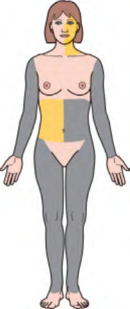
Fig. 101.1 Lipodystrophy syndromes and localized forms. Schematic representation of the predominant sites of lipoatrophy and lipohypertrophy. Additional syndromes, including familial partial lipodystrophy with mandibuloacral dysplasia, are outlined in Table 101.1. Tables 63.9 and 63.10 review progeroid syndromes in which lipoatrophy can be seen. AI-CTD, autoimmune connective tissue disease; ART, antiretroviral therapy; IR, insulin resistance; PD, programmed cell death. AGPAT2, encodes 1-acylglycerol-3-phosphate O-acyltransferase 2 (triglyceride and phospholipid synthesis); BSCL2, encodes seipin (lipid droplet formation); CAV1, encodes caveolin 1 (binds fatty acids and translocates them to lipid droplets); CAVIN1/PTRF, encodes caveolae associated protein 1 (biogenesis of caveolae and expression of caveolins 1 and 3); CIDEC, encodes cell death-inducing DNA fragmentation factor-like effector C (adipocyte apoptosis); LIPE, encodes lipase E, hormone sensitive type (hydrolysis of triglycerides to free fatty acids); LMNA, encodes lamins A/C (structural integrity of the nuclear lamina); LMNB2, encodes lamin B2 (structural integrity of the nuclear lamina); PLIN1, encodes perilipin 1 (formation, maturation, and function of lipid droplets within adipocytes); POMP, encodes proteasome maturation protein; PPARG, encodes peroxisome proliferator-activated receptor-gamma (essential role in lipogenesis); PSMA3, encodes proteasome subunit alpha 3; PSMB4, -B8, -B9, -B10, encode proteasome subunit beta 4, 8, 9, 10; PSMG2, encodes proteasome subunit gamma 2.

Fig. 101.2 Lipoatrophia semicircularis (semicircular lipoatrophy). Bilateral slightly curved depressions on the anterolateral thighs. Courtesy Diane Thaler, MD.
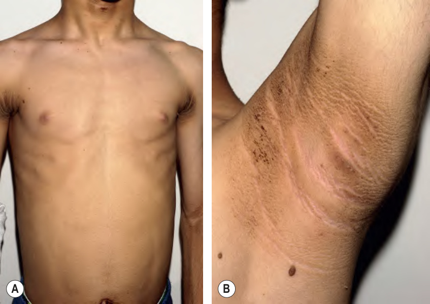
Fig. 101.4 Congenital generalized lipodystrophy syndrome (Berardinelli–Seip syndrome).A Lipoatrophy of the trunk leading to muscle definition. B Extensive acanthosis nigricans. Courtesy Edward Cowen, MD.

Fig. 101.5 Lipoatrophy of the lower extremities, leading to muscular prominence.Courtesy William D. James, MD.
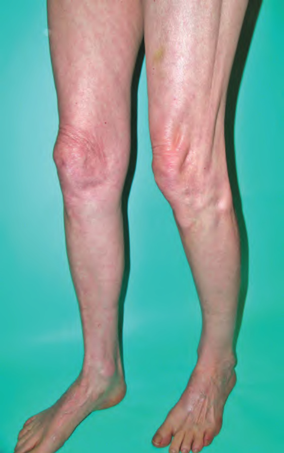
Fig. 101.6 Acquired generalized lipodys- trophy. There is loss of subcutaneous fat, resulting in a muscular appearance of the legs and accentuation of the tendons. Courtesy Jacqueline Junkins-Hopkins, MD.

Fig. 101.7 Acquired partial lipodystrophy syndrome in a patient with nephritic factor and renal disease.
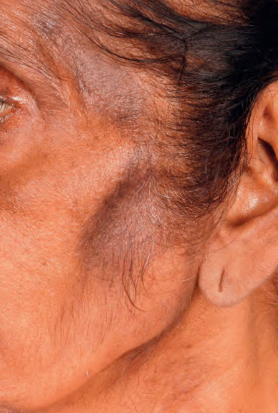
Fig. 101.8 Acquired partial lipodystrophy syndrome in a patient with Sjögren syndrome.
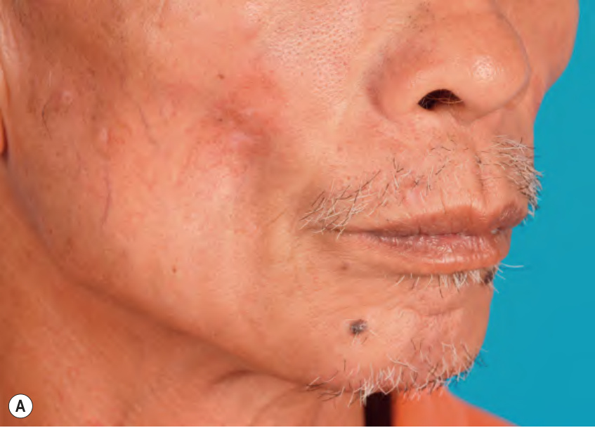
Fig. 101.9 Localized lipoatrophy secondary to lupus panniculitis.
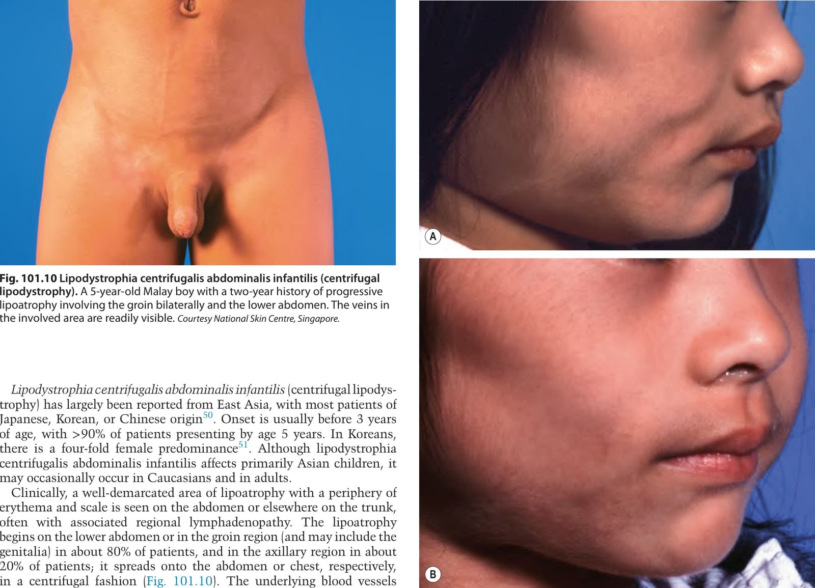
Fig. 101.10 Lipodystrophia centrifugalis abdominalis infantilis (centrifugal lipodystrophy). A 5-year-old Malay boy with a two-year history of progressive lipoatrophy involving the groin bilaterally and the lower abdomen. The veins in the involved area are readily visible. Courtesy National Skin Centre, Singapore.
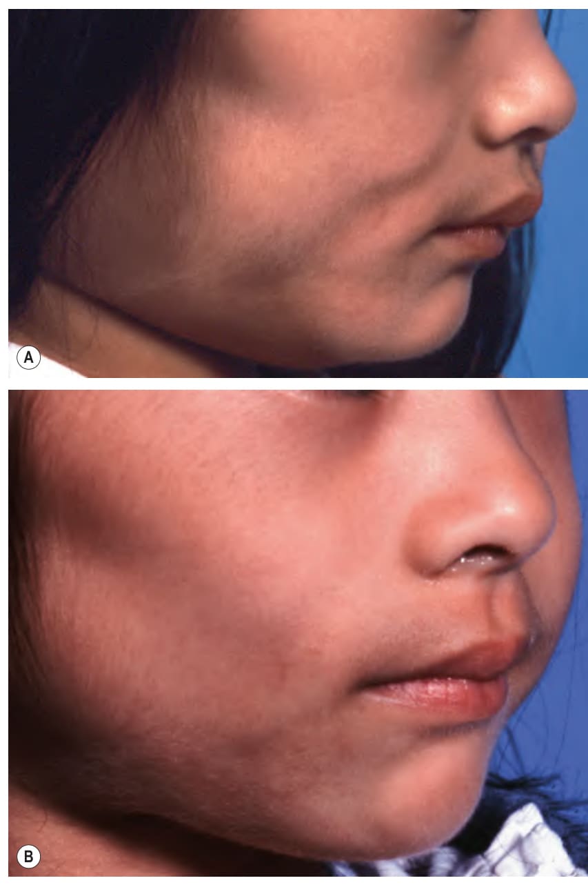
Fig. 101.11 Lipodystrophia centrifugalis infantilis (centrifugal lipodystrophy)
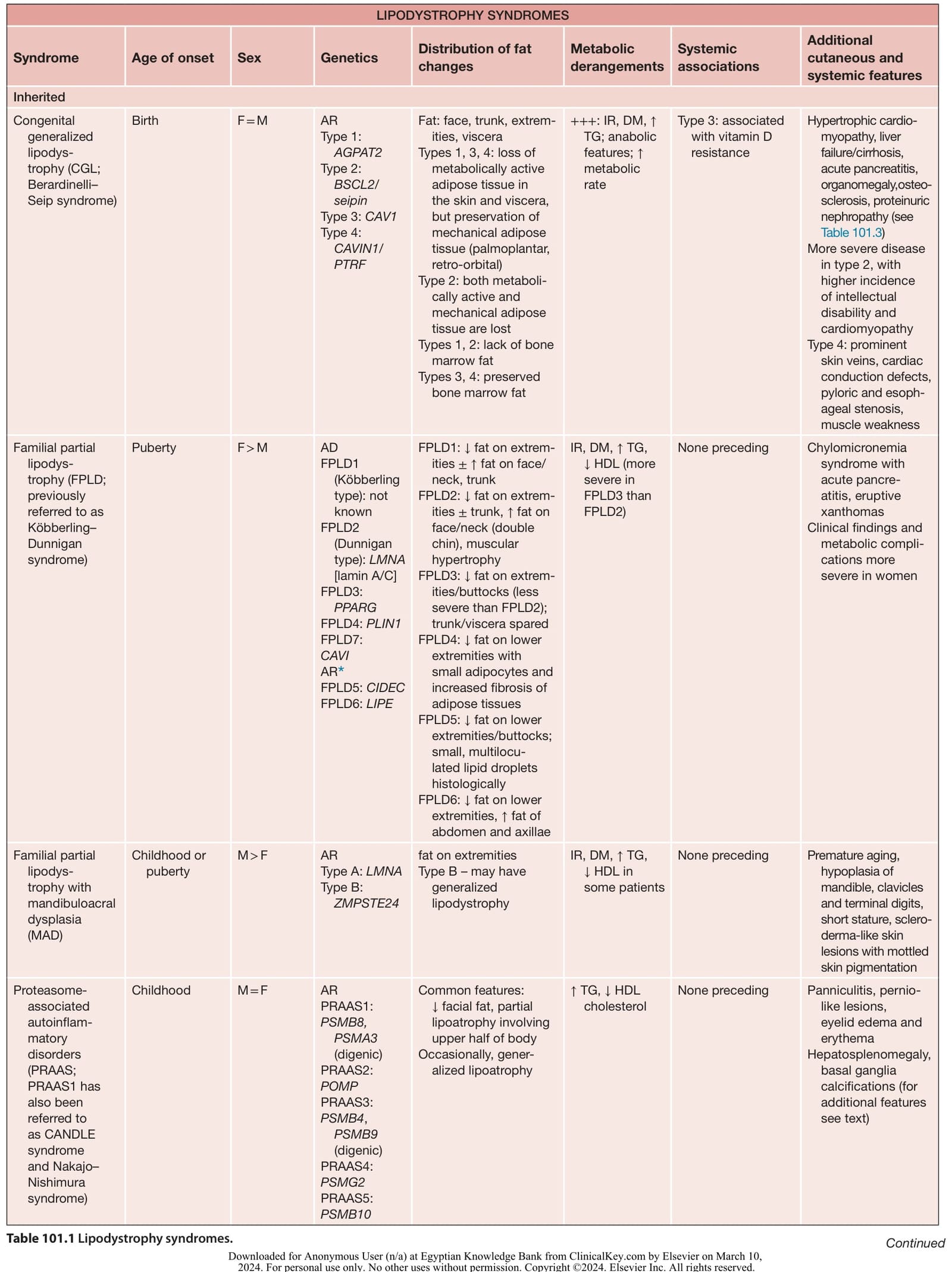
Table 101.1 Lipodystrophy syndromes. Continued
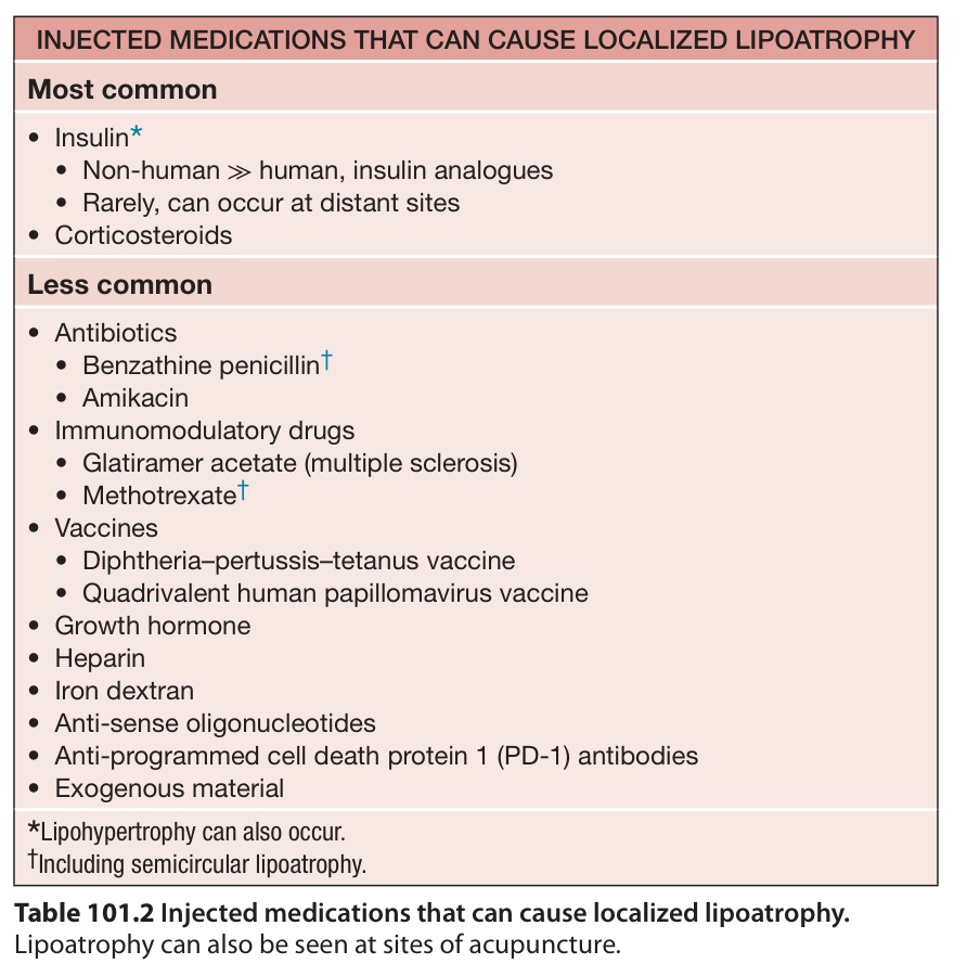
Table 101.2 Injected medications that can cause localized lipoatrophy.
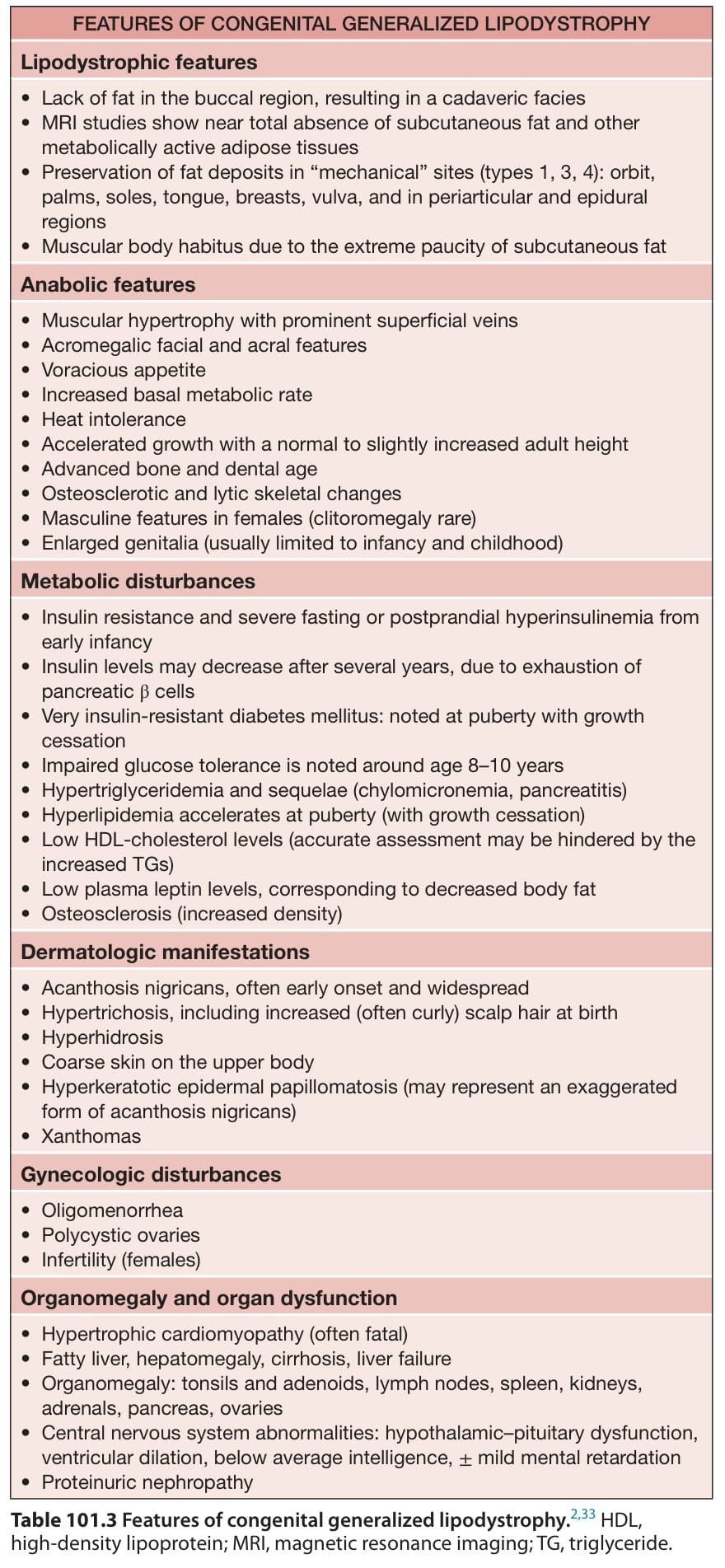
Table 101.3 Features of congenital generalized lipodystrophy. HDL, high-density lipoprotein; MRI, magnetic resonance imaging; TG, triglyceride.
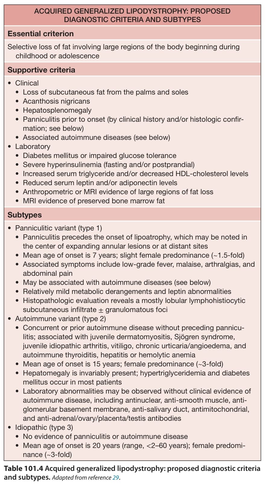
Table 101.4 Acquired generalized lipodystrophy: proposed diagnostic criteria and subtypes.Adapted from reference .
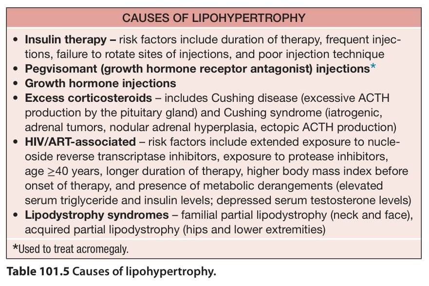
Table 101.5 Causes of lipohypertrophy.