PATHOLOGY
Generally speaking, the histologic picture of the various lipodystrophy syndromes (see Table 101.1) is non-inflammatory. In generalized lipodystrophy, there is complete or near-complete loss of subcutaneous fat, with the dermis and fascia in direct apposition. Less often,
biopsy specimens appear to have no overt abnormalities, except for an increased amount of collagen, which has replaced the subcutaneous fat. In contrast, biopsies of acquired generalized lipodystrophy, in particular type 1, may show a lobular panniculitis (Fig. 101.12; see Table 101.4).
In acquired generalized lipodystrophy and areas of lipoatrophy in some forms of partial lipodystrophy, two histologic patterns have been recognized: (1) non-inflammatory with involutional changes of the fat; and (2) inflammatory in the form of a lobular panniculitis with lymphocytes, lipophages, and plasma cells. Whether these two patterns are stage-related remains to be clarified, as even in patients without panniculitis clinically, biopsies of early lesions may show inflammation, but it is usually mild.
Involutional changes of the subcutaneous fat are characterized by small fat lobules with a reduction in the size and number of adipocytes (Fig. 101.13). Involutional lipoatrophy can be further divided into two histologic subtypes. In the first type, the lobules may be composed of faintly acidophilic, small fat cells that retract from one another as well as the surrounding connective tissue, leading to an appearance similar to embryonic fat; the lobules often have an eosinophilic appearance. These histologic findings are more prominent at the periphery of the clinically depressed area, with tiny acidophilic fat cells present more centrally. Inflammation is absent to sparsely mononuclear. The second type, with indistinguishable clinical features, shows small atrophic adipocytes with a normal fat cell membrane surrounded by prominent vasculature. At scanning magnification, the lobules are collapsed (with an orientation parallel to the skin surface) and surrounded by numerous capillaries. Acid mucopolysaccharide deposition within the fat lobules and fibrosis of the septae may be seen, as well as occasionally lipomembranous changes.
Although birefringent, non-crystalline material may occasionally be observed, foreign body giant cells and lipophages are typically absent in both subtypes of involutional lipoatrophy. CD68+ macrophages can be demonstrated with immunohistochemical staining. Ultrastructural studies have shown that these are lysosomally active macrophages adjacent to lipocytes and likely containing degenerated lipid remnants. In localized lipodystrophy, a loss of fatty tissue is seen, along with a variable degree of fibrosis. There is usually no inflammatory infiltrate, although panniculitis has been reported in a few cases. Deposits of immunoreactants can be seen within blood vessel walls or the basement membrane, particularly in the inflammatory type.
Histologically, well-developed areas of lipodystrophia centrifugalis abdominalis infantilis show a diminution of subcutaneous fat with scant or absent inflammation. The erythematous rim tends to correspond to a moderate or marked lymphohistiocytic infiltrate in the subcutaneous fat; a deep dermal and peri-eccrine lymphocytic infiltrate
is an uncommon finding. There may be fibrosis and thickening of the septae within the subcutis. The few adipocytes that are present may have myxoid changes.
Other localized variants such as lipoatrophia semicircularis (semicircular lipoatrophy) and drug-induced lipoatrophy usually show a loss of fatty tissue and replacement by collagen, with no features of panniculitis. There are often scattered lipophages. However, at sites of glatiramer acetate injections, panniculitis that resembles lupus profundus histologically can be seen. Lastly, HIV/ART-associated lipodystrophy resembles the non-inflammatory pattern of lipodystrophy.
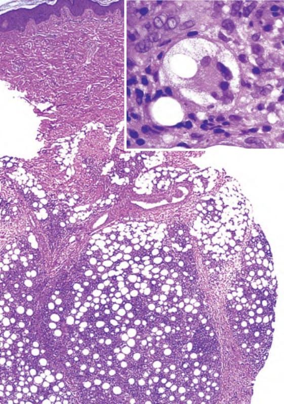
Fig. 101.12 Histopathologic features of the preceding panniculitis in type 1 acquired generalized lipodys- trophy. Lobular panniculitis with infiltrate of lymphocytes, histiocytes, multinucleated giant cells, and lipophages. There is also cytoplasmic vacuolization of the fat cells (inset). See Table 101.4 for three types. Courtesy Jacqueline Junkins- Hopkins, MD.
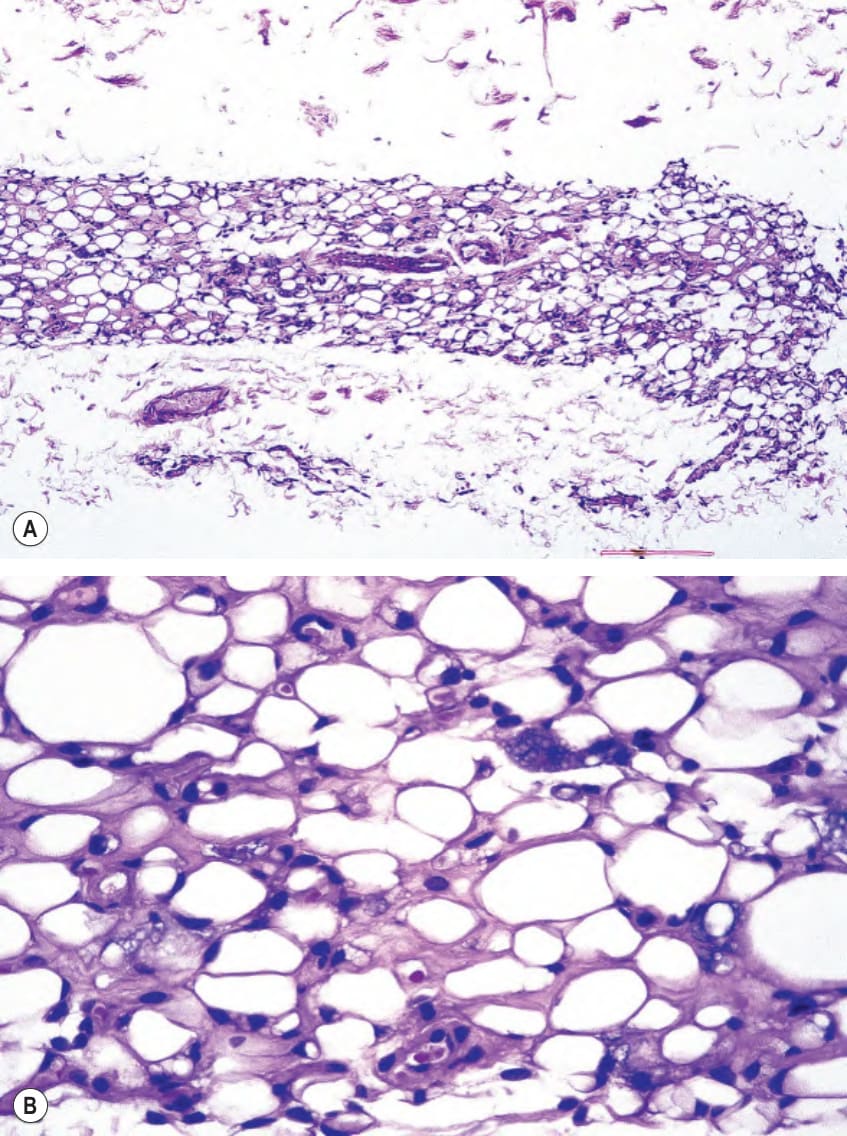
Fig. 101.13 Histopathologic features of involutional lipoatrophy secondary to corticosteroid injection.A There is marked diminution and collapse of the fat lobules, without inflammation. B Higher power demonstrates decreased size of fat cells with eosinophilic thickened cell walls with a hyalinized and mucinous stroma. Courtesy Jacqueline Junkins-Hopkins, MD.
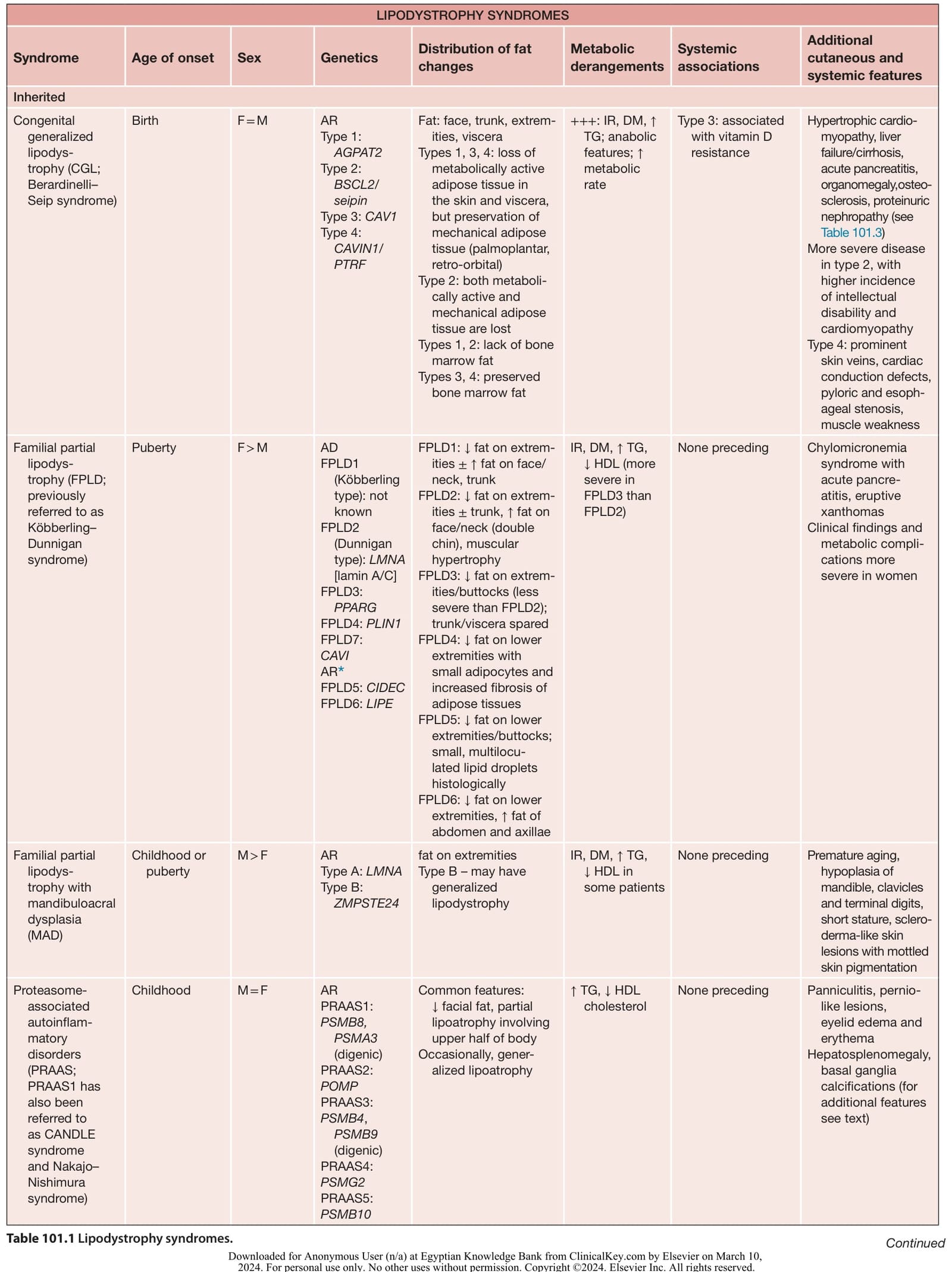
Table 101.1 Lipodystrophy syndromes. Continued
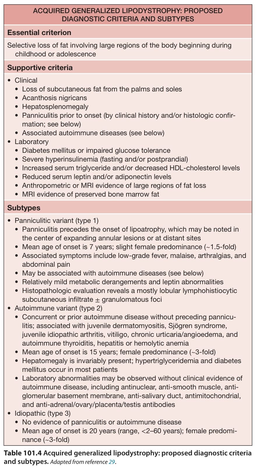
Table 101.4 Acquired generalized lipodystrophy: proposed diagnostic criteria and subtypes.Adapted from reference .