CLINICAL FEATURES
Presentation
Most IHs have a typical presentation and growth pattern. Lesions usually become apparent during the first few weeks of life, although the proportion of hemangiomas that are “congenital” in published series has ranged from 15% to 60%. IHs evident at birth most often appear as precursor lesions, which are apparent in up to 65% of IHs, but occasionally they present as relatively well-formed typical IHs that subsequently demonstrate variable proliferation then slow involution. Precursor IH lesions include telangiectasias surrounded by a vasoconstricted halo, areas of pallor, pink macules, and blue bruise-like patches (Fig. 103.1). Pink macules and patches may mimic a capillary malformation, and repeat examination over the ensuing weeks is essential to determine the diagnosis (Fig. 103.2). Rapidly involuting, partially involuting, and non-involuting congenital hemangiomas (RICH/PICH/ NICH) represent distinct entities that are discussed below.
Occasionally, an area of ulceration in the diaper area, in other skin folds, or on the lip may herald the onset of an IH, and in a newborn infant these lesions may be confused with bacterial or viral infections. Continued observation usually clarifies the diagnosis. With dermoscopy, the characteristic findings of red lacunae separated by white septae in superficial IHs and violaceous to blue lacunae in deeper IHs may be helpful when the diagnosis is not clear.
Hemangiomas may occur anywhere on the skin and mucosal surfaces. Although overall they most commonly develop on the trunk, ~50% of the IHs that present to a pediatric dermatologist are located on the head and neck. The clinical appearance of an IH is influenced by its location within the skin and subcutaneous tissues. Superficial hemangiomas are situated in the upper dermis and are bright red in color during their proliferating phase. The surface is finely lobulated, and the term “strawberry hemangioma” has been used to describe them (Fig. 103.3).
Deep hemangiomas are located in the deep dermis and/or subcutis. They usually are not apparent in the immediate newborn period, often
becoming evident a few weeks or even months after birth. They present as warm, ill-defined, light blue–purple masses with minimal or no overlying skin changes, making them more difficult to diagnose than superficial or mixed hemangiomas (Fig. 103.4). The presence of dilated veins or telangiectasias overlying a deep hemangioma provides a clue that the lesion is of vascular origin. Larger deep hemangiomas often have significant arterial blood supply during the proliferative phase, and detection of high flow by Doppler ultrasonography can help to confirm the diagnosis. Mixed hemangiomas have both superficial and deep components, often presenting during the proliferative phase as a well-delineated red vascular plaque overlying a larger, poorly circumscribed violaceous or light blue nodule.
Superficial hemangiomas are the most common type, accounting for ~50%–60% of IHs. An additional 25%–35% of hemangiomas are mixed and 15% are deep. Approximately 25% of patients have multiple
Lesions can be focal (A,B) or have a broader segmental distribution (C). Note the bright red color and finely lobulated surface, irrespective of size. Involvement may be diffuse or discontiguous. Courtesy Julie V. Schaffer, MD.
lesions, which are occasionally associated with hemangiomas in the liver or other internal organs (see below).
IHs can also be categorized based upon the pattern of involvement, which may help to predict prognosis. The two main pattern subtypes are: (1) focal – arising from a single localized nidus (see Fig. 103.3A,B); and (2) segmental – covering a broad area or developmental unit in a “plaque-like” manner (see Fig. 103.3C). Lesions that are difficult to classify are designated as indeterminate and may represent partial segmental lesions that respect anatomic boundaries similar to their larger segmental counterparts. The majority of IHs are focal lesions, and those on the face may show predilection for embryonic fusion lines. The distribution patterns of segmental hemangiomas on the face resemble classic embryonic facial prominences, differing the most on the upper face. Investigators have identified four primary segments for facial hemangiomas, S1 to S4 (Fig. 103.5). Involvement of these segments may be incomplete, and some hemangiomas encompass more than one segment. A C-shaped pattern extending from the posterior auricular area to the posterolateral scalp has also been observed.
Segmental hemangiomas often begin as broad patches of confluent or reticulated erythema and/or telangiectasias. Within a few weeks, bright red papules and plaques arise within this “field”. Segmental hemangiomas are more likely to be associated with regional extracutaneous anomalies, including PHACE(S) and LUMBAR syndromes (see below). On the extremities, smaller segmental hemangiomas typically spare the distal digits in a “biker glove” pattern (Fig. 103.6), while larger lesions that encompass the distal digits are associated with a higher risk of extracutaneous anomalies. Rhabdomyomatous mesenchymal hamartomas presenting as a papule or atrophic plaque in the midline anterior neck/trunk or sacrum in association with segmental hemangiomas have also been described.
Natural History
Studies have documented the typical growth pattern of IHs. The phases include early proliferation with a rapid increase in size, late proliferation with continued growth at a slower rate, plateau (existence as a distinct phase debated), and involution. Hemangiomas tend to “mark out their territory” early on, subsequently undergoing primarily volumetric rather than centrifugal growth. During the proliferative phase, hemangiomas frequently become warmer and firmer in texture, and the surface of superficial hemangiomas may appear tense. Mixed and deep hemangiomas in the proliferative phase often feel firmer and look larger when the infant is crying or active. Deep hemangiomas tend to
proliferate for a longer period of time than do superficial hemangiomas, and the deep component of mixed lesions often continues to grow even after the superficial component has plateaued (Fig. 103.7).
The majority of hemangiomas reach 80% of their final size by the end of the early proliferative phase, which occurs at a mean age of 3 months, and growth is usually most rapid from 5 to 8 weeks of age. The small minority of hemangiomas that grow after 9 months of age (rarely as late as ≥3 years of age) tend to have a deep component and/or segmental morphology, and IHs in the parotid gland area are particularly prone to this behavior.
A subset of hemangiomas referred to as IH with minimal or arrested growth (IH-MAG) (previously described as “abortive” or “reticular”) display little or no growth beyond patches of reticulated erythema with coarse and/or fine telangiectasias, often superimposed on a background of pallor (Fig. 103.8). If present, the proliferative component involves <25% of the surface area, often presenting as small red papules at the periphery. Development of eczematous dermatitis localized to a IH-MAG has been described. These hemangiomas favor the lower body and may develop recalcitrant ulceration or (if larger and segmental) be associated with LUMBAR or PHACE(S) syndrome (see below). They are GLUT1-positive and involute at a pace similar to classic IHs, although larger ectatic vessels may persist.
Telangiectasias and larger ectatic vessels are evident. Focal small bright red papules cover <25% of the surface area and are evident at the periphery (A); on the vulva and upper buttock (B); and behind the ear (C, arrow). Presence of these papules (which may be subtle or absent), reticulated erythema, and characteristic fine to coarse telangiectasias help to distinguish these lesions from capillary– venous malformations. Courtesy Julie V. Schaffer, MD.
Involution of IHs may begin as early as the first year of life and continues for several years. A color change from bright red to gray– purple (Fig. 103.9) and flattening of the surface are often the earliest signs of involution in superficial lesions. As the tumor involutes, deeper lesions become less firm.
Classic studies on the natural history of untreated hemangiomas demonstrated that 30% of lesions involute fully by 3 years of age, 50% by 5 years, 70% by 7 years, and >90% by 9 years. However, more recent studies have concluded that the median age of completed involution is 36 months, with >90% of children showing no further improvement after 4 years of age. Some hemangiomas involute completely, while others leave residual telangiectasias, atrophy/anetoderma, fibrofatty tissue, redundant skin, or (if previously ulcerated) scarring (Fig. 103.10). Predicting whether the residual lesion will be significant is a challenging aspect of hemangioma management. Risk factors for more prominent sequelae include an abrupt “step” border, “cobblestoned” surface, and mixed IH subtype.
Complications
The majority of hemangiomas are small lesions that require little or no intervention. However, some hemangiomas are problematic due to their size, location, or association with other anomalies. The age of the patient and the growth pattern of the hemangioma are additional factors that help to determine the risk of complications and need for systemic therapy (see Table 103.7).
Ulceration
Ulceration occurs in up to 10% of all IHs and represents the most common complication. Although hemangiomas in any location can be affected, those on the lip and in the anogenital region or other skin folds (e.g. the neck) have the greatest tendency to ulcerate (Fig. 103.11). Ulceration develops more commonly within large, mixed (superficial and deep), or segmental hemangiomas. For example, ulceration occurs in up to 30% of segmental extremity lesions (see Fig. 103.3C). In addition, IHs-MAG are prone to ulceration, which may be recalcitrant.
Ulceration of IHs occurs most commonly during the proliferative phase, with most ulcers developing within the first 6 months of life. Whitish discoloration of hemangiomas in infants younger than 3 months of age may be a sign of impending ulceration. In addition to causing pain, ulcers increase the risk of infection and result in scarring.
Bleeding rarely occurs and can usually be controlled with firm pressure. The management of ulcerated hemangiomas may be challenging (see below). Large hemangioma size, segmental morphology, and ulceration extending over a greater surface area are associated with longer healing times.
Disfigurement and interference with function related
Large hemangiomas can distort tissues, interfere with function, and lead to significant long-term sequelae. In addition, regional extracutaneous abnormalities may occur, primarily in association with segmental hemangiomas (see below). High-output congestive heart failure represents a rare complication that occurs primarily in patients with extensive hepatic hemangiomas.
Even small IHs may cause complications if they arise in vulnerable locations. Periocular hemangiomas can result in ophthalmologic complications (Fig. 103.12). Most commonly, periocular hemangiomas cause astigmatism by compressing the globe and deforming the cornea, which results in asymmetric refractive errors. They may also cause visual abnormalities by obstructing the visual axis or by invading the orbital musculature, which can lead to light-deprivation amblyopia and strabismus, respectively. Proptosis is a rare presentation of an orbital hemangioma, potentially leading to corneal exposure; the diagnosis of hemangioma may be difficult clinically when a cutaneous lesion is not present. Infants with periocular hemangiomas should be evaluated by an ophthalmologist at baseline with appropriate follow-up during the proliferative stage.
Hemangiomas on the nasal tip are particularly challenging (Fig. 103.13A). Deep and mixed hemangiomas can distort the under-lying cartilage and leave significant fibrofatty residua upon involution. The resultant “Cyrano-nose” deformity may require reconstructive surgery. In rare cases, superficial hemangiomas located along the columella may ulcerate and lead to destruction of underlying cartilage, which may be heralded by the appearance of a horizontal crease in the inferior columella.
Lip hemangiomas are often superficial or mixed lesions; painful ulceration is common as they proliferate, which leads to feeding difficulties. Local factors, including friction and moisture, may contribute to ulceration. Distortion of the vermilion border or lengthening of the lip can result in significant cosmetic residua, eventually requiring surgical correction (Fig. 103.13B).
Hemangiomas located on the pinna may ulcerate and become infected, increasing the risk of scarring and distortion of normal structures. Conductive hearing loss can result from obstruction of the external auditory canal by a hemangioma.
Hemangiomas located on the breast pose a particular challenge in girls. These lesions may affect the underlying breast bud, and residual masses may lead to the appearance of breast asymmetry. Early surgical intervention is not advised, as it may ultimately affect normal breast development.
Anogenital hemangiomas are often complicated by painful ulceration, and wound care may be difficult in this region. Large hemangiomas of the limbs may also ulcerate, and residual excess tissue and rarely limblength discrepancy can remain upon involution.
Extracutaneous involvement
Large hemangiomas on the head and neck, most commonly segmental lesions >5 cm in diameter, can pose challenges beyond the complications noted above. In particular, they are often associated with extracutaneous anomalies. These hemangiomas may present as segmental telangiectatic patches (e.g. IH-MAG), plaques, or deep masses. A relationship between facial hemangiomas and structural or vascular CNS anomalies was recognized >45 years ago. Since then, other associated congenital anomalies have been identified. In 1996, the acronym PHACE(S) syndrome was coined for this spectrum of findings: P, posterior fossa and other structural brain malformations; H, hemangioma; A, arterial anomalies of cervical and cerebral vessels; C, cardiac defects (especially coarctation of the aorta); E, eye anomalies; and S, sternal defects and supraumbilical raphe (Fig. 103.14). Impaired hearing, dysphagia, speech/language delay, dental anomalies, and endocrine abnormalities such as hypopituitarism and hypothyroidism are additional less commonly observed features. Diagnostic criteria for PHACE(S) syndrome were established by a multidisciplinary group of specialists in 2009 and updated in 2016 (Table 103.4).
In a prospective study of 108 infants (age <1 year) with large facial hemangiomas (≥22 cm2), 31% had PHACE(S) syndrome. The vast majority (91%) of affected individuals had more than one extracutaneous manifestation (major ± minor criteria), and the most common findings were cerebrovascular (91%), cardiovascular (67%), and structural brain (52%) anomalies. The cerebrovascular anomalies typically occur ipsilateral to the hemangioma and most commonly involve the internal carotid artery, highlighting the need for both head and neck imaging. Of note, in other studies, the prevalence of PHACE(S) in infants with segmental IHs of the head and neck was as high as 58%.
An approach to the evaluation of infants at risk for PHACE(S) syndrome is presented in Fig. 103.15, and there are consensus-derived recommendations for evaluation and ongoing care. Cerebrovascular and cardiovascular changes may be progressive, with potential complications including ischemic stroke and neurovascular sequelae in adulthood (e.g. atypical migraines, hearing loss). Affected individuals should be followed by specialists as clinically appropriate.
Lower facial or “beard” hemangiomas often serve as markers of airway hemangiomas, which typically involve the subglottis. The risk of airway hemangiomas can be estimated by the extent of cutaneous involvement in this region (Fig. 103.16), but occasionally they are associated with segmental hemangiomas that primarily involve the upper face or small hemangiomas in the “beard” area. The onset of respiratory symptoms such as noisy breathing or biphasic stridor ranges from weeks to months of age. Infants with lower facial hemangiomas of concern should be referred for otolaryngologic evaluation.
Hemangiomas located in the midline lumbosacral area (Fig. 103.17) are a marker for occult spinal dysraphism. The risk of spinal dysraphism in an infant or child with an isolated midline lumbosacral hemangioma or residuum of an involuted hemangioma >2.5 cm in diameter is ~35%. Factors that further increase the risk include a larger or ulcerated hemangioma and the presence of additional cutaneous markers such as a deviated gluteal cleft, lipoma, or skin appendage (see Ch. 64).
Large hemangiomas on the lower body, especially extensive segmental IHs, are associated with a broad spectrum of regional extracutaneous abnormalities, analogous to those of the upper body in PHACE(S) syndrome. Several acronyms have been proposed for this group of findings, and the most inclusive is LUMBAR syndrome: L, lower body/lumbosacral hemangioma and lipomas or other cutaneous anomalies (e.g. “skin tags”); U, urogenital anomalies and ulceration of the hemangioma; M, myelopathy (spinal dysraphism); B, bony deformities; A, anorectal and arterial anomalies; and R, renal anomalies (Fig. 103.18). Recommendations for evaluation of infants with hemangiomas in a midline lumbosacral location and/or a segmental distribution on the lower body are summarized in Fig. 103.15.
Multifocal lesions are found in 10%–25% of infants with hemangiomas and may be associated with visceral involvement. Most infants with both internal and skin involvement have many small, superficial cutaneous hemangiomas that range from a few millimeters to a few centimeters in diameter (Fig. 103.19). Historically, the terms “diffuse” and “benign” neonatal hemangiomatosis were used for infants who had multiple cutaneous hemangiomas with or without visceral lesions, respectively. However, some cases reported as diffuse neonatal hemangiomatosis actually represented other vascular anomalies (e.g. multifocal lymphangiomatosis with thrombocytopenia; see Ch. 114). To avoid confusion, multifocal infantile hemangiomas with or without extracutaneous hemangiomas is now the preferred term.
The liver is by far the most common site of internal hemangiomas in patients with multifocal IHs (Fig. 103.20). Because a larger number of cutaneous IHs increases the risk of hepatic involvement, evaluation
with abdominal ultrasonography is recommended when ≥5 skin lesions are present (see Fig. 103.15). Rarely, infants with a large IH or fewer than 5 small IHs have liver involvement.
Liver hemangiomas are classified as focal, multifocal, or diffuse. Focal hepatic hemangiomas are likely distinct from IHs, undergoing early involution similar to rapidly involuting congenital hemangiomas (RICH; see below). Multifocal and especially diffuse hepatic hemangiomas are associated with a risk of high-output cardiac failure due to arteriovenous or arterioportal shunts, as well as hypothyroidism (see below) and abdominal compartment syndrome due to massive hepatomegaly. Management of patients with hepatic hemangiomas requires interdisciplinary care, which typically includes serial ultrasounds, clinical assessments and laboratory evaluation, with treatment as indicated.
Extracutaneous hemangiomas occasionally occur in infants with a single large cutaneous hemangioma, usually a segmental facial lesion, with or without associated PHACE(S). For example, segmental cutaneous hemangiomas may be associated with segmental hemangiomas in the gastrointestinal tract, most often in the distribution of the superior mesenteric artery and potentially complicated by bleeding.
IHs within the CNS are very rare and typically represent intracranial or intraspinal extension of an overlying IH, which may be a segmental lesion. Associated hydrocephalus has been reported, but invasion into the CNS parenchyma does not typically occur. Additional sites of internal involvement rarely observed in association with segmental hemangiomas of the skin include the mediastinum and lungs.
Hemangiomas may also develop in a variety of other locations, including mucosal surfaces and the eyes. Extracutaneous hemangiomas may also occur in the absence of skin lesions. Typically, IHs in extracutaneous sites follow the same course of proliferation and involution as cutaneous lesions.
Increased levels of type 3 iodothyronine deiodinase, an enzyme that deactivates thyroid hormone, have been identified in tissue from proliferating hemangiomas. This can lead to hypothyroidism in infants with large-volume proliferative-phase lesions. The consumptive nature of the hypothyroidism often makes it difficult to correct, but it resolves as the tumor regresses. Screening for hypothyroidism in the immediate neonatal period is inadequate, as hemangiomas are typically only beginning to develop at that time. Although consumptive hypothyroidism is usually associated with hepatic hemangiomas, it has been described in patients with large hemangiomas in the parotid area, and type 3 iodothyronine deiodinase activity has been noted in cutaneous hemangiomas. Consequently, it has been recommended that evaluation for hypothyroidism be considered in infants with large cutaneous hemangiomas or hepatic hemangiomas.
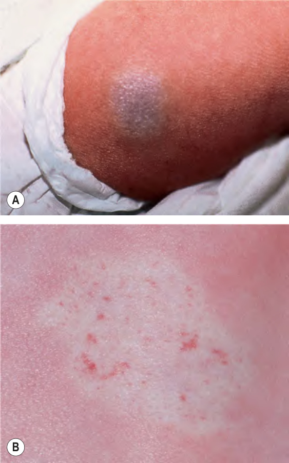
Fig. 103.1 Infantile hemangioma precursors. These premonitory lesions can have a “bruised” (A) or blanched (B) appearance. B, Courtesy Julie V. Schaffer, MD.
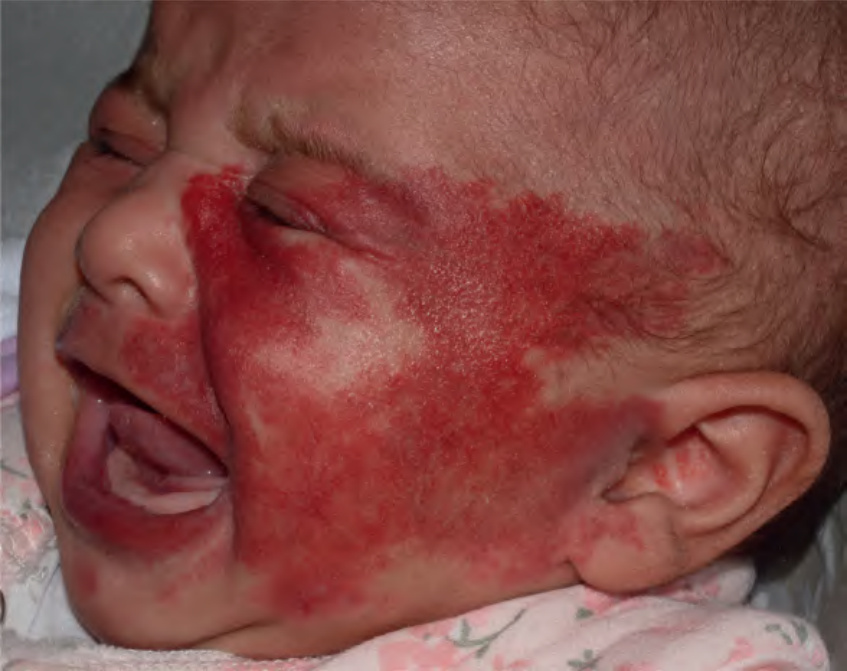
Fig. 103.2 Segmental superficial infantile hemangioma mimicking a port-wine birthmark. This 3-week-old girl is at risk of PHACE(S) syndrome. Courtesy Julie V. Schaffer, MD.
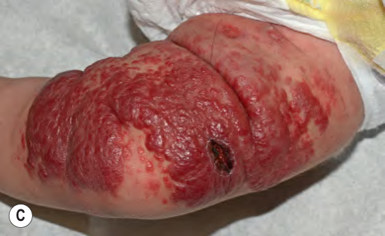
Fig. 103.3 Superficial infantile hemangiomas.
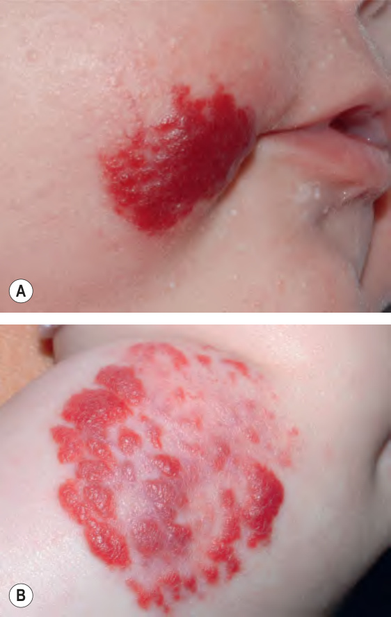
Fig. 103.3 Superficial infantile hemangiomas.
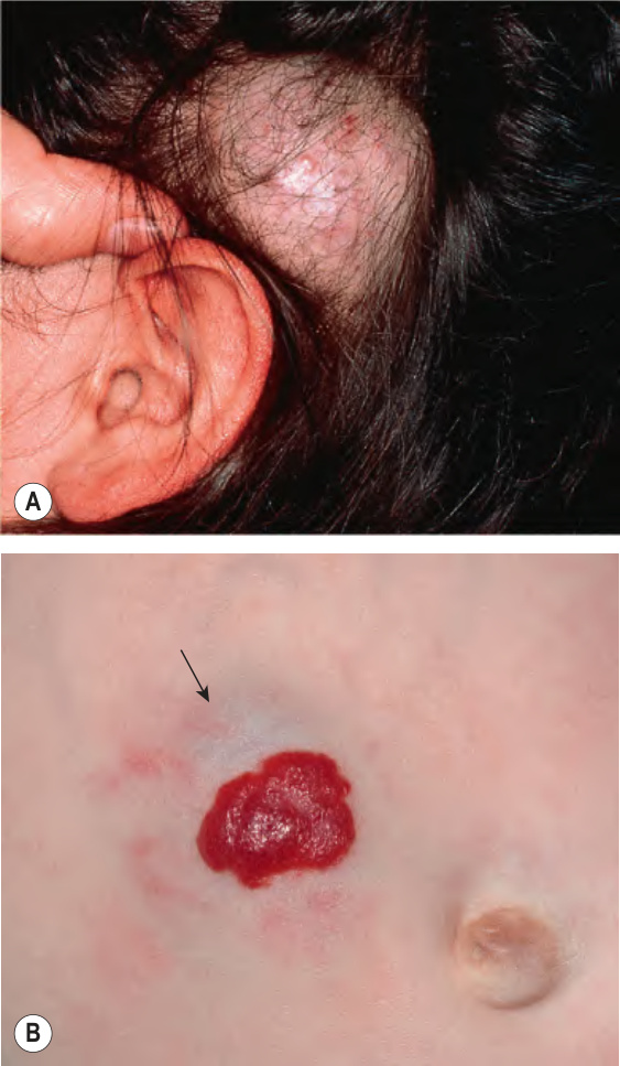
Fig. 103.4 Deep and mixed infantile heman- giomas.A Skin-colored deep hemangioma with overlying telangiectasias and alopecia on the scalp. B Mixed hemangioma composed of a well-demarcated, bright red superficial plaque plus a broader, ill-defined underlying deep nodule with a subtle light blue– purple hue (arrow) and scattered telangiectasias. B, Courtesy Julie V. Schaffer, MD.
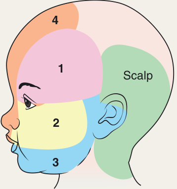
Fig. 103.5 Segmental hemangioma patterns. Based upon a prospective study (n = 165) of image analyses of hemangiomas. Adapted with permission from Endicott AA, Chamlin SL, Drolet BA, et al. Mapping of segmental and partial segmental infantile hemangiomas of the face and scalp. JAMA Dermatol 2021;157:1328–34.
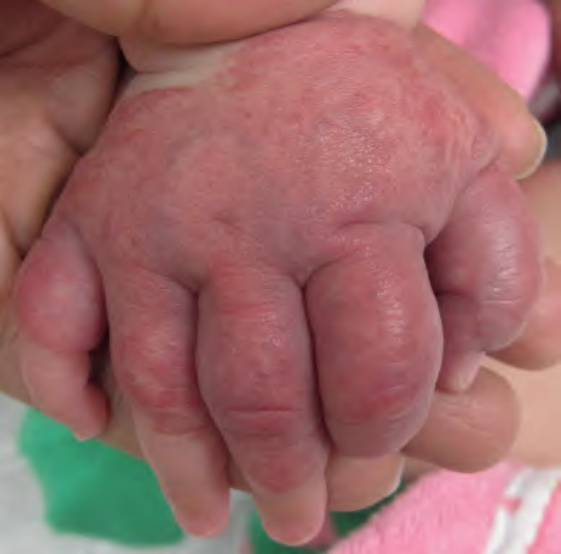
Fig. 103.6 “Biker glove” pattern of an acral infantile hemangioma with minimal or arrested growth. Note the characteristic sparing of the distal fingers.
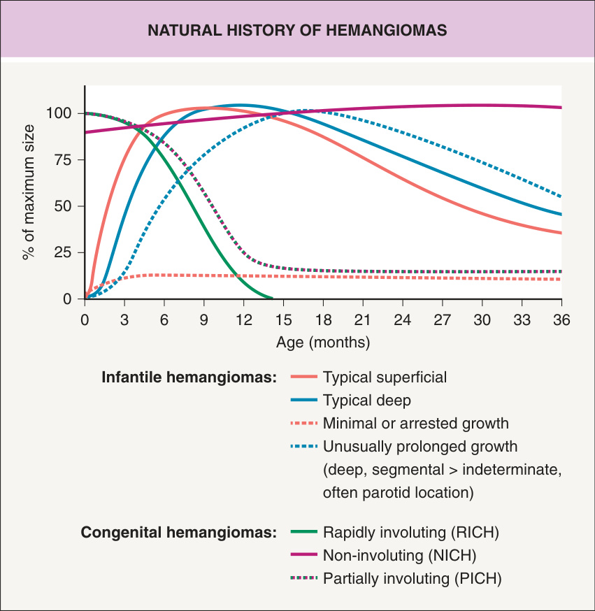
Fig. 103.7 Natural history of infantile and congenital hemangiomas.
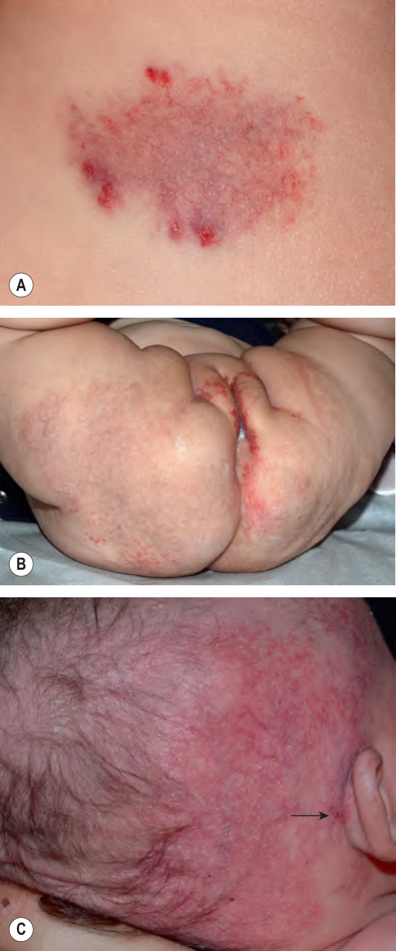
Fig. 103.8 Infantile hemangiomas with minimal or arrested growth (IH-MAG).
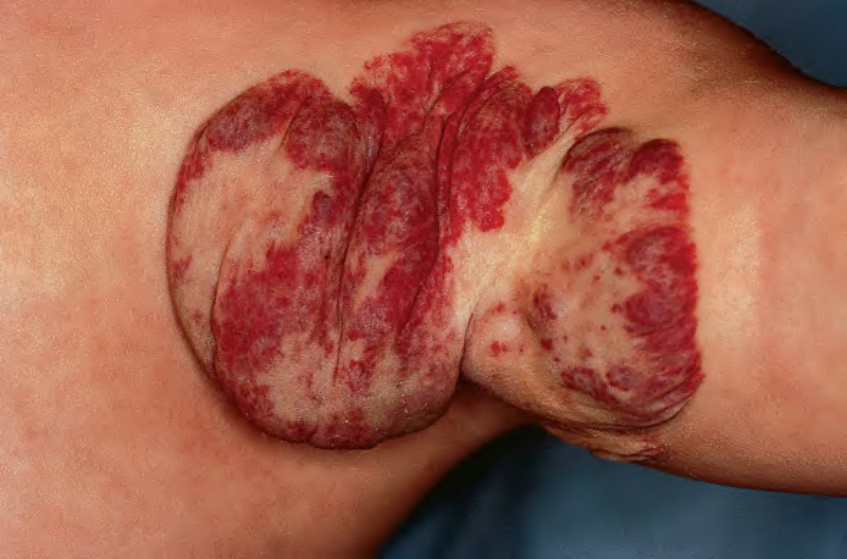
Fig. 103.9 Infantile hemangioma during involution. Note the patchy light-ening in surface color and softening.
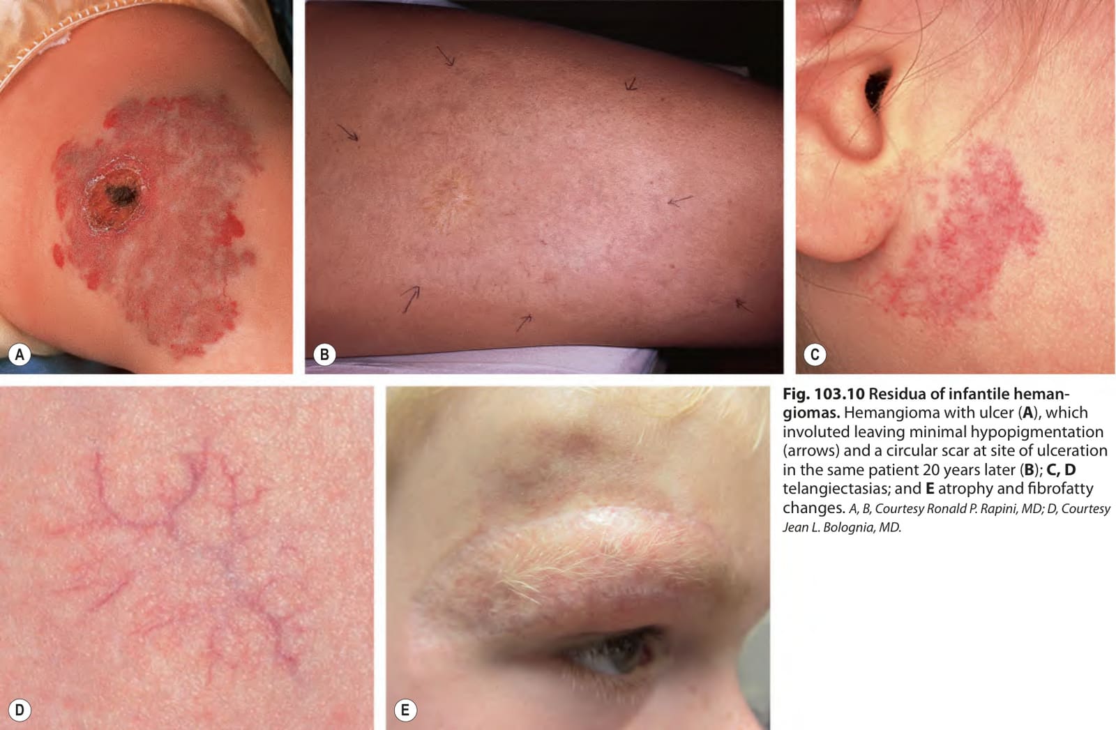
Fig. 103.10 Residua of infantile heman- giomas. Hemangioma with ulcer (A), which involuted leaving minimal hypopigmentation (arrows) and a circular scar at site of ulceration in the same patient 20 years later (B); C, D telangiectasias; and E atrophy and fibrofatty changes. A, B, Courtesy Ronald P. Rapini, MD; D, Courtesy Jean L. Bolognia, MD.
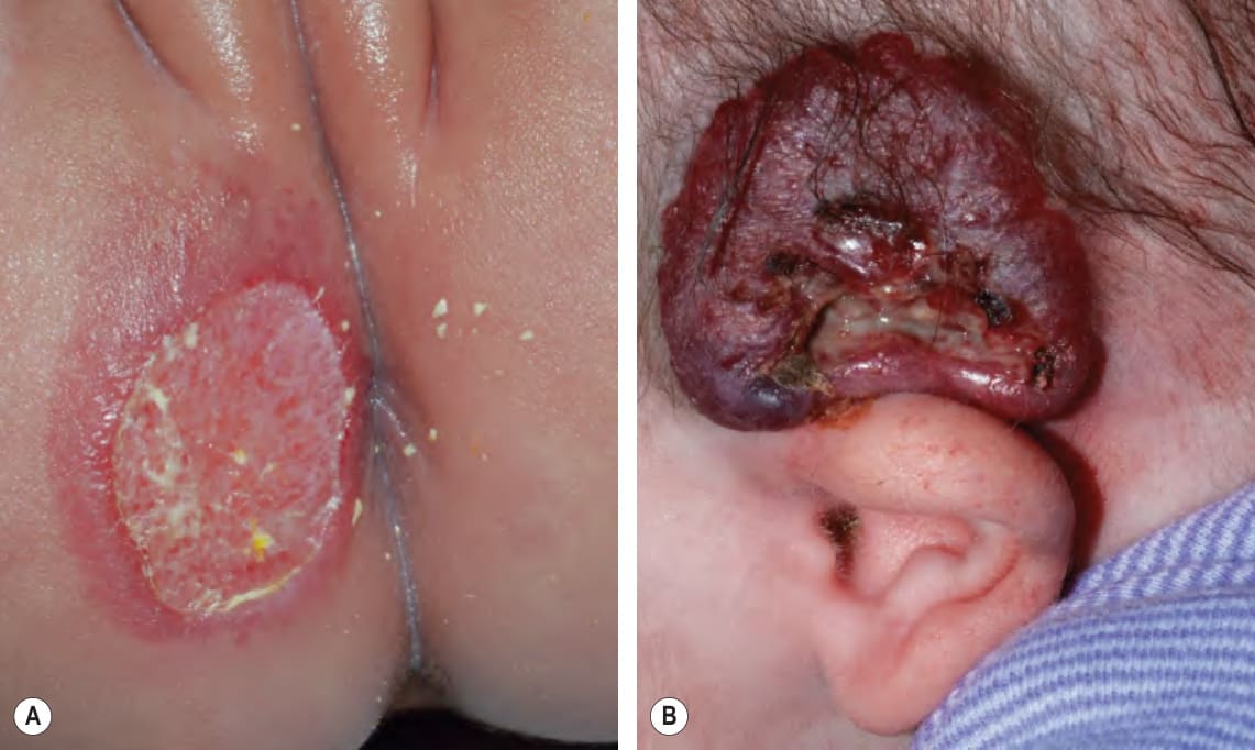
Fig. 103.11 Ulcerated hemangiomas.A Ulcerated early superficial hemangioma on the buttock. Because the hemangioma component may not be obvious, this diagnosis should be considered when an infant presents with an ulcer in the diaper area. B Ulcerated thick superficial hemangioma above the ear. Note the distortion of the pinna. Courtesy Julie V. Schaffer, MD.
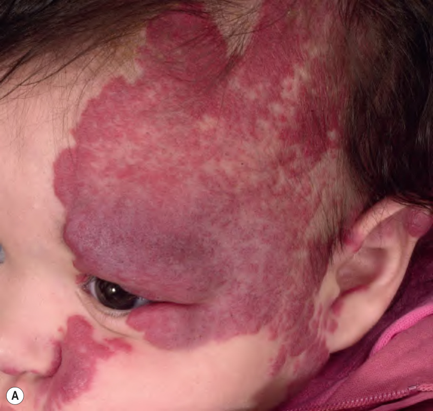
Fig. 103.12 Infantile hemangiomas affecting the eyelids. These superficial segmental (A) and deep (B) infantile hemangiomas are obstructing the visual axis. Courtesy Julie V. Schaffer, MD.
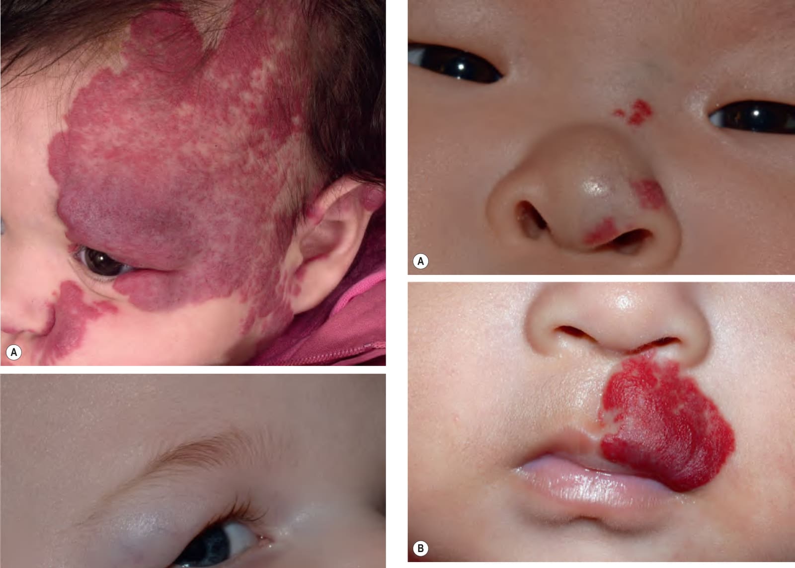
Fig. 103.13 Infantile hemangiomas in facial sites with increased risk of disfig- urement.A Nasal deformity can result from mixed or deep hemangiomas on the nasal tip. B The lip is a sensitive site, especially when lesions cross the vermilion border or involve the philtrum, and hemangiomas in this location are prone to ulceration. Courtesy Julie V. Schaffer, MD.
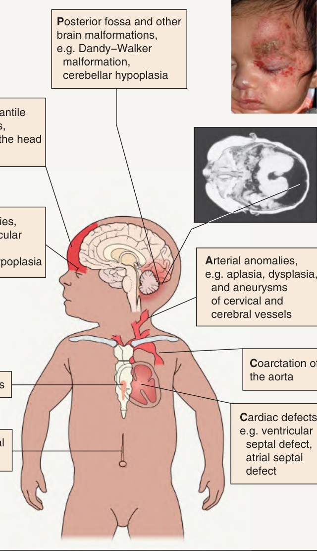
Fig. 103.14 PHACE(S) syndrome. Major clinical features are illustrated. Additional possible associated features include hearing impairment, dysphagia, speech/language delay, dental anomalies, and endocrine abnormalities such as hypopituitarism and hypothyroidism. Inset, courtesy Julie V. Schaffer, MD.
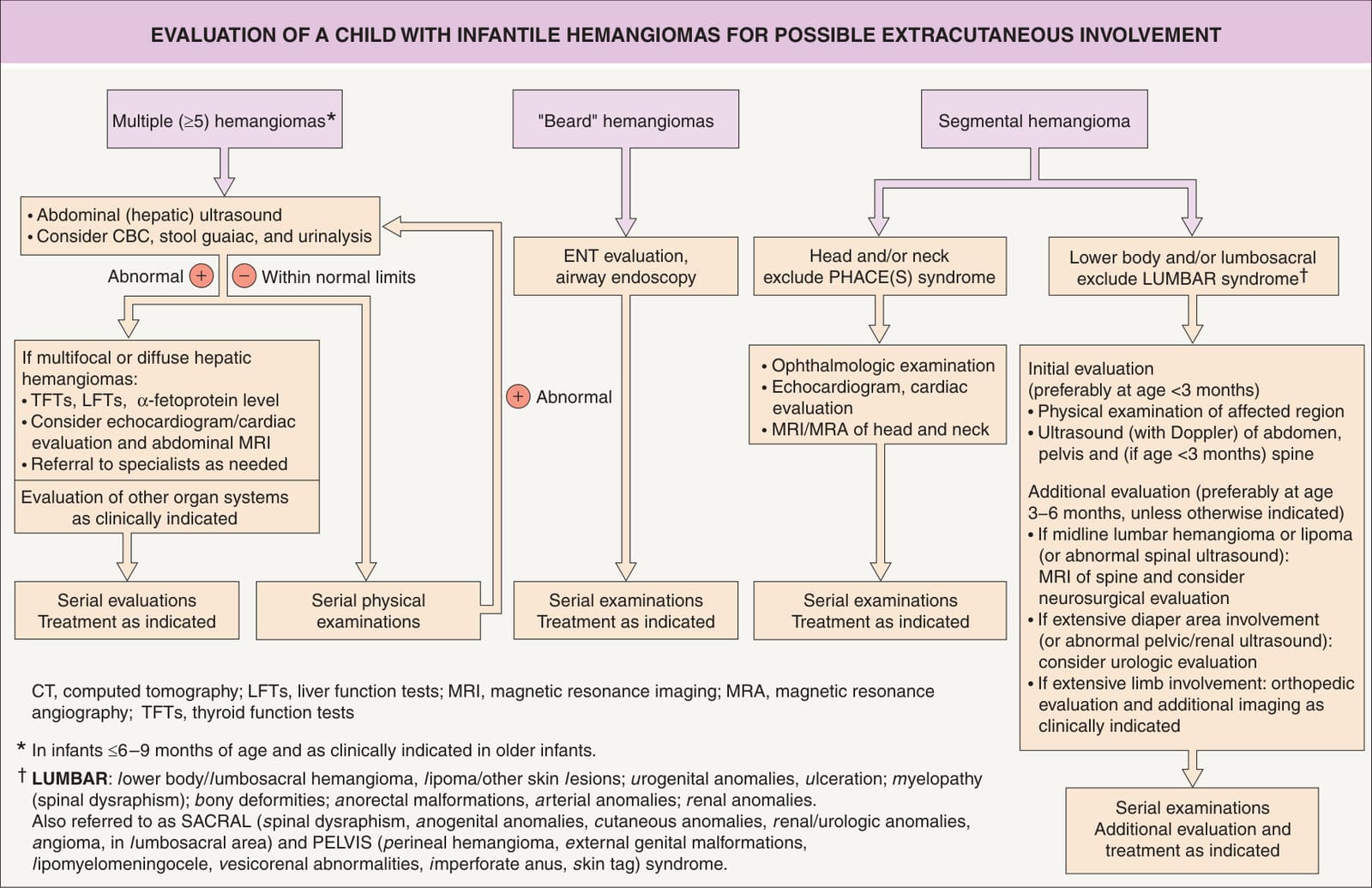
Fig. 103.15 Evaluation of a child with infantile hemangiomas for possible extracutaneous involvement.
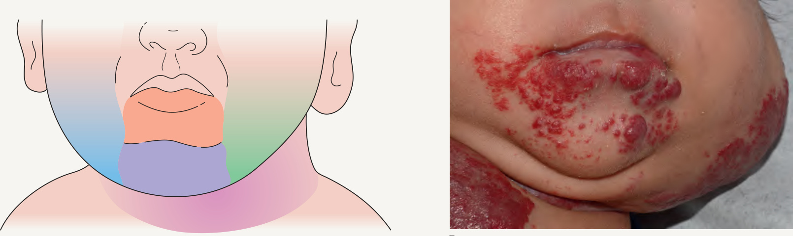
Fig. 103.16 Hemangiomas in a “beard” distribution.A Anatomic sites associated with risk of an airway hemangioma. B Infant with a laryngeal hemangioma in addition to cutaneous lesions in a “beard” distribution. A, Adapted, with permission, from ref. 71; B, Courtesy Julie V. Schaffer, MD.
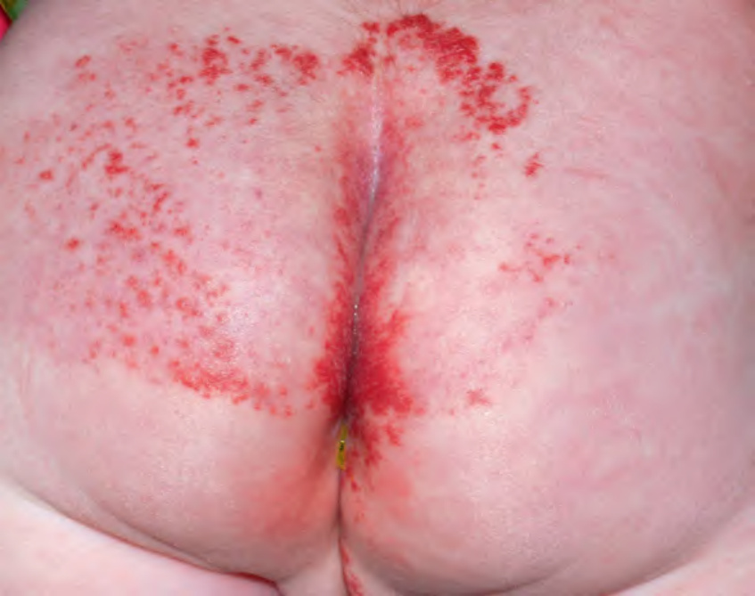
Fig. 103.17 Segmental lumbosacral hemangioma. This child is at risk for LUMBAR syndrome. Courtesy Julie V. Schaffer, MD.
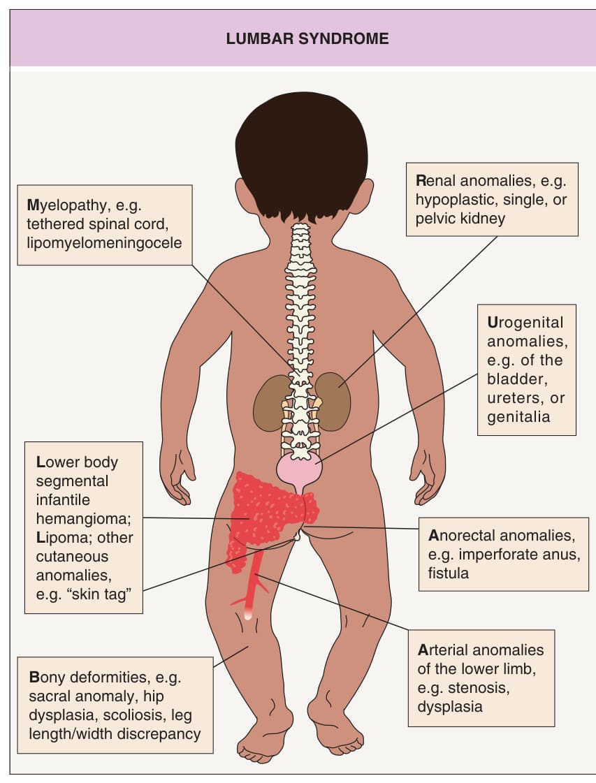
Fig. 103.18 LUMBAR syndrome. Major clinical features are illustrated. The segmental hemangioma is often ulcerated.
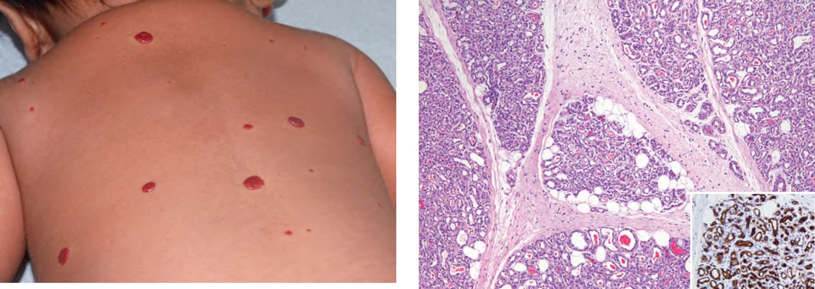
Fig. 103.19 Multifocal infantile hemangiomas with extracutaneous heman- giomas. In this child, many small, superficial skin lesions were present in a miliary pattern and hemangiomas were present in the liver. Historically, this was referred to as “diffuse neonatal hemangiomatosis”. Courtesy Julie V. Schaffer, MD.
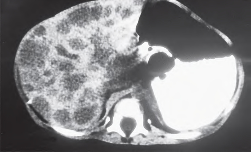
Fig. 103.20 Multifocal hepatic hemangiomas. CT scan demonstrating multiple hepatic hemangiomas in a child with multifocal cutaneous infantile hemangiomas (“diffuse neonatal hemangiomatosis”). Courtesy Amy Paller, MD.
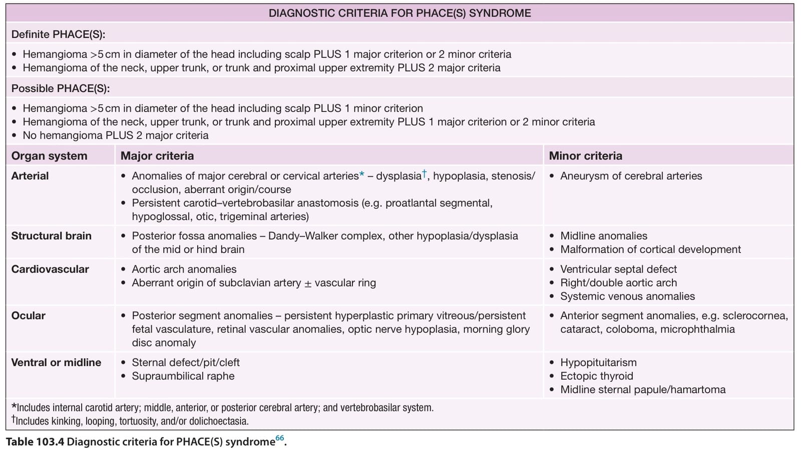
Table 103.4 Diagnostic criteria for PHACE(S) syndrome.
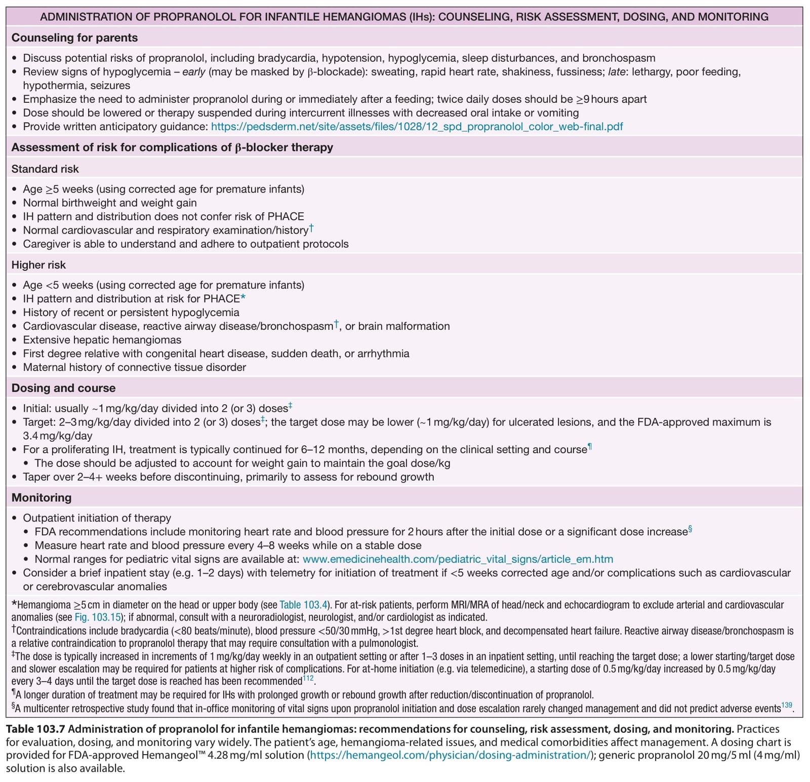
Table 103.7 Administration of propranolol for infantile hemangiomas: recommendations for counseling, risk assessment, dosing, and monitoring. Practices for evaluation, dosing, and monitoring vary widely. The patient’s age, hemangioma-related issues, and medical comorbidities affect management. A dosing chart is provided for FDA-approved Hemangeol™ 4.28 mg/ml solution (https://hemangeol.com/physician/dosing-administration/); generic propranolol 20 mg/5 ml (4 mg/ml) solution is also available.