PATHOLOGY
The histopathologic features of IH correlate with the stage of hemangioma development (see Table 103.1). IHs may extend from the super-ficial dermis to the deep subcutaneous tissue; in some locations, there may be involvement of surrounding tissues such as salivary glands and muscle. Biopsy specimens of proliferating hemangiomas demonstrate well-defined non-encapsulated masses composed of proliferating plump endothelial cells and pericytes. Small vascular lumens may be noted focally throughout the tumor, but lumen formation may be more difficult to appreciate in early proliferating lesions.
Later in the proliferating stage, lobules of endothelial masses separated by fibrous septae become more prominent (Fig. 103.21). Larger feeding and draining vessels are noted within the septae, and mitotic figures and apoptotic bodies may be present within the tumor mass. Although abnormal mitotic figures are not typical of proliferating hemangiomas, some authors have noted that mitotic activity and mild nuclear pleomorphism may be present in IHs and should not be a cause for concern if other typical histopathologic features are also present. An increased number of mast cells is often present within proliferating hemangiomas, and some studies have found even higher numbers of mast cells during early involution.
The involution phase is marked by flattening of the endothelium and reduced numbers of mitotic figures. Eventually, the vessels decrease in number and lose their tightly packed appearance, with fibrous and fatty tissue separating the vessels within and between lobules. The feeding and draining vessels within the septae may persist. Fully involuted lesions display fibrofatty tissue and a few persistent vessels at sites where tumor lobules and septae previously existed.
Immunohistochemical analysis is useful for confirming the diagnosis of IH. A high level of GLUT1 immunoreactivity is present in the endothelia within IHs of all phases (see Fig. 103.21). GLUT1 staining is absent in most other types of vascular tumors and vascular malformations. Wilms tumor 1 (WT1) expression is also observed in IH but not vascular malformations, with the exception of arteriovenous malformations. In addition, other placenta-associated vascular proteins, including FcγRII, merosin and Lewis Y antigen, are present in IHs but absent in vascular malformations and pyogenic granulomas. These placental markers are also absent in normal vessels of the skin and subcutis.
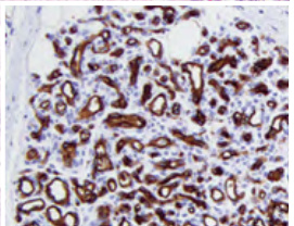
Fig. 103.21 Histologic features of a proliferating infantile hemangioma.
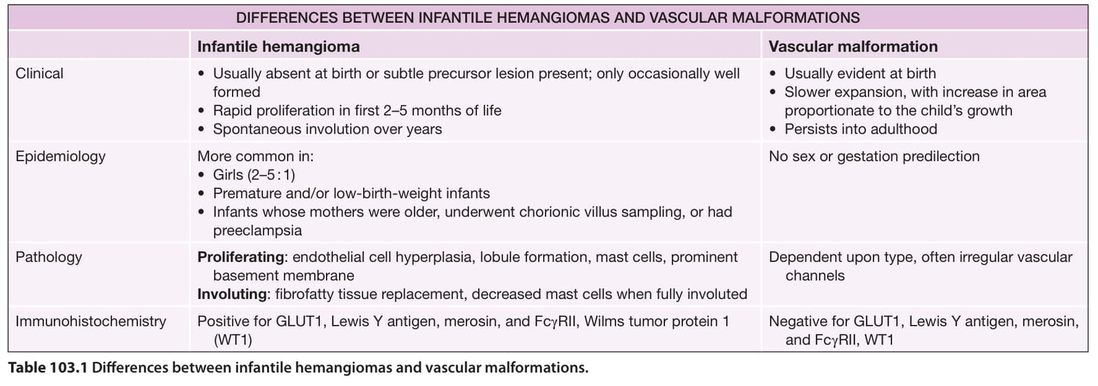
Table 103.1 Differences between infantile hemangiomas and vascular malformations.