DIFFERENTIAL DIAGNOSIS
Superficial and mixed hemangiomas are usually diagnosed based upon their characteristic clinical features. Hemangioma precursors and early proliferating lesions may sometimes be misdiagnosed as capillary malformations or telangiectasias. Pyogenic granulomas (PGs) occur commonly in children and may mimic a superficial hemangioma; however, PGs usually arise after the first few months of life, are typically pedunculated, and tend to bleed. Other vascular lesions of infancy that may be in the differential diagnosis of superficial hemangioma include tufted angioma, multifocal lymphangioendotheliomatosis with thrombocytopenia, infantile hemangiopericytoma, spindle cell hemangioma, verrucous venulocapillary malformation (“verrucous hemangioma”), and eccrine angiomatous hamartoma.
Deep hemangiomas provide a greater diagnostic challenge, and radiographic evaluation may be helpful. Other vascular anomalies that may be considered in the differential diagnosis of a deep hemangioma include a venous, lymphatic, or combined venous–lymphatic malformation (see Ch. 104) as well as a congenital hemangioma (see below; Table 103.5) or kaposiform hemangioendothelioma (see Ch. 114). Kasabach–Merritt phenomenon (KMP), a potentially life-threatening thrombocytopenic coagulopathy, typically presents during the first months to year of life with a rapidly enlarging, ecchymotic mass (see Ch. 114); KMP is now known to be primarily associated with kaposiform hemangioendotheliomas and less often tufted angiomas, but not IHs as historically assumed.
Congenital fibrosarcoma is often firm and blue–purple in color and can have ectatic superficial veins, which may lead to misdiagnosis as a hemangioma. In addition, some patients with this malignancy present with disseminated intravascular coagulation, which further confuses the clinical picture. Rhabdomyosarcoma, neuroblastoma,
primitive neuroectodermal tumor, lymphoblastic lymphoma, and dermatofibrosarcoma protuberans are other neoplasms that may have a vascular appearance due to overlying telangiectasias or a blue–red color. Additional entities that may be mistaken for a hemangioma include: infantile myofibromatosis – solitary or multiple red to pink plaques or nodules; lipoblastoma – an enlarging skin-colored or (less often) erythematous mass; and nasal glial heterotopia – classically a congenital blue–red mass on the bridge of the nose. Radiographic studies, especially MRI, may be useful in differentiating these tumors from a hemangioma. However, if the diagnosis is not clarified by clinical and radiographic examinations, or if the lesions are atypical, then histologic examination is indicated.
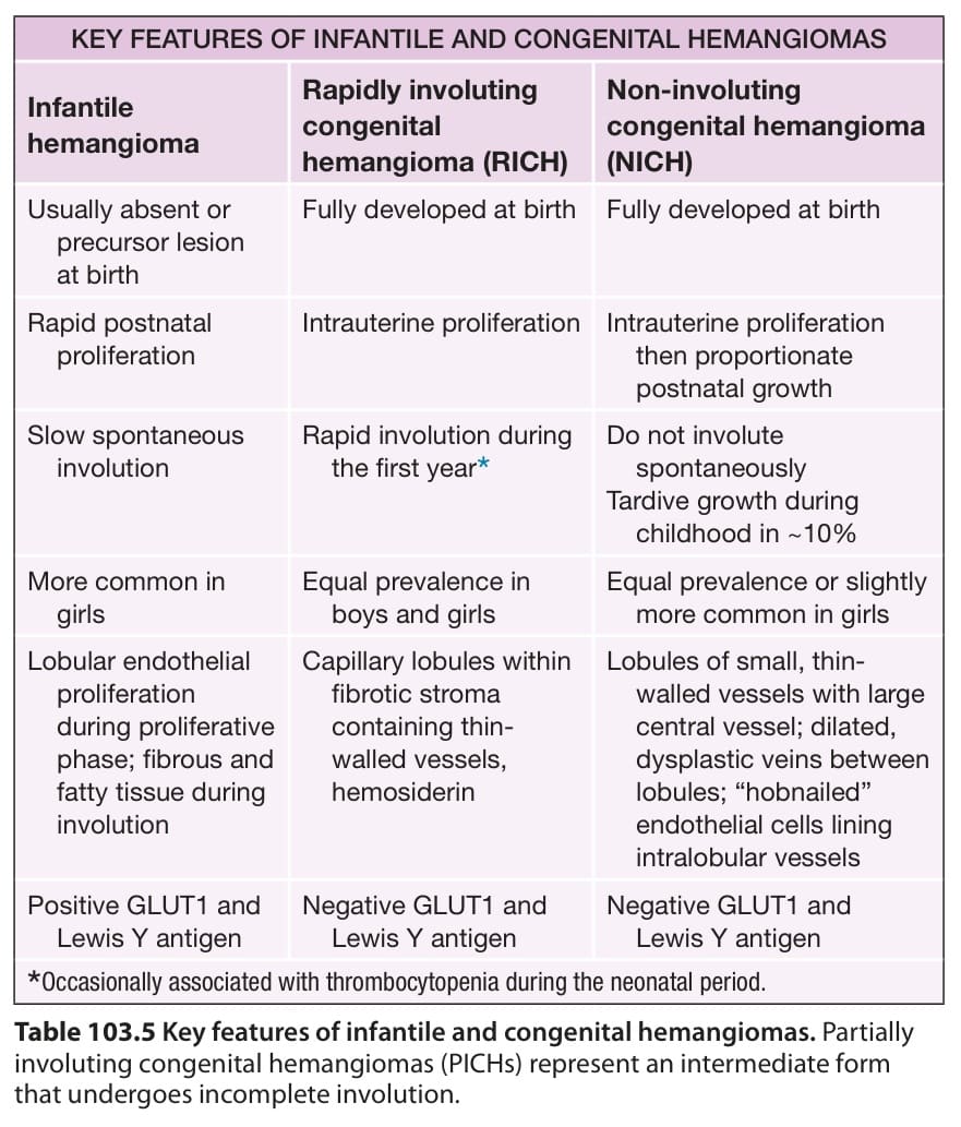
Table 103.5 Key features of infantile and congenital hemangiomas. Partially involuting congenital hemangiomas (PICHs) represent an intermediate form that undergoes incomplete involution.