TREATMENT
IHs manifest with a wide spectrum of clinical severity, and their management needs to be individualized. The Hemangioma Activity Scale and Hemangioma Severity Scale were developed to help objectively assess IHs and their response to therapy. Active intervention is recommended for IHs with a greater risk of developing local or systemic complications (Table 103.6). The major goals of management include: (1) preventing or reversing life- or function-threatening complications; (2) treating ulcerations; (3) preventing permanent disfigurement; (4) minimizing psychosocial distress to patients and their families; and (5) avoiding overly aggressive, potentially scarring procedures for lesions that have a strong probability of involuting without significant residua.
Active Non-Intervention
Small, uncomplicated hemangiomas carry an excellent prognosis and are often managed with active non-intervention, especially in extrafacial sites. However, physicians should recognize that even small, seemingly trivial lesions may be troubling to parents and cause significant distress. Active non-intervention includes addressing parental concerns as well as discussing the expected natural history of IHs, indications for treatment, types of therapy, and why such interventions are or are not appropriate for their child. The hemangioma can be followed with periodic clinical assessment and photography. Reviewing photographic examples of the evolution of hemangiomas may be helpful for families. Many parents are fearful of bleeding; therefore, reviewing the care for superficial bleeding, along with reassurance that it is rarely problematic, is worthwhile.
Management of Ulceration
Ulceration is the most frequent complication of IHs (see Fig. 103.11). Management should be directed at healing the ulceration, preventing infection, and reducing pain. Multiple modalities are often used concurrently, with no single therapy proven to be most effective. Components of management include wound care, treatment of infection (relatively uncommon), specific therapies (e.g. topical or systemic β-blockers), and pain control.
Wound care
Many small, superficial ulcerations can be managed with local wound care. Saline solution compresses may be used to gently debride thick
crusts. Although infection is uncommon, topical antibacterial ointments (e.g. mupirocin) can be applied for occlusive as well as antimicrobial effects. If clinical signs of infection develop, a bacterial culture should be obtained. Occlusive dressings (e.g. hydrocolloid, foam, siliconebased) aid in healing and pain control. They can be applied after the topical antibiotic or petrolatum at sites that are amenable (see Ch. 145). Thin hydrocolloid dressings are often favored because of the ease of application over curved surfaces and the ability to leave them in place for a few days. Inexpensive petrolatum-impregnated gauze is another option and it is especially helpful for perineal ulcers, but needs to be changed frequently.
Specific therapy
Modalities that are utilized to treat hemangiomas for other indications (see below) may be helpful in treating ulcerated lesions. Application of topical timolol can aid in healing of small ulcerated IHs, but caution must be taken regarding systemic absorption (see below).
Oral propranolol therapy is often useful for ulcers that are extensive or have failed local therapies. In one study of ulcers present for a mean of 7 weeks, the median time to healing after initiating propranolol was 4 weeks and most patients achieved pain relief within 15 days. Although early propranolol therapy may help to prevent ulceration of IHs in high-risk locations, precipitation or worsening of ulceration in such lesions has also been described with propranolol administration. Initiation of propranolol at a low dose with slow escalation is recommended for IHs with ulceration or at high risk of ulceration – a recent retrospective study found that ulcer healing time was shorter with a propranolol dose of ≤1 mg/kg/day compared to higher doses.
Pulsed dye laser has been used for the treatment of ulcerated hemangiomas, especially in combination with other modalities. Some uncontrolled studies demonstrated healing of ulcerations and resolution of pain after two to three treatments. However, others observed mixed results, with 50% showing improvement and 5% experiencing worsening of the ulceration. Excisional surgery may be considered for small or pedunculated ulcerated lesions.
Pain control
An important consideration in the treatment of ulcerated hemangiomas is pain control. Local wound care, and especially occlusive dressings, can provide some pain relief. Oral acetaminophen and topical lidocaine 2%–5% ointment may also help to alleviate the discomfort. Lidocaine ointment should be used sparingly (e.g. maximum of a pea-sized amount) to prevent systemic lidocaine toxicity. Eutectic mixture of local anesthetics is not recommended for the treatment of ulcerated hemangiomas because it should not be applied to eroded or ulcerated skin. In severe cases, consultation with a pain management specialist may be required.
Local Therapies
Topical β-blockers
Topical application of timolol maleate, a non-selective β-blocker, can be of benefit in the treatment of IHs, especially small superficial lesions that are <1 mm in thickness (Fig. 103.22). Timolol is available in several ophthalmic formulations, including 0.5% solution and 0.5% gel-forming solution (GFS; extended-release), with one drop (0.25 mg) twice daily of the latter most commonly utilized to treat IHs. A randomized control study found this dosing of timolol 0.5% GFS to be an effective and safe therapy for small (<2.5 cm), thin, non-ulcerated cutaneous IHs in infants 5–24 weeks of age, with benefit seen after 8–16 weeks of treatment. A pharmacokinetic study found that as hemangiomas with a prominent superficial component increased in thickness, higher plasma concentrations of topically applied timolol were observed. Although topical timolol treatment guidelines have not been established, it is recommended that the dose be limited to a maximum of 2 drops of timolol 0.5% GFS daily or <0.25 mg/kg/day. Despite systemic absorption, topical timolol treatment is generally well tolerated and serious adverse events are rare.
Intralesional corticosteroids
Historically, intralesional corticosteroids were commonly used to treat localized IHs; however, this modality is now utilized infrequently
A An ulcerated superficial infantile hemangioma on the neck of a 10-week-old girl. B Four weeks after institution of topical timolol 0.5% gel, with one small drop applied twice daily. Note the healing of the ulcer, which occurred within two weeks, as well as the lighter color and decreased thickness of the hemangioma. Courtesy Julie V. Schaffer, MD.
considering the efficacy of β-blocker therapy. Intralesional corticosteroids (e.g. triamcinolone <3 mg/kg per treatment session) may be employed for selected small, focal hemangiomas. However, serious complications, including retinal and ophthalmic artery occlusion leading to permanent vision loss, as well as local atrophy and necrosis, have limited the use of this modality in the periorbital area.
Systemic Therapies
Systemic β-blockers
The serendipitous discovery of the non-selective β-blocker propranolol as a treatment for IH in 2008 has revolutionized the management of this common tumor, and it currently represents the first-line systemic therapy for IHs. In 2014, propranolol (Hemangeol™) became the first FDA-approved treatment for IHs. Oral β-blockers are effective in halting growth and expediting regression of cutaneous, ocular, airway, and hepatic IHs as well as for complications such as ulceration.
The mechanisms of action of propranolol are thought to be multi-faceted. The color change from red to purple and softening that are observed within the first few days of treatment are likely related to immediate vasoconstrictive effects via β-adrenergic receptors on hemangioma pericyte cells. Subsequent progressive reduction in volume and redness may be due to decreases in serum concentration of VEGF, bFGF, and renin as well as induction of endothelial cell apoptosis.
Compared to systemic corticosteroids, propranolol has superior efficacy and a more favorable side-effect profile. Potentially serious adverse effects such as hypotension, bradycardia, hypoglycemia, and bronchospasm are uncommon. β-blockers may mask early signs of hypoglycemia, so infants should be fed in conjunction with medication administration. More common side effects include sleep disturbances (e.g. restless sleep, nightmares), cold extremities, diarrhea, and somnolence (especially in the first few days of therapy). There are rare reports of hyperkalemia associated with propranolol treatment of large ulcerated hemangiomas. Long-term studies have found no evidence of cognitive, developmental, or growth impairment in 4- to
7-year-old children who received propranolol treatment for hemangiomas during infancy.
Table 103.7 outlines recommendations for propranolol administration, although clinical practices regarding evaluation, dosing, and monitoring vary. Patients at risk of PHACE(S) syndrome should undergo evaluation for cardiovascular and cerebrovascular anomalies that may merit caution given the theoretical risk for hypotension. Most infants with PHACE(S) syndrome can be successfully treated with propranolol. However, depending on their extracutaneous manifestations, these patients may need to be managed together with cardiologists and/or neurologists, proceeding cautiously with dose escalation in individuals at increased risk for complications such as stroke. A history of airway reactivity or asthma may not be an absolute contraindication to propranolol therapy, but such patients require closer monitoring and consultation with a pulmonologist.
Propranolol dosing is weight-based, usually with a target dose of 2–3 mg/kg/day (see Table 103.7). In infants with ulcerated hemangiomas, doses of ≤1 mg/kg/day may reduce healing time. Treatment
is typically continued for 6–12 months, followed by gradual tapering. Approximately 25% of IHs undergo rebound growth after cessation of propranolol, with a higher risk if the lesion is deep or therapy is discontinued before 9 months of age.
β-blockers that do not cross the blood–brain barrier because they are hydrophilic (rather than lipophilic like propranolol) may potentially produce less CNS effects such as sleep disturbance. These agents include atenolol (β-selective) and nadolol (non-selective), neither of which is currently FDA-approved for IH treatment. Randomized controlled studies of atenolol and nadolol treatment of IH have demonstrated efficacy and safety similar to that of propranolol, with a faster response to nadolol. Because nadolol is excreted primarily in the feces, bowel movement frequency should be monitored and the medication held if substantial constipation. A recent study found that the R(+) enantiomers of propranolol and atenolol, which do not have β-blocker activity, inhibited formation of IH-like blood vessels from hemangioma stem cells in a murine xenograft model, suggesting the possibility of these agents’ utility for IH treatment.
Systemic corticosteroids
Systemic corticosteroids, usually prednisolone or prednisone, were traditionally employed for the treatment of life- or function-threatening hemangiomas (see Table 103.6). Corticosteroids suppress VEGF production by hemangioma-derived stem cells and inhibit vasculogenesis in a murine model. Although no longer a first-line therapy, systemic corticosteroids are still occasionally utilized when β-blocker therapy is contraindicated or in combination with β-blockers, e.g. for airway or diffuse hepatic hemangiomas. Initial doses equivalent to 2–3 mg/kg/day of prednisone are typically administered, with variable duration of therapy, tapering schedules, and monitoring. Adverse effects of systemic corticosteroids include cushingoid facies, irritability, disruption of sleep, gastrointestinal symptoms, hypertension, hypothalamic–pituitary–adrenal (HPA) axis suppression, and increased susceptibility to opportunistic infections (see Ch. 125).
Other systemic therapies
Sirolimus (rapamycin) inhibits the mTOR signaling pathway, which plays an important role in cell growth and proliferation (see Fig. 61.12). Oral sirolimus is emerging as a second-line systemic treatment for recalcitrant life- or function-threatening IHs. There have been reports of its successful use for large facial IHs that failed to respond to β-blocker therapy. Potential adverse effects include mucositis, hyperlipidemia, headaches, hepatotoxicity, and neutropenia. Laboratory monitoring is necessary including complete blood counts, serum creatinine, liver function tests and lipid panels, as well as sirolimus serum levels.
Vincristine and recombinant interferon-α (2a and 2b) represent historical therapies for severe, recalcitrant IHs that are no longer used because of potential complications such as peripheral neuropathy and spastic diplegia, respectively.
Surgical Therapy
Pulsed dye laser (PDL) therapy (585–600 nm wavelength, usually 0.45–1.5 ms pulse duration) is effective for the treatment of capillary malformations. Although PDL therapy may have some benefit for superficial IHs, there is risk for ulceration when it is used early in the growth phase. Multiple treatment sessions, usually every few weeks, are required during the proliferative phase to produce improvement. Considering the superior efficacy of β-blockers, PDL is currently not a first-line therapy for proliferating IHs. However, PDL therapy is often helpful for residual telangiectasias following propranolol therapy and/or involution of the IH. Potential adverse effects from PDL therapy include pigmentary alteration and atrophic scarring as well as ulceration. The Nd:YAG laser has also been used to treat IHs, either alone or in combination with the PDL; however, the higher risk of scarring limits its utility.
The fractional CO laser has been used to improve the texture of residual atrophy and scarring following IH involution. Multiple treatments are typically required and may not be well tolerated without sedation.
Surgical excision may be employed for involuted or partially involuted lesions to remove fibrofatty tissue and redundant skin. The optimal timing of surgery depends upon multiple factors, including the location, size, and morphology (e.g. pedunculated vs sessile) of the lesion. In a retrospective study of 112 IHs resected by a single surgeon, 87% of the lesions were located on the head or neck, 38% of patients had received other treatments during the proliferative phase, and most excisions were performed at 2–3 years of age.
Surgical excision during the proliferative phase is controversial and usually reserved for situations in which: (1) a function-threatening hemangioma, e.g. a periocular lesion blocking the visual axis or lip lesion interfering with feeding or speech, failed to respond to pharmacologic therapy; or (2) pharmacologic therapy is believed to pose a greater risk to the patient than surgical excision. Other settings where early surgery may be appropriate include pedunculated lesions for which resection will be inevitable and small, persistently ulcerated lesions that have failed conservative therapy. The ultimate management goal is to achieve as normal an appearance as possible, considering the expected surgical scar as well as the predicted time course and outcome of involution. Therefore, just as with other treatment modalities, the surgical management of IHs needs to be individualized.
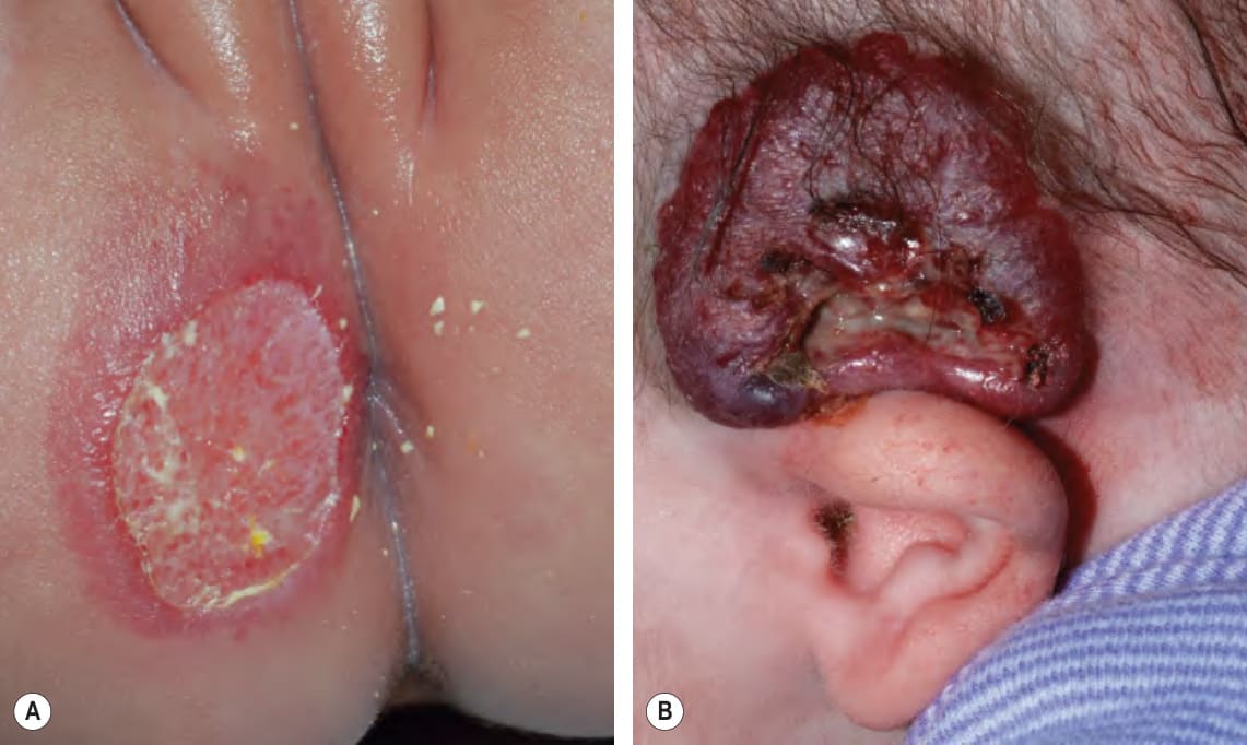
Fig. 103.11 Ulcerated hemangiomas.A Ulcerated early superficial hemangioma on the buttock. Because the hemangioma component may not be obvious, this diagnosis should be considered when an infant presents with an ulcer in the diaper area. B Ulcerated thick superficial hemangioma above the ear. Note the distortion of the pinna. Courtesy Julie V. Schaffer, MD.
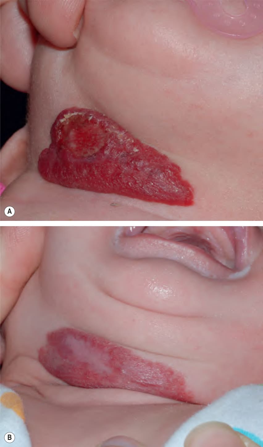
Fig. 103.22 Ulcerated superficial hemangioma treated with topical timolol.
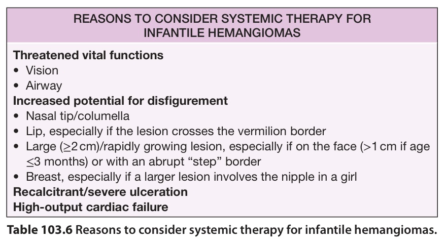
Table 103.6 Reasons to consider systemic therapy for infantile hemangiomas.
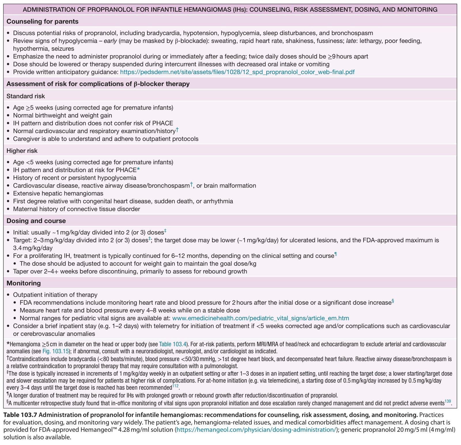
Table 103.7 Administration of propranolol for infantile hemangiomas: recommendations for counseling, risk assessment, dosing, and monitoring. Practices for evaluation, dosing, and monitoring vary widely. The patient’s age, hemangioma-related issues, and medical comorbidities affect management. A dosing chart is provided for FDA-approved Hemangeol™ 4.28 mg/ml solution (https://hemangeol.com/physician/dosing-administration/); generic propranolol 20 mg/5 ml (4 mg/ml) solution is also available.