INTRODUCTION
According to the classification system adopted by the International Society for the Study of Vascular Anomalies (ISSVA), there are two types of vascular anomalies: vascular tumors (most commonly the infantile hemangioma) and vascular malformations (Table 104.1; see Table 103.2). Vascular malformations are localized defects of vascular morphogenesis, likely caused by dysfunction in pathways regulating the formation of vascular channels during embryonic development, including effects on endothelial cell growth and survival. True angiogenesis may occur in some of these lesions, potentially explaining their propensity to thicken and even expand over time. However, in general, vascular malformations do not exhibit the markers of endothelial cell proliferation seen in infantile hemangiomas during their proliferative phase (Fig. 104.1). The suffix “oma” (meaning “tumor”) has therefore been deemed inaccurate for vascular malformations, and terms such as “angioma”, “lymphangioma”, and “hemangioma” should not be used for these lesions.
In the current ISSVA classification system, which was updated in 2018, vascular malformations are categorized depending on the predominant type(s) of anomalous vessels:
●capillary (CM), classically a port-wine birthmark (PWB), but also including telangiectasias, cutis marmorata telangiectatica congenita (CMTC), and nevus simplex
●venous (VM): a misnomer for this entity is “cavernous hemangioma”, a term with a long history of use to describe deep infantile hemangiomas
●lymphatic (LM): this category includes microcystic and macrocystic lesions, which were previously termed lymphangioma and cystic hygroma (respectively), along with more extensive forms with visceral or bone involvement. Central conducting lymphatic anomalies and primary lymphedema are also included in this category.
●arteriovenous (AVM): fast-flow lesions characterized by malformed arteries, veins, and capillaries with direct communications that produce arteriovenous shunting.
Vascular malformations are further divided into four groups: (1) simple – containing only one type of vessel or an AVM; (2) combined – composed of ≥2 types of vessels, e.g. capillary–venous (CVM), capillary–lymphatic (CLM), capillary–lymphatic–venous (CLVM), lymphatic–venous (LVM), capillary–arteriovenous (C-AVM), or capillary–lymphatic–arteriovenous (CL-AVM); (3) major named vessels involved; and (4) associated with other anomalies as a part of a complex syndrome, which have classically been referred to with eponyms such as Sturge–Weber and Klippel–Trenaunay syndromes (see Table 103.2). In a few scenarios, vascular tumors and malformations occur together, such as spindle cell hemangiomas and VMs in patients with Maffucci syndrome or pyogenic granulomas superimposed on a CM or AVM. Co-segregation of infantile hemangiomas and vascular malformations within families has also been observed.
Vascular malformations may occur in any body part or organ system. They are most easily identified in the skin and mucous membranes, with some of these lesions invading deep into underlying muscles, bones, or joints. Others are found in visceral locations. Vascular malformations can appear as localized lesions of varying sizes, occur within a segmental distribution, or be dispersed as multifocal or disseminated lesions.
Vascular malformations are persistent and some worsen over time if not treated. Categorizing a lesion as a CM, LM, VM, AVM, or combined lesion is important, since diagnostic modalities as well as treatment differ depending on the type(s) of vessels involved. The genetic bases of multiple types of vascular malformations and associated syndromes have been elucidated (Table 104.2, Fig. 104.2). This has been incorporated in the ISSVA classification and provides the foundation for targeted approaches to therapy. In many patients, a multidisciplinary team is required to provide optimal management. Cure is rarely achievable. A realistic aim is improvement and minimization of cosmetic or functional consequences. Many patients require longitudinal care from infancy through adulthood.
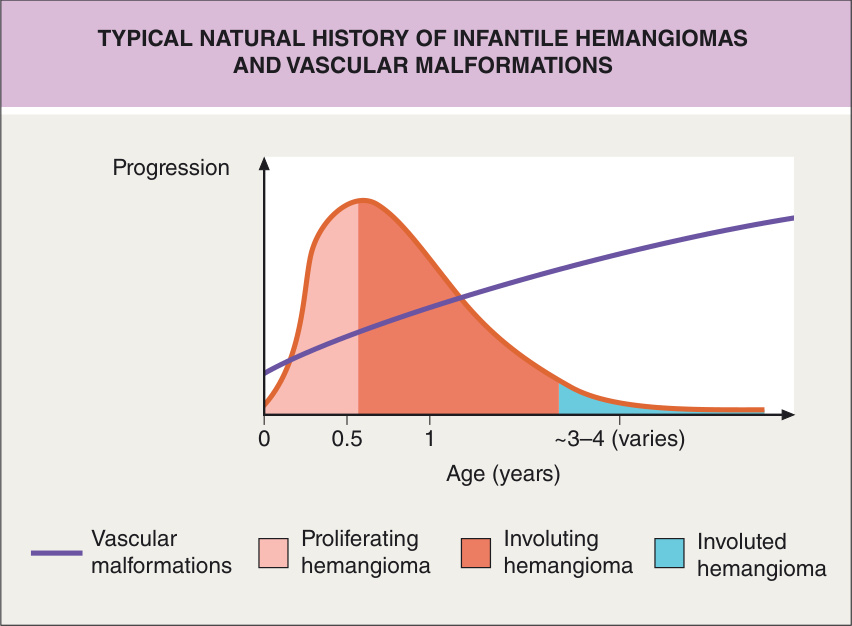
Fig. 104.1 Typical natural history of infantile hemangiomas and vascular malformations.
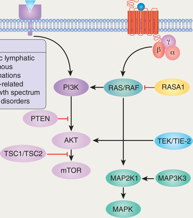
Fig. 104.2 Signaling pathways implicated in vascular malformations. MAPK, mitogen-activated protein kinase; MAP2K1, MAPK kinase 1; MAP3K3, MAPK kinase kinase kinase 3; mTOR, mechanistic target of rapamycin; PIK3CA, phosphatidylinositol-4,5-biphosphate 3-kinase catalytic subunit α.
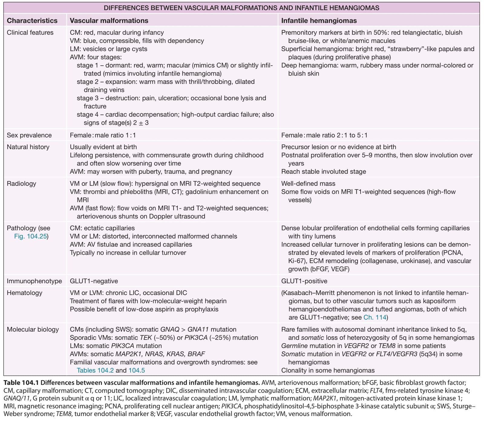
Table 104.1 Differences between vascular malformations and infantile hemangiomas. AVM, arteriovenous malformation; bFGF, basic fibroblast growth factor; CM, capillary malformation; CT, computed tomography; DIC, disseminated intravascular coagulation; ECM, extracellular matrix; FLT4, fms-related tyrosine kinase 4; GNAQ/11, G protein subunit α q or 11; LIC, localized intravascular coagulation; LM, lymphatic malformation; MAP2K1, mitogen-activated protein kinase kinase 1; MRI, magnetic resonance imaging; PCNA, proliferating cell nuclear antigen; PIK3CA, phosphatidylinositol-4,5-biphosphate 3-kinase catalytic subunit α; SWS, Sturge– Weber syndrome; TEM8, tumor endothelial marker 8; VEGF, vascular endothelial growth factor; VM, venous malformation.
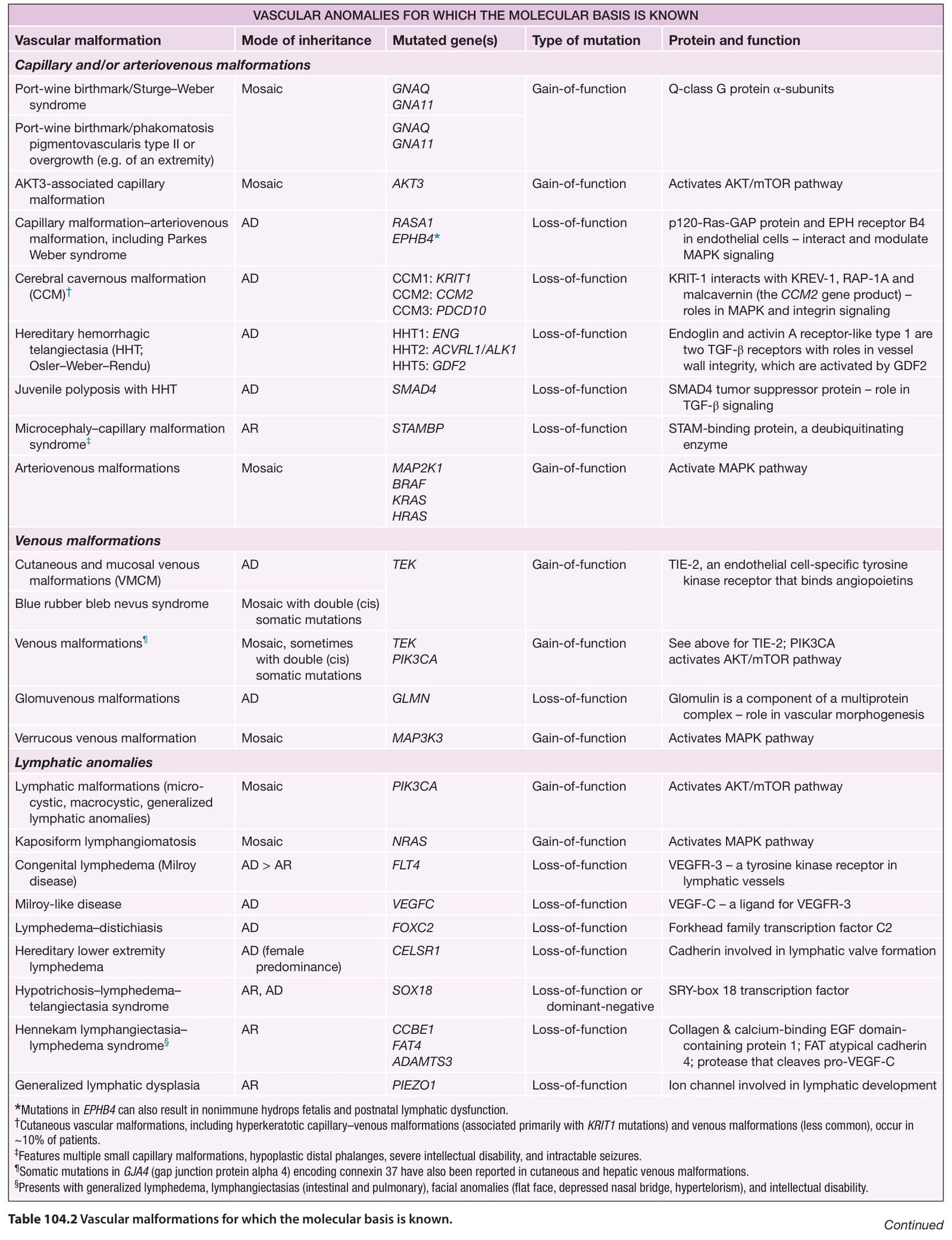
Table 104.2 Vascular malformations for which the molecular basis is known. Continued