PATHOGENESIS
An initial embryologic process, vasculogenesis, creates the primitive vascular plexus. Then angiogenesis, the secondary sprouting of
mesoderm-derived endothelial cells, forms new vessels from existing ones, thereby generating most blood and lymphatic vessels. Endothelial cells differentiate and recruit smooth muscle cell precursors to ensheathe them and build vessel walls. Changes in channel size, morphology, and rheology then create capillaries, veins, and arteries.
Vascular malformations constitute a heterogeneous group of disorders that result from alterations in the formation of blood or lymphatic channels. Dysfunction in signaling processes that regulate migration, differentiation, maturation, adhesion, and survival of the cells of vascular walls is thought to have a pathogenic role. Markers of cellular proliferation are not elevated in vascular malformations. In the cephalic region of the embryo, mural cells associated with endothelial cells come from the neural crest; therefore, a vascular malformation complex involving the head, such as Sturge–Weber syndrome (SWS), is caused by a somatic mutation in the embryo’s anterior neural crest or adjacent cephalic mesenchyma. Identification of the genetic defects under-lying various types of vascular malformations and the functions of the encoded proteins has provided insights into regulatory pathways critical to vascular morphogenesis (see Table 104.2).
Sporadic vascular malformations may be caused by somatic mutations in the same or different genes than those implicated in the less common familial forms of the same type of malformation. For example, somatic
TEK mutations that cause constitutive activation of the endothelial cell tyrosine kinase receptor TIE-2 are found in lesional tissue (but not the peripheral blood) in approximately half of patients with sporadic VM. Individuals with blue rubber bleb nevus syndrome and a subset of those with sporadic VMs (multifocal > unifocal lesions) have two somatic gain-of-function TEK mutations within the same allele (double [cis]). While familial cutaneous and mucosal VMs (VMCM) result from a germline gain-of-function TEK mutation, a second-hit mutation in the same or other TEK allele has been documented in a few individuals. Advances in our understanding of the pathogenesis of vascular malformations has provided the basis for targeted treatment strategies.
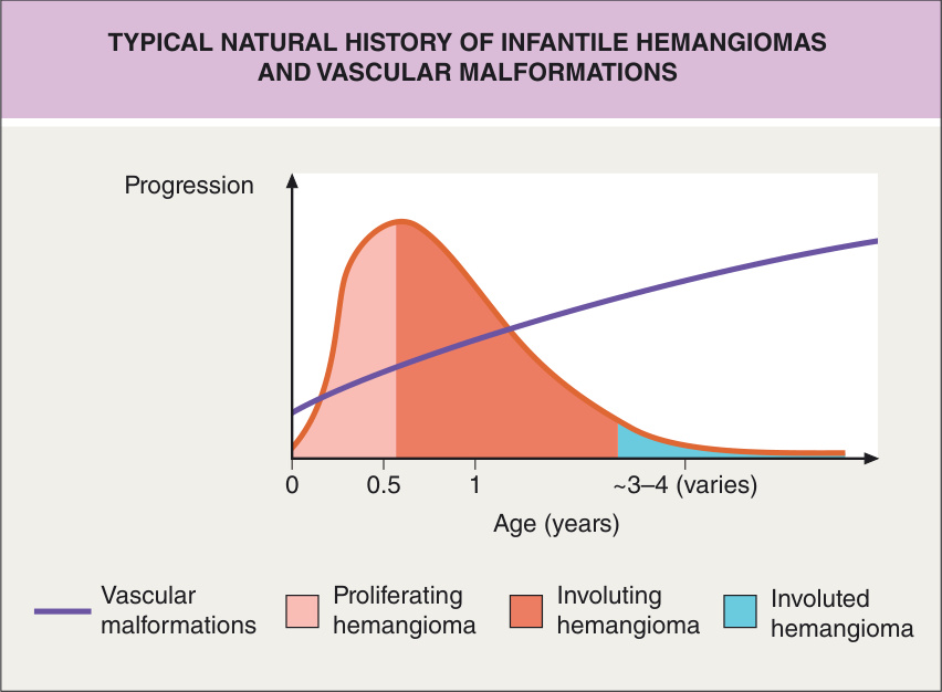
Fig. 104.1 Typical natural history of infantile hemangiomas and vascular malformations.
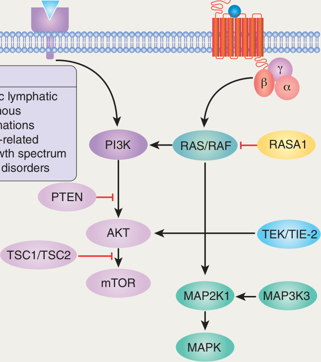
Fig. 104.2 Signaling pathways implicated in vascular malformations. MAPK, mitogen-activated protein kinase; MAP2K1, MAPK kinase 1; MAP3K3, MAPK kinase kinase kinase 3; mTOR, mechanistic target of rapamycin; PIK3CA, phosphatidylinositol-4,5-biphosphate 3-kinase catalytic subunit α.
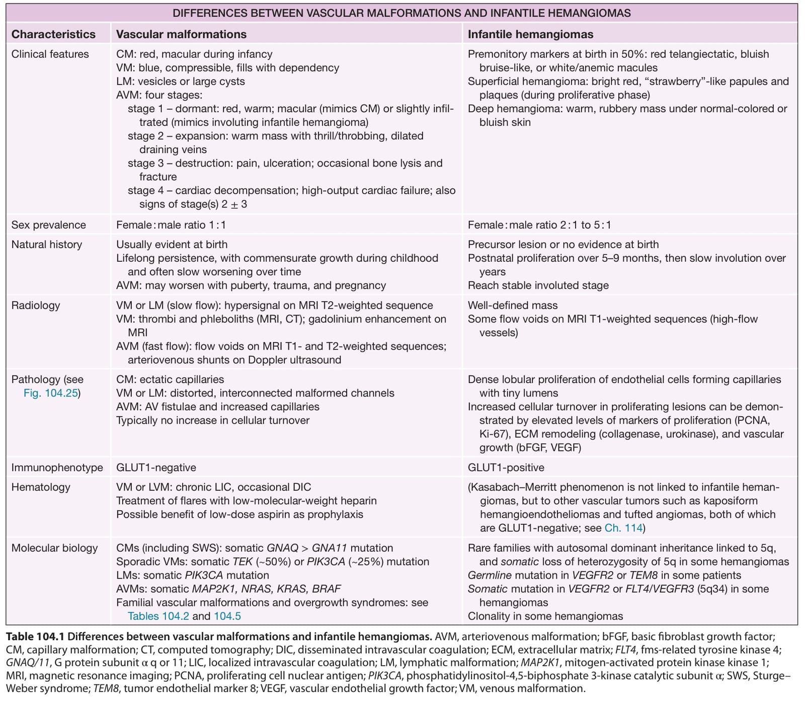
Table 104.1 Differences between vascular malformations and infantile hemangiomas. AVM, arteriovenous malformation; bFGF, basic fibroblast growth factor; CM, capillary malformation; CT, computed tomography; DIC, disseminated intravascular coagulation; ECM, extracellular matrix; FLT4, fms-related tyrosine kinase 4; GNAQ/11, G protein subunit α q or 11; LIC, localized intravascular coagulation; LM, lymphatic malformation; MAP2K1, mitogen-activated protein kinase kinase 1; MRI, magnetic resonance imaging; PCNA, proliferating cell nuclear antigen; PIK3CA, phosphatidylinositol-4,5-biphosphate 3-kinase catalytic subunit α; SWS, Sturge– Weber syndrome; TEM8, tumor endothelial marker 8; VEGF, vascular endothelial growth factor; VM, venous malformation.
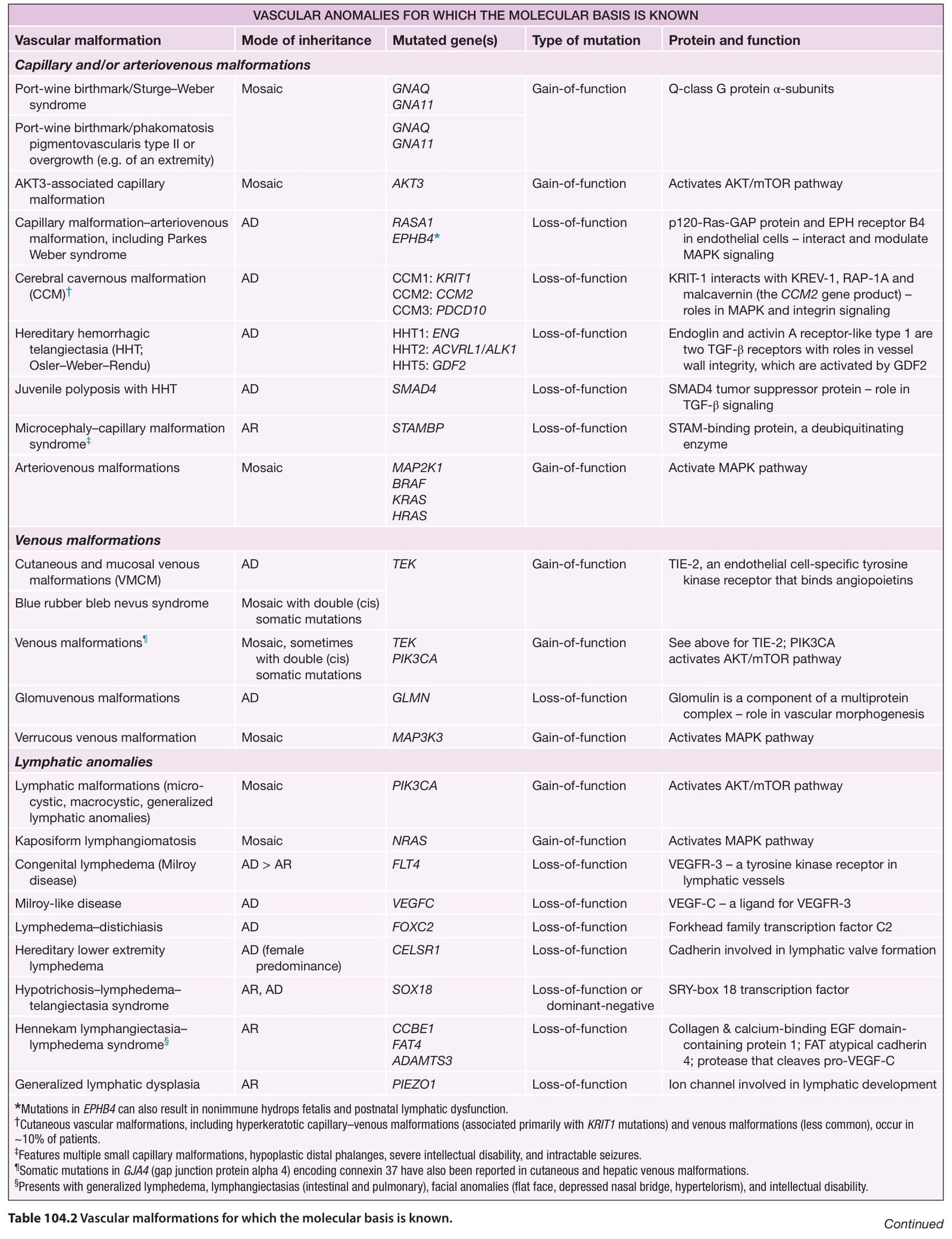
Table 104.2 Vascular malformations for which the molecular basis is known. Continued