CLINICAL FEATURES
Most vascular malformations can be correctly categorized based on their clinical features. This helps in selection of the most appropriate investigative tools (Table 104.3), avoiding redundant and unnecessary diagnostic imaging procedures. In addition, genetic analysis may be of utility in patients with vascular malformations associated with clinical manifestations that may potentially benefit from targeted systemic therapy as well as those with familial transmission. A sample of affected tissue is typically required for genetic testing in conditions caused by a somatic mutation, and massively parallel/next-generation sequencing
(NGS) panels are available for overgrowth syndromes and lymphedema (see Appendix).
Capillary Malformations
The ISSVA CM category includes nevus simplex, PWBs, reticulate CMs, CMTC, CM of CM-AVM, and telangiectasias; additional clinical variants such as “geographic” and “diffuse” CMs have also been described. Occasionally, CMs represent the most obvious sign of a complex syndrome. An approach to the evaluation of a patient with a presumed CM of the head and neck region is shown in Fig. 104.3.
Nevus simplex
Nevus simplex (salmon patch) is a very common congenital vascular birthmark evident in 30%–80% of neonates. Although classified as a CM by the ISSVA, nevus simplex is thought to represent remnants of the fetal circulation rather than a mosaic condition due to a somatic mutation. These pink-to-red macules and patches with somewhat indistinct borders have characteristic locations on the forehead/glabella in a V-shape (“angel kiss”), eyelids, philtrum, occiput, nape (“stork bite”), and lumbosacral area (Fig. 104.4A,B). They often become more prominent with crying or vigorous activity. The term nevus simplex complex has been proposed for extensive lesions.
Facial nevus simplex tends to fade spontaneously between 1 and 3 years of age, but extrafacial and some glabellar lesions are more persistent. During infancy, eczematous dermatitis may preferentially or solely develop within a nevus simplex. The vast majority of children with a nevus simplex do not have associated abnormalities; however, a prominent or persistent midfacial nevus simplex is a feature of several syndromes, including Beckwith–Wiedemann and megalencephaly–CM. Although there has been controversy regarding whether a lumbosacral nevus simplex represents a possible sign of occult spinal dysraphism, most authors do not recommend spinal imaging in the absence of additional cutaneous findings (see Ch. 64).
Port-wine birthmark (PWB) and variants
A somatic activating mutation in GNAQ (Q-class G protein subunit α) or, occasionally, GNA11 (G protein subunit α 11) in affected skin (especially the blood vessels) ± regional extracutaneous tissues (e.g. brain, eyes) underlies isolated PWBs and Sturge–Weber syndrome (SWS). These mutations (most often of R183 residues) lead to increased mitogenactivated protein kinase (MAPK) signaling (see Fig. 104.2), which results in increased cell proliferation and decreased apoptosis.
PWBs typically present at birth as well-demarcated, bright or deep red macules and patches, with their color resembling that of port wine. Fainter red or pink birthmarks have been termed “nevus roseus” because their color resembles that of rosé wine. Some light red–pink birthmarks are not solid but finely reticulated, often with a blotchy appearance and indistinct borders; these reticulated CMs are distinct from CMTC, which has a more well-defined, purplish net- or tram track-like pattern and is often associated with cutaneous atrophy. Reticulated CMs can be due to somatic gain-of-function mutations in GNA11 and, less commonly, GNAQ or PIK3CA.
The growth of PWBs is commensurate with the child’s growth. They can be localized, have a segmental pattern, or be multifocal and widespread. The distribution patterns of facial PWBs are thought to reflect the prominences that form during embryonic craniofacial development and their associated vasculature. Because these regions resemble the dermatomes of the trigeminal nerve, the following areas were classically recognized: V1 – forehead and upper eyelid; V2 – maxillary region (Fig. 104.4C); and V3 – mandibular region.
Over time, PWBs, especially those in the maxillary and mandibular areas, often develop a deeper red hue, changing from pinkish-red at birth to purplish-red by adulthood. Affected skin may thicken and become nodular (Fig. 104.5), and superimposed pyogenic granulomas occasionally appear. In a study of 173 patients with PWBs, thickening was observed in 11% (median age 32 years), nodularity in 24% (median age 44 years), and both in 6% (median age 45 years). These changes are rarely observed in the lighter “nevus roseus” or in PWBs located on the trunk and limbs. Overgrowth of the soft tissues and facial bones underlying a PWB can also occur, creating problems such as an open-bite deformity. Affected gums and lips may enlarge, potentially resulting in epulides, gingival bleeding, macrocheilia, and lip incompetence. As with nevus simplex, eczematous dermatitis may have a predilection for areas of skin affected by the PWB.
PWBs are congenital in the vast majority of patients. However, acquired port wine-like lesions have been described in adolescents and adults, and onset of such lesions may be precipitated by trauma. Of note, early morphea, especially the linear variant, occasionally presents with a red vascular patch that mimics an acquired port wine-like lesion.
A CM can be admixed with a network of blanched macules representing a nevus anemicus (nevus vascularis mixtus; Fig. 104.6). The association of a CM with dermal melanocytosis (Fig. 104.7) or a nevus spilus (speckled lentiginous nevus) is referred to as phakomatosis pigmentovascularis (PPV) (Table 104.4). Patients with PPV type II (cesioflammea) have the same activating mutation, either in GNAQ or GNA11, in both the PWB and dermal melanocytosis. Likewise, mosaic mutations in PTPN11 (protein tyrosine phosphatase nonreceptor type
11) have been found to underlie both the nevus roseus and nevus spilus in patients with PPV type III23a. Hypertension due to renal artery stenosis has been described in patients with extensive PWBs.
Sturge–Weber syndrome
SWS is a sporadic neurologic disorder in which a facial PWB is associated with ipsilateral leptomeningeal/brain and ocular vascular anomalies. Although all three components constitute a diagnosis of “complete” SWS, patients with a PWB plus CNS involvement alone are also typically diagnosed as having SWS, whereas those with a facial PWB
plus ocular involvement alone are often classified separately. The brain and/or ocular manifestations of SWS rarely occur without an associated PWB. Mosaicism due to a somatic activating mutation in GNAQ (or rarely GNA11) represents the cause of both SWS and non-syndromic facial PWBs (see above), with the former resulting from a mutation that arises earlier in development and affects the vasculature of the eye and CNS as well as the skin.
Facial PWBs associated with SWS were classically considered to involve the V1 region, which includes the forehead and upper eyelid, often with extension unilaterally or bilaterally over more of the face (Fig. 104.8). More recent studies have shown that involvement of the “forehead area”, which includes the upper eyelid and is delineated inferiorly by a line drawn from the outer canthus to the top of the ear, represents the best clinical predictor for SWS (Fig. 104.9). This risk area encompasses portions of V2/V3 as well as V1 and is thought to correspond with the embryonic frontonasal prominence. The latter includes neural crest-derived vasculature and develops in conjunction with the forebrain, which gives rise to the cerebral cortex and optic vesicles. A somatic GNAQ mutation occurring in the frontonasal area prior to migration of embryonic neural crest derivatives, which also include the leptomeninges and choroid, may therefore explain the forehead location of CMs associated with cerebral and ocular vascular malformations. Some SWS patients also have PWBs on the extremities and trunk. In addition, SWS occasionally occurs in the setting of PPV type II (see Table 104.4).
(cesioflammea). A large port-wine birthmark and extensive dermal melanocytosis are seen.
Ocular involvement in SWS can result in enlarged venous vessels affecting the conjunctiva, episclera, retina, and/or choroid. The most common ocular manifestation is glaucoma, which affects 30%–60% of patients with a PWB on the forehead and/or eyelids; choroidal hemorrhage and retinal detachment are rare complications. Ocular involvement is more frequent when the PWB affects both the V1 and V2 regions. Glaucoma may be detected at birth because of buphthalmos (enlargement of the globe), and acute glaucoma with a cloudy cornea can be an infantile emergency. Usually, however, increased eye pressure develops slowly. As a result, glaucoma may become evident during late childhood, adolescence, or even adulthood. Thus, periodic lifelong assessment of visual function and pressure of both eyes, as contralateral glaucoma occasionally occurs, is mandatory.
Neurologic symptoms can result from hypoperfusion of the ipsilateral brain due to CVMs within the pia mater, absence of superficial cortical veins, and dilated deep draining veins. Over time, chronic hypoxia may lead to cerebral hemiatrophy and calcifications, with the occipital region being the most frequent location. The most common neurologic manifestation is seizures, which are most often focal motor, either affecting the side of the body opposite to the vascular anomaly or generalized. The seizures typically develop in the first two years of life and affect ≥75% of children with SWS; they may be difficult to control with anticonvulsants, especially early on. Additional findings can include stroke-like episodes with contralateral hemiparesis or hemiplegia; developmental delay affecting motor and cognitive skills; emotional and behavioral problems; attention deficit disorder; and migraine headaches. Endocrine dysfunction such as central hypothyroidism and growth hormone deficiency may also occur, even in patients with normal neuroimaging.
The risk of ocular and/or neurologic manifestations of SWS is ~25%–50% when a unilateral PWB affects most of the forehead area; it is lower when the PWB involves a smaller portion of this area and higher (>50%) for a hemifacial lesion or a bilateral PWB covering the forehead and both upper eyelids (see Fig. 104.9). One prospective study also found an increased risk of SWS in patients with PWBs on the median forehead (a distribution reminiscent of nevus simplex), whereas linear PWBs on the forehead were not associated with SWS.
A recent consensus statement on the management of SWS recommended referral of any infant or child with a high-risk facial PWB (see above) to a pediatric neurologist and ophthalmologist for baseline evaluation and periodic follow-up (see Fig. 104.3). In newborns and infants with a high-risk PWB but no history of seizures or neurologic symptoms, neuroimaging should be considered in selected individuals (e.g. with an extensive bilateral facial PWB or if presymptomatic treatment is contemplated) rather than on a routine basis. In such patients, a non-sedated, non-contrast screening MRI including T1 and T2 (± fluid attenuation inversion recovery [FLAIR]) and susceptibilityweighted imaging (SWI) ± diffusion-weighted imaging (DWI) is recommended. These modalities can demonstrate early leptomeningeal
(SWS). Involvement of the “forehead area” (A), which includes the upper eyelids and is delineated inferiorly by a line drawn from the outer canthus to the top of the ear, is associated with risk of SWS. Hemifacial and median forehead (B) as well as bilateral forehead lesions are associated with highest risk.
changes with enlargement of transmedullary and periventricular veins; although sensitivity in asymptomatic patients is limited in the first few months of life, leptomeningeal involvement can be excluded by ≥1 year of age. In patients with neurologic symptoms, pre- and post-contrast MRI that includes high-resolution volumetric sequences in addition to SWI and post-contrast FLAIR imaging is recommended. Gyriform calcifications and atrophy develop during childhood in SWS patients and are visible by CT scans and other neuroimaging modalities.
Early diagnosis of SWS is important, as prophylactic aspirin administration may reduce the frequency of stroke-like episodes and seizures. Some infants and children with SWS have cognitive deficits that are more severe than anticipated based upon their limited cortical involvement on conventional MRI studies. Functional cerebral imaging, e.g. SPECT (single photon emission computed tomography) that evaluates regional cerebral blood flow or PET (positron emission tomography) that demonstrates metabolism of glucose, can provide additional prognostic information. Quantitative electroencephalography (EEG) may also aid in screening for brain involvement in asymptomatic infants at risk of SWS.
Capillary malformations associated with overgrowth,
PWBs and reticulated CMs on the trunk and extremities may be associated with regional overgrowth and other syndromic features (Table 104.5). Mosaicism for mutations that result in activation of the phosphatidylinositol 3-kinase (PI3K)/AKT pathway have been found to underlie several of these conditions (see Figs. 104.2 and 113.1), including those in the PIK3CA-related overgrowth spectrum (PROS). The various terms and eponyms used for overgrowth syndromes are likely to be refined as the molecular etiologies are further clarified.
Diffuse CM with overgrowth (DCMO) is a term proposed for a widespread reticulated CM associated with proportional soft tissue and/or bony hypertrophy (Fig. 104.10A). Unlike Klippel–Trenaunay syndrome (KTS), the overgrowth is not progressive and does not correlate with the location of the CM; in addition, although some DCMO patients have clinically prominent subcutaneous veins, they do not have a true VM or LM. In contrast to megalencephaly–CM, DCMO patients are normocephalic and do not have associated neurologic abnormalities. The pale red–pink, reticulated CM in DCMO involves multiple contiguous anatomic regions, often with a sharp midline demarcation on the anterior trunk, and may lighten over the first few months of life; approximately half of patients have additional areas with a confluent CM, usually in acral sites. Somatic activating mutations in GNA11 and PIK3CA (phosphatidylinositol-4,5-biphosphate 3-kinase catalytic subunit α) have been reported to cause diffuse CM associated with mild and more significant overgrowth, respectively.
Klippel–Trenaunay syndrome (KTS) is currently defined by the triad of a PWB, anomalous veins (representing a VM), and progressive overgrowth of the affected extremity. However, this eponym has also been erroneously used for any capillary malformation associated with limb hypertrophy. The vascular malformation in KTS is a combined CVM or CLVM, which may be localized or more extensive. Somatic mutations in PIK3CA have been identified in CLVMs associated with KTS. In addition, somatic mutations in PIK3R1 (phosphoinositide-3- kinase regulatory subunit 1) have been identified in patients with C(L) VMs and overgrowth of the affected extremity (see Table 104.5).
A well-demarcated, dark red-to-purple birthmark with irregular borders, referred to as a geographic CM, favors the lateral aspect of the thigh and is usually associated with a lymphatic component. Superimposed purple papules or hemorrhagic vesicles frequently develop and, especially if a persistent lateral marginal vein is evident on the outer aspect of the knee, a geographic CM predicts more severe, progressive overgrowth and a higher likelihood of complications such as cellulitis (Fig. 104.11A). Other children have pink-to-red, blotchy capillary birthmarks, which are associated with a better prognosis (Fig. 104.11B); in the absence of a VM, these patients may be more appropriately classified as having DCMO (see above) or simply a PWB with associated overgrowth. Foot and hand malformations in KTS are usually associated with deep venous system anomalies.
With lower extremity KTS, leg length discrepancy is progressive and requires ongoing orthopedic care. Lymphedema can lead to massive limb overgrowth and cellulitis. Some severely affected individuals have capillary–venous or lymphatic anomalies affecting the anogenital area, bladder, pelvis, retroperitoneum, and gastrointestinal tract that may bleed and result in anemia. A protein-losing enteropathy has also been described.
Like simple VMs (see below), the anomalous venous network in KTS may be associated with a chronic intravascular coagulopathy with high D-dimer and variably low fibrinogen levels. Patients with specific anomalies such as the presence of the lateral marginal vein, insufficient perforator veins, or intramuscular involvement are at higher risk of deep vein thrombosis, chronic thromboembolic pulmonary hypertension, and even life-threatening pulmonary embolism. Prophylactic anticoagulation should be considered in KTS patients, especially those with evidence of hypercoagulability, when they have additional risk factors for venous thromboembolism such as surgery, trauma, or pregnancy. Periodic echocardiography to assess for pulmonary hypertension has also been recommended by some authors.
Duplex ultrasonography is useful in investigating the vascular anomalies underlying KTS (see Table 104.3). Multidetector computed tomography (MDCT) and three-dimensional magnetic resonance venography (MRV) can help to determine the extent of musculoskeletal, thoracic, and abdominopelvic involvement as well as to characterize bone density changes, limb asymmetry, and the presence/patency of the deep venous system. Lymphoscintigraphy is rarely indicated. In patients with GI bleeding, colonoscopy or capsule endoscopy may reveal capillary patches, extensive dilated venous channels, and microcystic LM in the intestines.
This “Proteus-like” syndrome is characterized by asymmetric, progressive, infiltrative congenital lipomatous overgrowth that is typically truncal; vascular anomalies; epidermal nevi; scoliosis/ skeletal abnormalities that include broad hands and feet; spinal/ paraspinal arteriovenous lesions; and seizures. CLOVES syndrome is caused by somatic activating mutations in PIK3CA, and the clinical findings overlap considerably with those of other forms of PROS, such as KTS and megalencephaly–CM (see Table 104.5). LMs are often present within the lipomatous truncal mass of CLOVES, with variable extension into the retroperitoneum and mediastinum. Well-demarcated, dark red, geographic CMs/CLMs usually overlie the mass, which is also typically associated with both aberrant super-ficial veins and phlebectasia of the thoracic or major central veins; the latter result in increased risk of pulmonary embolism, especially when undergoing surgery.
Megalencephaly–CM, which was previously known as macrocephaly– CM and macrocephaly–CMTC, features asymmetric overgrowth, progressive (hemi)megalencephaly, and a widespread reticulated CM, often with a characteristic midline demarcation on the abdomen (Fig. 104.10B). Additional features include a persistent midfacial nevus simplex, frontal bossing, neonatal hypotonia, developmental delay, syndactyly (especially 2nd–3rd toes), polydactyly, joint laxity, and hyperelastic skin. Neuroimaging characteristically shows ventriculomegaly, acquired cerebellar tonsillar ectopia ± herniation, polymicrogyria, and cerebral asymmetry. Megalencephaly–CM is caused by postzygotic mutations in PIK3CA.
CLAPO syndrome features a CM of the lower lip and LM of the tongue and neck, together with asymmetry and partial overgrowth of the face and extremities. The CM of the lower lip is always midline and symmetrical; it may affect the entire lip and usually extends to the adjacent skin (Fig. 104.12). The LM may not be apparent in affected neonates, becoming evident later in life. Somatic mutations in PIK3CA are responsible for CLAPO syndrome.
Proteus syndrome is characterized by progressive overgrowth over time and a mosaic distribution of anomalies (see Ch. 62). It is caused by somatic activating mutations in AKT1, which encodes a protein that positively regulates cellular growth and proliferation. Manifestations are minimal at birth, typically becoming apparent at 6–18 months of age. Asymmetric, disproportionate bone overgrowth leads to consequences such as megaspondylodysplasia with scoliosis, knee deformities, asymmetric macrodactyly, and cranial hyperostosis. In addition to vascular malformations with variable capillary, venous, and lymphatic components, cutaneous features include highly characteristic cerebriform connective tissue nevi of the palms and soles (Fig. 104.13), epidermal nevi, lipomatous overgrowth, and regional absence of fat. Bullous pulmonary degeneration, specific neoplasms (e.g. ovarian cystadenoma, parotid monomorphic adenoma), and a facial phenotype with ptosis and anteverted nares represent other findings. Deep vein thrombosis results in premature death in up to 20% of patients. The differential diagnosis of Proteus syndrome includes PROS as well as PTEN hamartoma tumor syndrome (see Table 104.5).
Cutis marmorata telangiectatica congenita
CMTC is characterized by a dark purple to red–purple, broad reticulated vascular pattern intermingled with telangiectasias and occasionally prominent veins (Fig. 104.14). It commonly affects one or more limbs and the corresponding quadrant(s) of the trunk. Unlike physiologic cutis marmorata, CMTC persists upon warming an affected infant. Atrophic depressions may be evident within the net-like pattern (see Fig. 104.14C), especially over joints, and can result in ulceration and scarring. CMTC often lightens after the first year of life, but some residual violaceous network is common. Up to 50% of patients have hypoplasia (girth > length) of the affected limb(s) or additional vascular malformations. Other skeletal (e.g. syndactyly), ocular (e.g. glaucoma), and neurologic defects have been reported, especially in patients with generalized CMTC; however, these associations may reflect misdiagnosis of CM-associated syndromes as CMTC. Adams–Oliver syndrome
features CMTC or a reticulated CM together with distal transverse limb defects, variable cardiac malformations, and scalp/skull defects (aplasia cutis congenita; see Ch. 64).
A somatic AKT3 mutation has been identified in patients with CMs reminiscent of CMTC but more sharply delineated, linear, and located on the lateral side of the trunk or an extremity. Associated soft tissue atrophy and variable megalencephaly were also observed. Lastly, relatively short-lived skin findings resembling CMTC have been observed in patients with neonatal lupus.
Telangiectasias
Telangiectasias are dilated capillary-type blood vessels (see Ch. 106). These punctate, stellate, or linear red lesions may have a localized, segmental, or widespread distribution. In some disorders, they tend to affect particular anatomic sites. For example, angioma serpiginosum features clusters of tiny punctate telangiectasias in serpiginous patterns with a predilection for the extremities, whereas unilateral nevoid telangiectasia usually refers to a segmental configuration favoring the face, neck, chest, and arms. The lesions of hereditary benign telangiectasia (HBT) may mimic those of hereditary hemorrhagic telangiectasia
(HHT; see below), but the former is not associated with epistaxis or visceral hemorrhage. In addition, some kindreds initially reported as having HBT actually had capillary malformation–arteriovenous malformation (CM-AVM; see below).
Hereditary hemorrhagic telangiectasia (HHT; Osler–Weber–Rendu disease) is an autosomal dominant disorder characterized by visceral AVMs and mucocutaneous telangiectasias, which actually represent small AVMs; these AVMs have a propensity to bleed. Phenotypes vary considerably, even within a given family. The first manifestation of HHT is usually epistaxis during childhood or adolescence, beginning at a mean age of 12 years. Multiple telangiectasias of the skin and oral mucosa usually appear after puberty and may not be evident until adulthood, with ~50% of affected children <10 years of age having at least one evident telangiectasia. The telangiectasias most commonly affect the face, lips, tongue, palms and fingers, including periungual areas and the nail bed. These dark red lesions are either round, slightly elevated papules or ill-defined, stellate macules.
It is important to screen individuals suspected to have HHT for visceral AVMs (Table 104.6). In particular, AVMs in the lungs and brain are often asymptomatic prior to the sudden development of life-threatening complications. Pulmonary AVMs can result in hypoxemia, hemorrhage, and cerebral abscesses or strokes due to paradoxical emboli. Transcatheter embolotherapy of pulmonary AVMs is recommended. Lethal intracranial hemorrhage from cerebral AVMs can occur as early as infancy, and acute paraplegia due to spinal AVMs has also been described in children with HHT. Treatment strategies for AVMs of the CNS include embolization, microsurgery, and stereotactic radiation. Gastrointestinal or (less often) genitourinary tract bleeding typically occurs in mid adulthood and often presents as iron deficiency anemia. Lastly, hepatic AVMs may lead to high-output heart failure, portal hypertension, and biliary disease.
Classic HHT is due to heterozygous mutations in ENG (endoglin; HHT1) or ACVRL1 (activin A receptor-like type 1, also known as ALK1; HHT2). Both genes encode glycoprotein components of endothelial transforming growth factor-β (TGF-β) receptors. A genotype–phenotype correlation has been established, with a higher risk of pulmonary (especially in women) and cerebral AVMs in HHT1 and a higher risk of liver AVMs in HHT2. A few reported patients with HHT-like manifestations but somewhat larger and more widespread cutaneous telangiectasias/AVMs had underlying mutations in GDF2 (growth differentiation factor 2), which encodes a protein that binds to endoglin and ACVRL1. In addition, mutations in SMAD4, which encodes a protein that transmits signals from the TGF-β receptor, underlie a disorder that features juvenile gastrointestinal polyposis and HHT. Genetic testing can establish the diagnosis of HHT in children and young adults who do not yet meet clinical criteria (see Table 104.6). Information on medical centers and laboratories that specialize in HHT is available at curehht.org.
Ataxia–telangiectasia is an autosomal recessive disorder that occurs in approximately 1 in 40 000 births and is due to mutations in ATM (see Ch. 60). Ataxia is the initial symptom and usually presents in toddlers. Telangiectasias typically begin to appear at 4–6 years of age, primarily on the conjunctivae, face, and ears. Immunoglobulin deficiencies (e.g. IgA, IgG) and defective cell-mediated immunity explain the frequent sinopulmonary infections. Patients are at high risk for the development of lymphoma and leukemia. Elevated levels of circulating α-fetoprotein are found in affected individuals. Heterozygote carriers of a mutated ATM gene also have an increased risk of breast cancer and hematologic malignancies.
Angiokeratomas
Angiokeratomas are currently categorized by ISSVA as a provisionally unclassified vascular anomaly. They consist of ectasias of dermal vessels plus an acanthotic and hyperkeratotic overlying epidermis (see Ch. 114). Immunohistochemical studies have shown that most angiokeratomas stain positively with lymphatic markers. These dark red-topurple, papular vascular anomalies vary considerably in size, depth, and location. The two most common types are solitary papular angiokeratoma and angiokeratomas of the scrotum and vulva. The former is often found on the lower extremity, and clinically it may be mistaken for a melanoma.
In angiokeratoma circumscriptum, clusters of ectasias form a plaque or linear array, usually on an extremity and often present at birth. A congenital hyperkeratotic vascular anomaly resembling angiokeratoma circumscriptum, but with a deeper dermal component consisting of thick-walled blood vessels larger than capillaries, may represent a verrucous venulocapillary malformation (“verrucous hemangioma”; see below and Ch. 114), which can be caused by somatic MAP3K3 (mitogen-activated protein kinase kinase 3 gene) mutations. Grouped tiny lesions are also seen in angiokeratoma of Mibelli, which favors the toes, fingers, knees, and elbows.
Angiokeratoma corporis diffusum is characterized by more widespread lesions, often in a bathing trunk distribution; it can be associated with several hereditary lysosomal storage disorders such as Fabry disease, an X-linked recessive condition due to deficiency of α-galactosidase A, and α-fucosidase deficiency (see Table 63.7). Angiokeratoma-like lesions as well as microcystic lymphatic lesions may develop on the surface of geographic CMs in KTS (see above).
Venous Malformations
There are two major types of VMs: (1) common and familial VMs, together accounting for ~95% of VMs; and (2) glomuvenous malformations (GVMs). VMs may be confused with deep infantile hemangiomas or, when they involve a large portion of an extremity, KTS. Common and familial VMs are recognized clinically by their blue hue, softness, compressibility, and tendency to fill with dependency (Fig. 104.15). They are most often focal or segmental but may have a widespread distribution in forms with autosomal dominant inheritance. Somatic activating mutations in the TEK and PIK3CA genes account for ~50% and ~25% of sporadic common VMs, respectively; VMs due to mutations in the former gene are more likely to affect the surface of the skin. VMs are best imaged using T2-weighted MRI (see Table 104.3).
Fig. 104.16 provides an approach to the evaluation and management of patients with VMs.
Cephalic venous malformation
These lesions frequently lead to cosmetic and functional problems that worsen over time. In addition to the skin, they may affect the lips, oral mucosa, and deeper structures such as muscles, the infratemporal fossa, and the orbit (see Fig. 104.16). Cephalic VMs expand when the head is in dependent position. With time, distortion of facial features often becomes conspicuous. Patients with parapharyngeal and laryngeal VMs should be monitored for sleep apnea, which can put them at risk of sudden death during sleep. Approximately 20% of patients with extensive cephalic VMs have defects of the underlying skull, which must be identified before performing sclerotherapy to avoid embolization through the defect.
Approximately 25% of patients with extensive cephalic VMs have developmental venous anomalies, previously referred to by the misnomer “venous angiomas”, compared to <1% of the general population. These are uncommon functional trajectories of venous drainage from the brain, usually with enlarged deep venous channels. Developmental venous anomalies may cause headaches, but they do not confer risk of cerebral hemorrhage and treatment is not needed. Of note, patients with cephalic VMs are not at increased risk of CCM-type cerebral vascular malformations (see below).
Trunk and limb venous malformation
VMs of the trunk and limbs often have cosmetic and functional consequences (see Fig. 104.17). VMs form spongy masses of saggy ectatic venous channels, which are easily emptied by elevating and massaging the affected region (see Fig. 104.16). However, permeation deep into muscles, joints, and bones is common. Muscle involvement is associated with episodes of pain after motion and in the morning. The pain is linked to thromboses inside the low-flow channels, leading to phlebolith formation (round calcifications). Half of patients with large VMs have a chronic localized intravascular coagulopathy with elevated D-dimer levels. Joint involvement usually becomes symptomatic before 10 years of age, with complications such as effusions and hemarthrosis. Limb VMs are commonly misdiagnosed as KTS, but undergrowth of a limb affected by a simple VM is more common than overgrowth.
Sinusoidal “hemangioma” is thought to represent a distinctive type of venous malformation that typically presents in adults, especially
middle-aged women, with deep bluish nodules that favor the breast and extremities (see Ch. 114). Diagnosis is based on histologic features: well-circumscribed lobules composed of densely packed, large, thinwalled, blood-filled venous channels. Sinusoidal patterns have also been observed in congenital VMs.
FAVA presents between birth and early adulthood with a painful intramuscular mass, most often on the calf and in association with a contracture. Histopathologically, there is a combination of fat, abnormal venous channels with perivascular fibrosis, and lymphoplasmacytic aggregates within atrophied skeletal muscle. Treatment options include excision or image-guided percutaneous cryoablation. Underlying somatic mutations in PIK3CA have been reported.
Verrucous VM (see Ch. 114) is included in the ISSVA classification as a subtype of VM, and an underlying somatic mutation in MAP3K3 has been identified in a subset of these lesions (see Fig. 104.2). This malformation presents as a purple red birthmark, often with a segmental distribution on an extremity, and hyperkeratosis develops over time. The clinical appearance overlaps with that of angiokeratoma circumscriptum and the hyperkeratotic cutaneous capillary–venous malformation associated with familial cerebral cavernous malformation. Red vascular dots are seen with dermoscopy, which helps to distinguish verrucous VM from a CM or LM.
Syndromes associated with venous malformations
Familial VMCM is an autosomal dominant condition in which multiple VMs affect the skin, oral mucosa, and muscles. Visceral VMs (e.g. of intestines, lungs, CNS) and cardiac malformations have been observed in a few families. The disorder is caused by heterozygous germline mutations in TEK that result in hyperphosphorylation and constitutive activation of TIE-2, a vascular endothelium-specific tyrosine kinase receptor (see Fig. 104.2); the downstream signaling of this receptor is also altered. Interestingly, approximately half of sporadic VMs have somatic activating TEK mutations within lesional tissue.
BRBNS is a sporadic disease characterized by the progressive appearance of dark blue papules and nodules and skin-colored compressible protuberances (“rubber blebs”) in a widespread distribution, in addition to larger VMs affecting subcutaneous tissues and muscle (Fig. 104.17). Some patients have a large congenital VM with a characteristic darker purple–blue fern-shaped pattern on the surface. Gastrointestinal lesions bleed and result in iron deficiency anemia. Other sites of visceral involvement (CNS, lungs, heart) are uncommon. BRBNS was recently found to be caused by double (cis) somatic activating TEK mutations, i.e. two mutations within the same allele of the gene.
Maffucci syndrome is a sporadic condition that features VMs, which present as blue to skin-colored nodules, and enchondromas similar to those of Ollier disease, which can lead to orthopedic consequences
(Fig. 104.18). It most commonly affects the extremities; however, cephalic lesions with severe neuro-ophthalmologic complications may also occur. Histologically, the cutaneous nodules demonstrate features of both a VM and a vascular tumor, the spindle cell hemangioma (see Ch. 114).
Heterozygous somatic mutations in IDH1 (NADP(+)-dependent isocitrate dehydrogenase 1) have been identified in affected tissues, with the same mutation in the spindle cell hemangiomas and enchondromas; the latter lesions also rarely have underlying IDH2 mutations.
GVMs (previously known as glomangiomas) represent a variant of VM with rows of glomus cells around distorted venous channels (see Ch. 114). They occur as small solitary lesions, widely scattered blue–purple nodules, or (less commonly) large segmental plaques (Fig. 104.19). GVMs may be evident at birth, especially larger plaques, or appear from childhood to adolescence; they often enlarge over time. In contrast to classic VMs, GVMs tend to be painful when palpated, partially (rather than fully) compressible, and hyperkeratotic with a cobblestone-like appearance (especially on the extremities). GVMs do not typically affect viscera or joints, and they are not associated with a coagulopathy. Mucosal involvement with deep intraoral lesions and superficial invasion of muscles occasionally occurs. Approximately two-thirds of patients with GVMs, but only 1% of those with classic VMs, have a family history of similar lesions. GVMs result from a heterozygous germline mutation in the glomulin gene (GLMN), which is passed from generation to generation in an autosomal dominant manner, plus a somatic “second hit” (loss of heterozygosity) in this gene within lesional skin (see Table 104.2).
A distinctive hyperkeratotic cutaneous capillary–venous malformation (HCCVM) occurs in a subgroup of patients with familial cerebral cavernous malformation (CCM). Neurologic manifestations, which develop at a mean age of 30 years (range, 2–72 years), may include headaches, seizures, and cerebral hemorrhage. This autosomal dominant disorder can be caused by mutations in three different genes (see Table 104.2): KRIT1 (CCM1), CCM2 (malcavernin), and PDCD10 (CCM3). Among the ~10% of familial CCM patients who have cutaneous capillary and/or venous malformations, ~90% have a KRIT1 mutation.
HCCVMs occur almost exclusively in patients with KRIT1 mutations, with one report to date in an individual with a PDCD10 mutation. These irregularly shaped, dark crimson or reddish-purple plaques with surrounding bluish discoloration are typically congenital and located on the extremities; GLUT1-positivity has been described. Smaller reddish-brown macules with peripheral telangiectatic puncta have also been observed. Less frequently, children and adults with mutations in any of the three genes develop dark blue cutaneous papules and nodules reminiscent of the blue rubber bleb nevus syndrome, which represent VMs. The differential diagnosis includes a verrucous VM.
Lymphatic Malformations
The spectrum of LMs includes primary lymphedema, common LMs, generalized lymphatic anomaly (GLA), and central conducting lymphatic anomalies (CCLAs; channel-type LMs). The targetoid hemosiderotic lymphatic malformation (hobnail “hemangioma”) is discussed in Chapter 114. Lymphedema results from inadequate drainage of lymph due to hypoplasia, aplasia, or disruption of lymphatic channels. Lymphedema is divided into primary forms due to abnormal lymphatic development and secondary forms due to acquired disruption of lymphatic drainage (see Ch. 105). In contrast, LMs are due to hyperplasia of the lymphatic network. This hyperplasia most commonly results in cysts, including smaller microcystic and/or larger macrocystic lesions; these cysts can develop within the skin, mucous membranes, muscles, bone, or occasionally viscera. Somatic activating mutations in PIK3CA underlie most common LMs. CCLAs affecting structures such as the cisterna chyli and thoracic duct result in lymphatic leakage that leads to pleural or pericardial effusions and ascites.
Primary lymphedema
Patients with primary lymphedema usually accumulate lymph fluid in the extremities (lower > upper); cephalic and genital involvement occasionally occurs. Affected individuals are at risk for bacterial infections and septicemia. Generalized lymphedema may be associated with intestinal or pulmonary lymphangiectasias, exudative enteropathy, and pleural effusions.
Primary lymphedema may be classified according to its age of onset into congenital, peripubertal (praecox), and late-onset (tarda; >35 years of age) forms, but since the time of onset can vary even within the same family, genetic bases are now more commonly used for classification (see Table 105.7). Table 104.2 outlines the multiple genes that have been implicated in isolated and syndromic forms of lymphedema. Many of the proteins encoded by these genes act in the vascular endothelial growth factor receptor-3 (VEGFR3) signaling pathway (see Ch. 102).
The most common congenital form, classically referred to as Milroy disease, presents with lymphedema below the knees, prominent veins, and upslanting toenails. Lymphedema–distichiasis syndrome due to FOXC2 mutations features lymphedema of peripubertal onset, congenital distichiasis (aberrant eyelashes arising from the meibomian glands lead to a double row of eyelashes), venous varicosities, and occasionally yellow nails. Lymphedema, chylothorax, and chylous ascites are also variably seen in Turner syndrome (see Ch. 55) and RASopathies including Noonan, cardio-facio-cutaneous, Costello, and rarely CM-AVM syndromes (see below and Table 61.3).
Macrocystic lymphatic malformation (“cystic hygroma”)
Macrocystic LMs represent collections of large, interconnected lymphatic cysts lined by a thin endothelium. The most common locations are the neck, axilla, and lateral chest wall (Fig. 104.20A). Prenatal detection by ultrasound is possible as early as the first trimester of pregnancy. Some patients who are diagnosed prenatally have malformation syndromes caused by chromosomal abnormalities or other genetic disorders (e.g. Turner syndrome, Noonan syndrome). A macrocystic LM appears as a large, soft, translucent mass under normal skin. The diagnosis can be confirmed by ultrasound, CT, or MRI (see Table 104.3) as well as by direct puncture and cytologic analysis of the fluid. Hemorrhage inside a cyst can create sudden swelling, with the mass becoming tender, tense, firm, and purple to yellowish in color.
Microcystic lymphatic malformation (“lymphangioma
These ill-defined aggregates of relatively small, abnormal lymphatic channels are the most common type of LM. They favor the proximal limbs and chest but can occur in any cutaneous site or in the mouth,
including the tongue, buccal mucosa, lips, and oral floor. Plaques with crops of clear or hemorrhagic vesicles scattered on their surface vary over time, both in size and in the number and color of superimposed vesicles (Fig. 104.20B–E). Additional clinical findings are intermittent swelling, hemorrhage, and leakage of lymph from superficial vesicles. Lesions are often much more extensive than clinically expected from the number of vesicles. Complications include erysipelas-like reactions following minor injuries, other inflammatory flares, and infections.
Combined microcystic and macrocystic lymphatic
Extensive cervicofacial combined LMs commonly have bony involvement. This can lead to mandibular overgrowth and prognathism, resulting in a long or asymmetric face, bite deformities, and abnormal occlusal planes. Intraoral combined LMs are frequently complicated by inflammatory flares or spontaneous bleeding in the setting of upper respiratory or dental infections. This can result in sudden expansion of the lesions, particularly those in the tongue. Local infections can lead to cellulitis or even septicemia. In some patients with massive oropharyngeal combined LMs, airway compromise (extrinsic or intrinsic) requires tracheostomy. Hypersalivation and dental caries are common, with the potential for loss of teeth. Speech, mastication, and swallowing may be impaired. In addition to swelling, pain, and infections, specific complications of combined LMs in the orbital region include chemosis, strabismus, amblyopia, proptosis, and visual loss.
On the trunk and limbs, combined LMs tend to worsen as the child grows. When a minor wound on the affected extremity becomes infected, the inflammatory reaction results in expansion of the LM, as also occurs in patients with limb lymphedema. Perineal and gluteal cleft LMs, which are particularly prone to fluid leakage and infection, severely impair quality of life. Extensive combined LMs, in particular those with a venous component, may be associated with a chronic localized intravascular coagulopathy with elevated D-dimer levels (see above).
Generalized lymphatic anomaly (GLA)
GLA, previously referred to as “lymphangiomatosis”, represents an extensive, multifocal abdominal and thoracic LM that extends into viscera such as the spleen, liver, intestines, and pleura/lung; the bones are often involved, but the cortex is typically spared. GLA may have a lethal outcome due to expansion, chylous effusions, visceral complications, and vertebral compression.
Kaposiform lymphangiomatosis (KLA)
KLA is currently considered to represent an aggressive subtype of GLA that is characterized by progressive involvement of the media stinum, lungs, pleura, retroperitoneum, spleen, bones, soft tissue, and skin. Histologically, it features clusters or sheets of hemosiderotic, spindled lymphatic endothelial cells oriented in a parallel fashion amid abnormal dilated lymphatic channels. Circulating angiopoietin-2 levels are elevated in KLA and may serve as a biomarker.
Patients present at a median age of 6 years (range, birth to fifth decade) with respiratory symptoms, thrombocytopenia, hypofibrinogenemia, and life-threatening hemorrhagic pericardial and pleural effusions. An enlarging subcutaneous mass may be evident, sometimes with an overlying red–purple birthmark and lymphatic vesicles. An underlying somatic NRAS mutation is present in most affected individuals. The reported 5-year survival is ~20%–50%, and successful treatment with sirolimus (rapamycin) and MEK inhibitors has been described (see Fig. 113.1).
Gorham–Stout disease
In Gorham–Stout (disappearing bone) disease, a LM creates progressive radiolucent bone lesions with cortical involvement and eventually massive osteolysis. This results in pathological fractures and deformities. Involvement of the thoracic skeleton may be associated with pulmonary lymphangiectasia. A somatic activating KRAS mutation has been identified in affected tissue from a patient with Gorham-Stout disease.
Central conducting lymphatic anomalies (CCLAs;
CCLAs are characterized by dilation, dysfunction, or obstruction of major thoracic or abdominal lymphatic channels. The manifestations depend on the site of the anomaly and can include chylothorax, chylous ascites, protein-losing enteropathy, peripheral lymphedema, lymphorrhea, and cutaneous vesicles. Underlying mutations have been identified in genes encoding various components of the RAS–mitogenactivated protein kinase (MAPK) pathway (germline or mosaic; see Fig. 55.4) as well as other proteins with roles in lymphatic development (e.g. MyoD family inhibitor domain containing protein, piezo-type mechanosensitive ion channel component 1).
Arteriovenous Malformations
AVMs are fast-flow vascular malformations with direct communications (AV shunting) between arteries and veins, creating an AV nidus. They are rare and constitute the most dangerous group of vascular anomalies. Schobinger staging was adopted by the ISSVA in order to classify AVMs according to their clinical severity. The stages are: (1) quiescent/ dormant – macular or slightly infiltrated, red, warm lesions mimicking a CM (Fig. 104.21A,B); (2) expansion – warm masses with throbbing and thrills over dilated draining veins (Fig. 104.21C); (3) destruction – necrosis, hemorrhage, ulceration and (occasionally) lytic bone lesions (plus findings of stage 2) (Fig. 104.21D); (4) cardiac decompensation (plus findings of stage 2 ± 3). Initial evaluation with ultrasonography and MRI studies can confirm the diagnosis and delineate the extent of the lesion (see Table 104.3). Somatic activating mutations in MAP2K1 and less commonly KRAS, NRAS, or BRAF have been found to underlie AVMs (see Fig. 104.2).
In 40% of patients, AVMs are visible at birth; the remainder appear later in life. Features that can help to distinguish an early-stage AVM from a CM include heterogeneous color saturation, peripheral pallor, sharply demarcated “archipelago-like” borders (see Fig. 104.21A), and warmth to touch as well as associated soft tissue swelling/overgrowth (see Fig. 104.21B) and prominent veins. The most frequent location is cephalic (~70% of patients; Fig. 104.22). Puberty (75% of patients), pregnancy (25% of women), and trauma may result in worsening of AVMs. In an analysis of 65 patients with cephalic AVMs (Odile Enjolras, personal data), 39 were centrofacial and 7 were hemifacial; 10 affected the forehead, 10 the ear, and 4 the scalp.
Stage 3 cephalic AVMs are disfiguring and function- or even lifethreatening lesions, and those on an ear or extremity may lead to amputation. Multifocal AVMs affecting an extremity can lead to cardiac overload and distal ischemia. Attempted treatment with arterial or venous ligatures, partial excision, or proximal (rather than selective distal) embolization often precipitates complications. Stewart–Bluefarb syndrome represents acroangiodermatitis (pseudo- Kaposi sarcoma; see Ch. 105) occurring in association with an AVM of the lower extremity.
Syndromes associated with arteriovenous malformations
This rare sporadic condition features cutaneous, intraspinal (intramedullary ± meningeal), and vertebral AVMs in the same segment. These findings can occur as a manifestation of CM-AVM syndrome due to RASA1 mutations (see below). Congenital red or red–brown vascular birthmarks mimicking a CM, but actually representing stage 1 AVMs (Fig. 104.23), or throbbing masses with dilated veins (stage 2 AVMs) are present in 20% of patients with spinal AVMs. Neurologic deficits typically develop during young adulthood due to a mass effect of the expanding AVM on the spinal cord or subarachnoid hemorrhage. Neurologic signs range from back pain and radiculalgia to rectal and bladder dysfunction to paraparesis and paraplegia. MRI and angiography establish the presence of the spinal AVM. Endovascular embolization of the spinal AVM may improve the prognosis.
This rare segmental AVM extends from the craniofacial region to the orbit and brain. Incomplete forms of the disease also exist. The brain AVM may remain asymptomatic or manifest as seizures or hemiparesis/hemiplegia. In 12 BDBS patients between 5 and 51 years of age, the facial AVMs (stage 1, 2, or 3) were midfacial in 3, hemifacial in 3, and both midfacial and hemifacial in 6 (Odile Enjolras, personal observations). The presence of an orbital AVM was an inconsistent finding (4 patients), but brain AVMs of the chiasm, choroid plexus, or thalamus
were present in all 12 individuals and led to cerebral hemorrhage in 4 patients. In a second series of 15 patients with BDBS, orbital AVMs were present in 14 and frequently affected the optic nerve (13 patients) and retina (11 patients); symptoms in this group included reduced visual acuity or fields, blindness, recurrent epistaxis, nasal obstruction, and gingival hemorrhage.
In this rare fast-flow vascular malformation syndrome, limb overgrowth (in length and girth) occurs secondary to arteriovenous fistulae (AVF); this must be differentiated from the purely slow-flow malformations of KTS. A red CM, excess fat, and lymphatic anomalies may also be observed in Parkes Weber syndrome. Lytic bone lesions and cardiac failure often develop, and prognosis is poor after puberty. Over 50% of patients with Parkes Weber syndrome have a germline RASA1 or EPHB4 mutation plus an early somatic “second hit” mutation in the same gene underlying their larger AVM/AVF (see Ch. 62); these individuals also display the characteristic features of the CM-AVM syndrome (see below).
This autosomal dominant disorder is caused by mutations in RASA1 (∼50% of patients; CM-AVM1) and less often in EPHB4 (CM-AVM2) (see Table 104.2 and Fig. 104.2). Multiple, scattered small cutaneous CMs are evident at birth and continue to appear during childhood and adolescence. These CMs present as pink to red–brown macules, often with a narrow blanched border or in CM-AVM2 a blanched central region, as well as hypotrichosis of the affected skin (Fig. 104.24). Some macules are warmer than adjacent uninvolved skin, and pulsed Doppler may show decreased peripheral vascular resistance, suggesting a pre-AVM lesion. Histologically, the macules have numerous thickwalled arterioles in the superficial dermis, suggestive of an incipient AVM rather than a CM.
Patients sometimes have numerous punctate red macules, each surrounded by a white halo, located primarily on the extremities. Patients with CM-AVM2 may also have telangiectasias favoring the lips, perioral area, and upper trunk. Approximately 35% and 15%–20% of individuals with CM-AVM1 and 2, respectively, also have high-flow lesion(s). Among 138 patients from 68 families with CM-AVM1, >90% had multifocal small CMs, 6% had a solitary “atypical” CM, 18% had an AVM/AVF of the skin/subcutaneous tissue (extremities > face/neck), 5% had Parkes Weber syndrome, and 15% had an intracranial or intraspinal AVM. The CNS AVMs may lead to neurologic symptoms, which typically develop in the first decade of life and can include headaches, seizures, and sensorimotor deficits. MRI of the brain/spine is recommended for asymptomatic infants and young children with CM-AVM as well as patients of any age with neurologic manifestations.
PHTS due to PTEN mutations encompasses the autosomal dominant Bannayan–Riley–Ruvalcaba and Cowden syndromes as well as the type 2 mosaic segmental overgrowth, lipomatosis, AVMs, and epidermal nevus (SOLAMEN) syndrome (see Ch 63). Affected individuals are often macrocephalic, and they are predisposed to the development of malignancies, especially of the breast, thyroid, and endometrium. Cutaneous manifestations with onset at birth or in childhood include genital pigmented macules, segmental excess of hypervascularized fat, and vascular malformations. The latter are typically multifocal, intramuscular, fast-flow lesions associated with ectopic fat and focal segmental dilatation of draining veins. By both imaging and histopathologic features, these vascular malformations differ from sporadic AVMs and they have been renamed PTEN hamartomas of soft tissue. Oral sirolimus has been used to treat these lesions. Cerebral developmental venous anomalies are also common in PHTS patients, and additional mucocutaneous lesions in affected adolescents and adults include tricholemmomas, acral keratoses, sclerotic fibromas, neuromas, and oral papillomas.
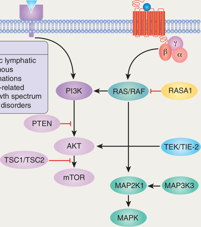
Fig. 104.2 Signaling pathways implicated in vascular malformations. MAPK, mitogen-activated protein kinase; MAP2K1, MAPK kinase 1; MAP3K3, MAPK kinase kinase kinase 3; mTOR, mechanistic target of rapamycin; PIK3CA, phosphatidylinositol-4,5-biphosphate 3-kinase catalytic subunit α.
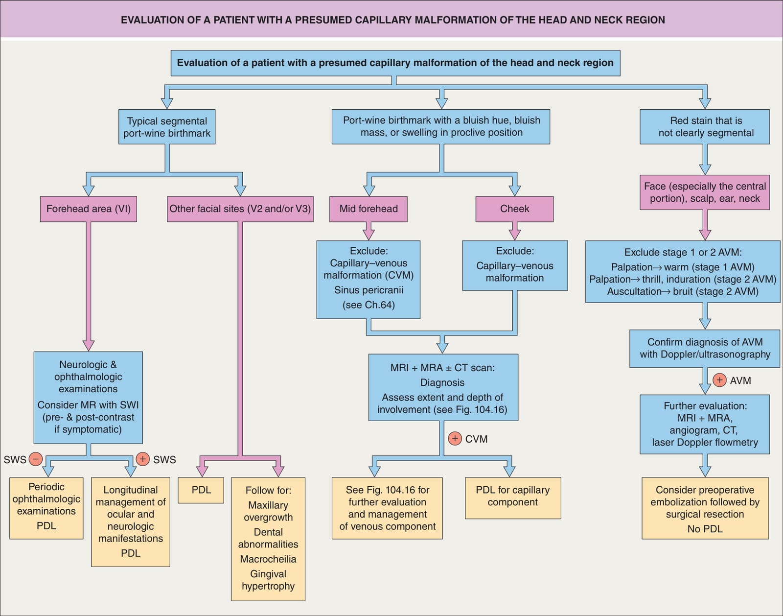
Fig. 104.3 Evaluation of a patient with a presumed capillary malformation of the head and neck region. AVM, arteriovenous malformation; CT, computed tomography; PDL, flashlamp-pumped pulsed dye laser; MRA, magnetic resonance arteriography; MRI, magnetic resonance imaging; SWI, susceptibility-weighted imaging; SWS, Sturge–Weber syndrome.
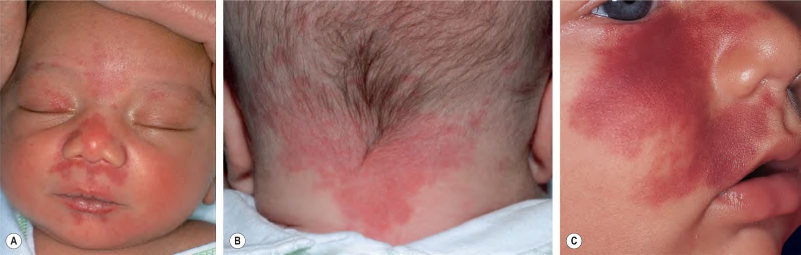
Fig. 104.4 Nevus simplex versus port-wine birthmark (PWB).A Involvement of the central face in a symmetric pattern is typical of a nevus simplex (salmon patch); such lesions usually fade over the first few years of life but are sometimes misdiagnosed as a PWB. B Nevus simplex in the nape area (“stork bites”) tend to be more persistent. C In this young infant with a PWB in the maxillary region, the affected skin is smooth. This lesion will be persistent. B, Courtesy Julie V. Schaffer, MD.
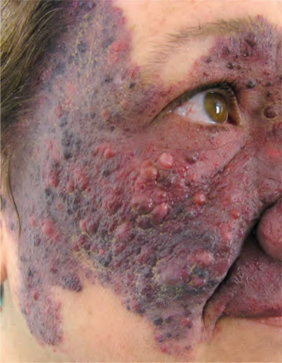
Fig. 104.5 Facial port-wine birthmark in the maxillary region in an adult. Hyperplasia and nodularity are prominent. Courtesy Pablo Boixeda, MD.
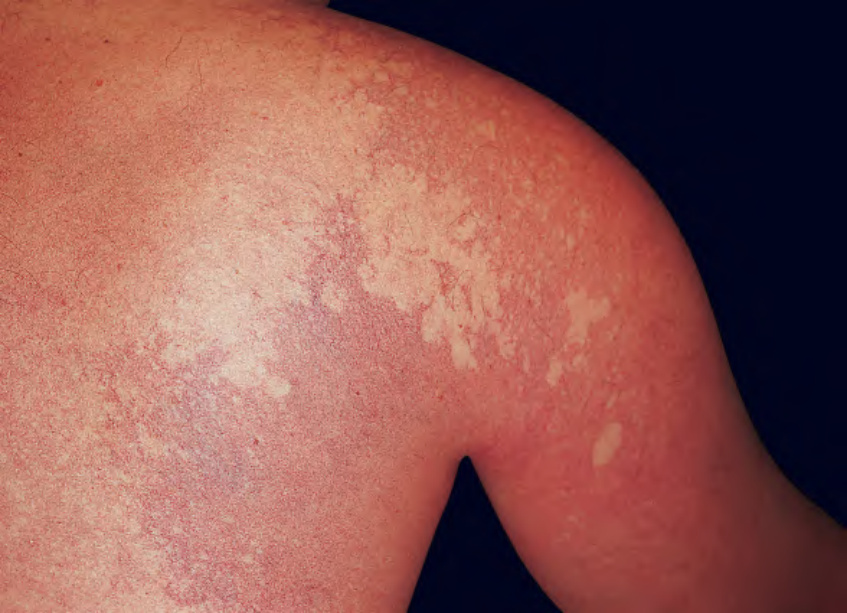
Fig. 104.6 Port-wine birthmark intermingled with a nevus anemicus on the shoulder. This man had phakomatosis pigmentovascularis type IIb, with additional features including an upper facial port-wine birthmark, Sturge– Weber syndrome, and extensive dermal melanocytosis.
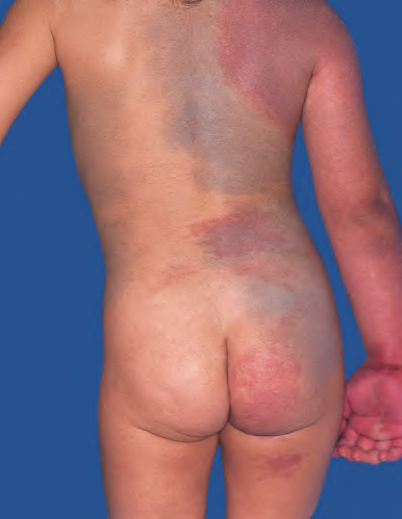
Fig. 104.7 Phakomatosis pigmentovascularis type IIa
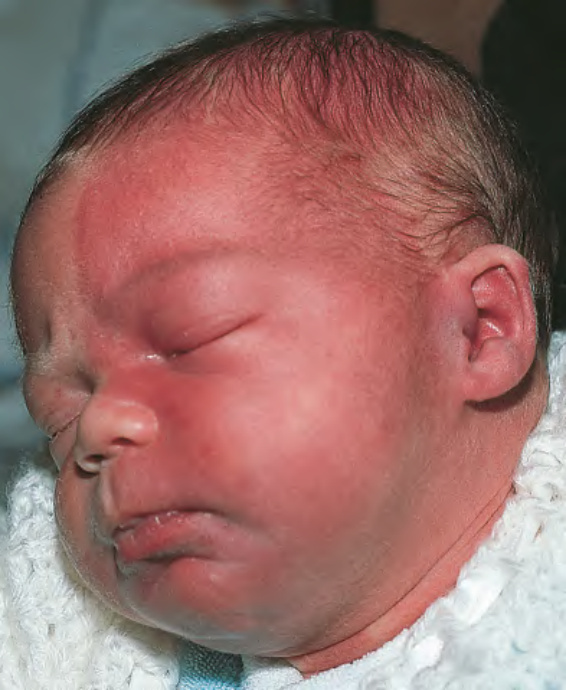
Fig. 104.8 Infant at risk for Sturge–Weber syndrome (SWS). The port-wine birthmark covers most of the left side of the face.
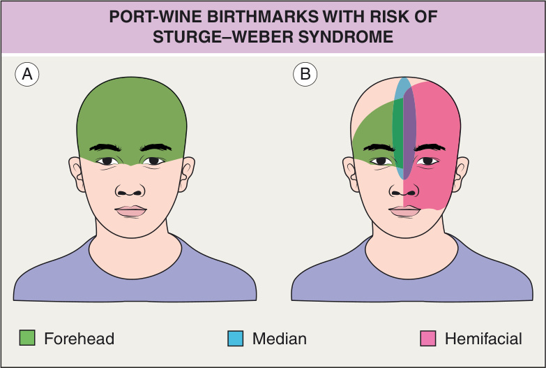
Fig. 104.9 Port-wine birthmarks (PWBs) with risk of Sturge–Weber syndrome
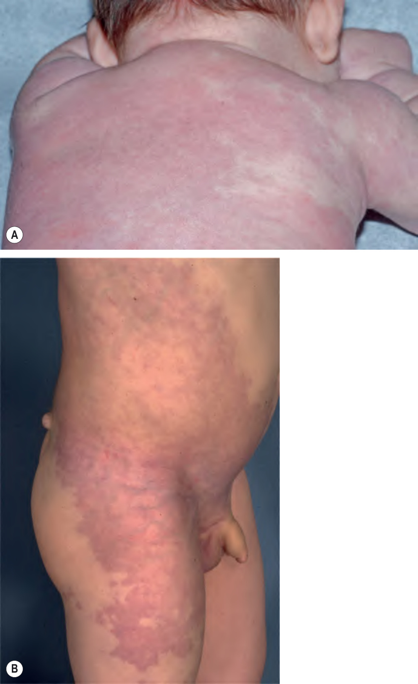
Fig. 104.10 Reticulated capillary malformations.A Diffuse capillary malformation with overgrowth. This normocephalic infant has an extensive reticulated capillary malformation involving the trunk, one arm, and both legs. B Megalencephaly–capillary malformation syndrome. This boy has hemihypertrophy of the side of the body opposite to the capillary malformation. Courtesy Julie V. Schaffer, MD.
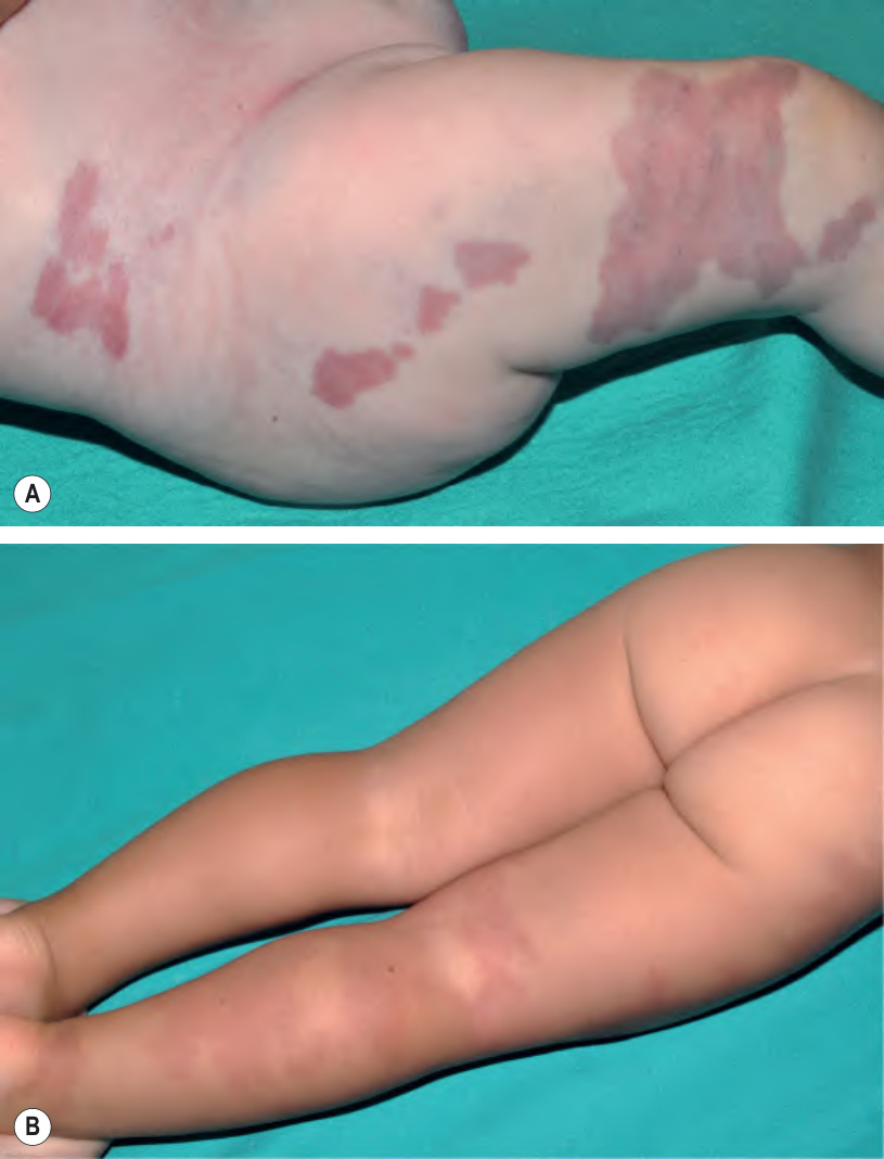
Fig. 104.11 Klippel–Trenaunay syndrome.A Darker red–purple, well-demarcated geographic capillary birthmark on the thigh associated with dilated veins and an enlarged lower limb. B Light red, blotchy capillary birthmark associated with enlargement of the lower extremity. Compared to blotchy CMs, geographic CMs are more likely to be associated with lymphatic malformations as well as tissue overgrowth.
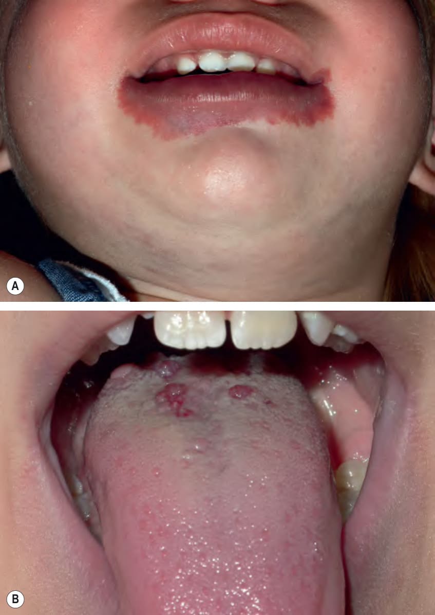
Fig. 104.12 CLAPO syndrome.A Capillary malformation on the lower lip and chin, with swelling of the neck due to a lymphatic malformation. B Microcystic lymphatic malformation on the tongue. A, Courtesy Julie V. Schaffer, MD.
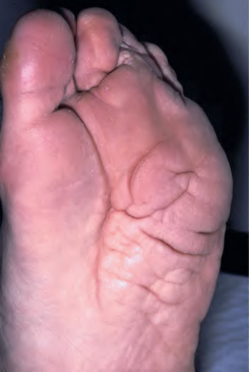
Fig. 104.13 Plantar cerebriform connective tissue nevus in a man with Proteus syndrome.
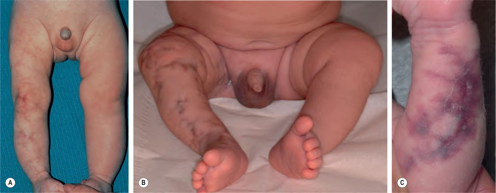
Fig. 104.14 Cutis marmorata telangiectatica congenita (CMTC). Note the hypoplasia of the affected limb (A,B) and associated cutaneous atrophy leading to a depression (C). The color of the broad vascular network can be red–purple (A,C) or, as seen in this infant of Caribbean descent, brownish-purple (B). C, Courtesy Richard Antaya, MD.
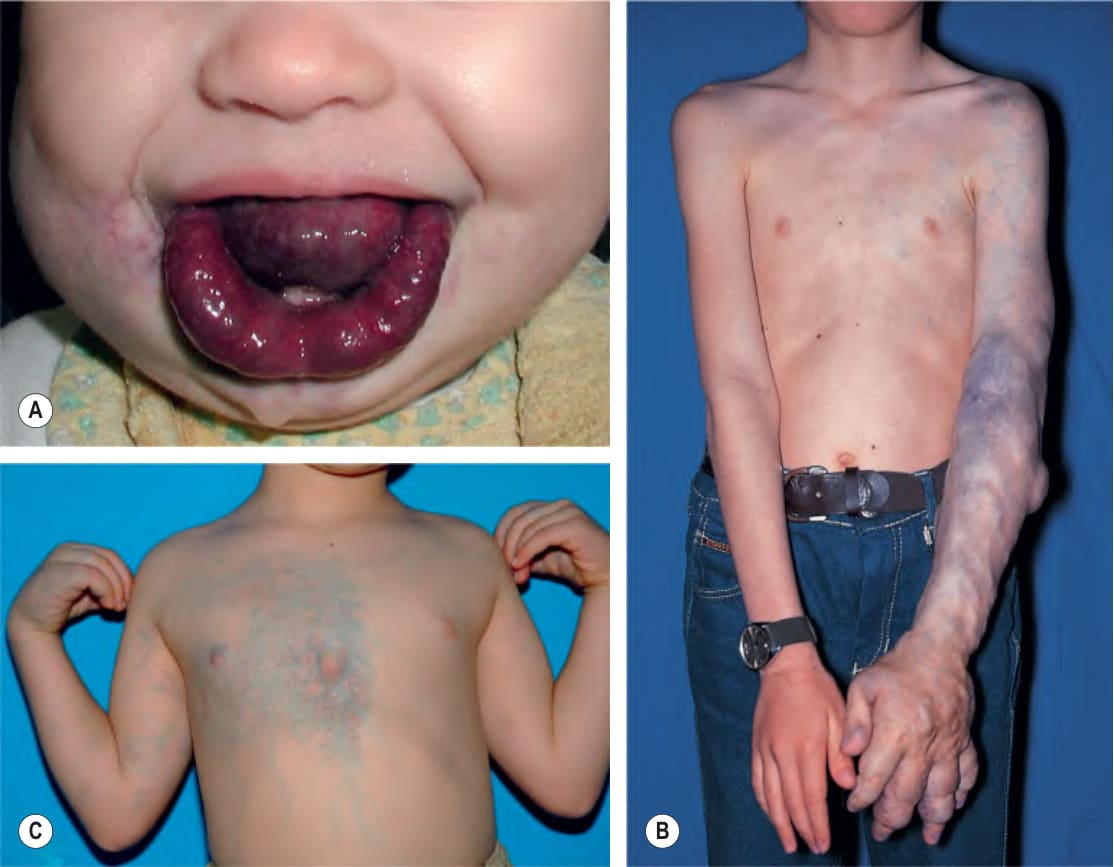
Fig. 104.15 Venous malformations (VMs).A Distortion of the tongue and lower lip has led to an open bite. B The skin and muscles of the entire arm are affected, with soft bluish nodules and obvious swelling in the dependent position. C This VM has a segmental distribution on the right trunk and involvement of the ipsilateral arm.
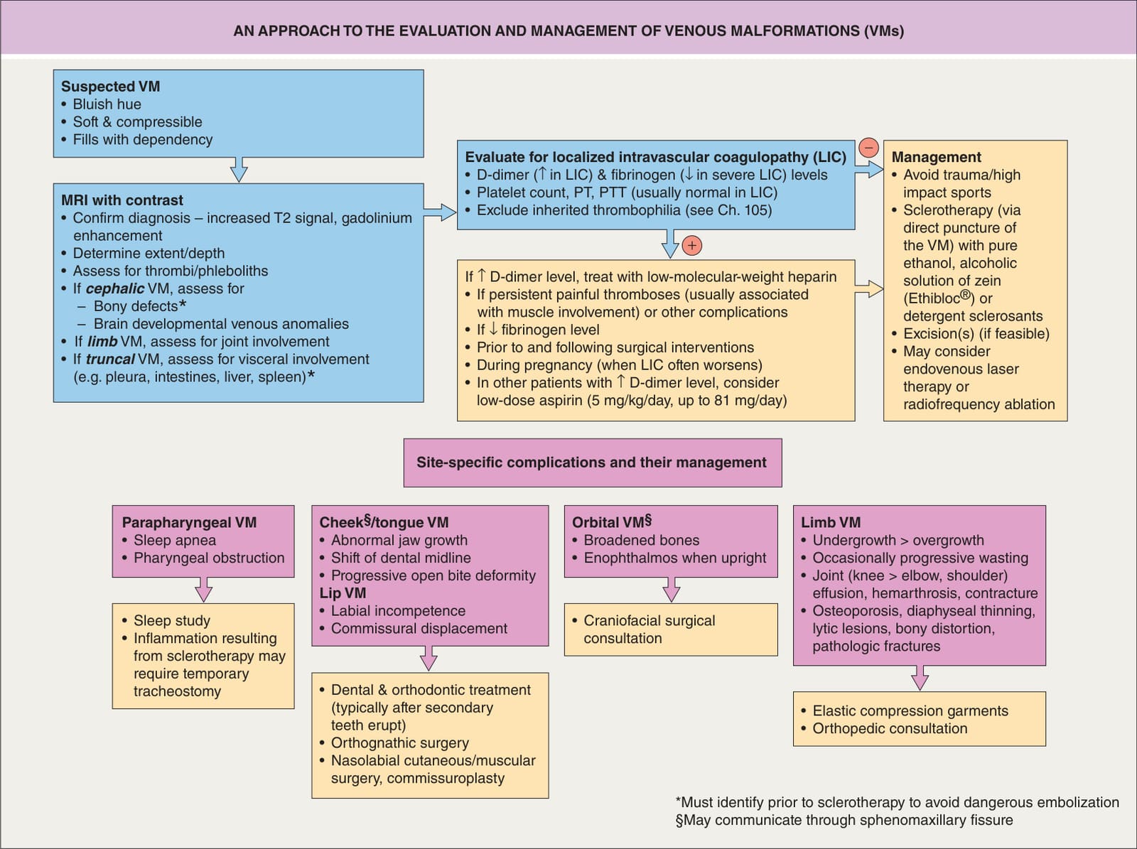
Fig. 104.16 An approach to the evaluation and management of venous malformations (VMs).
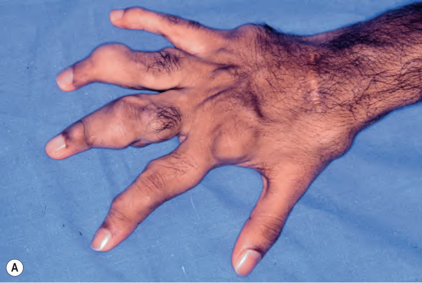
Fig. 104.17 Blue rubber bleb nevus syndrome. Multiple scattered small blue venous nodules, several of which are superimposed on a larger subcutaneous venous malformation. Courtesy Juan Carlos López, MD.
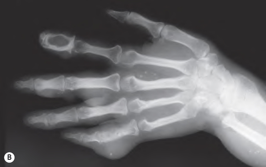
Fig. 104.18 Maffucci syndrome.A Both nodular venous malformations and enchondromas distort the hand. B A radiograph demonstrates an enchondroma and multiple phleboliths.
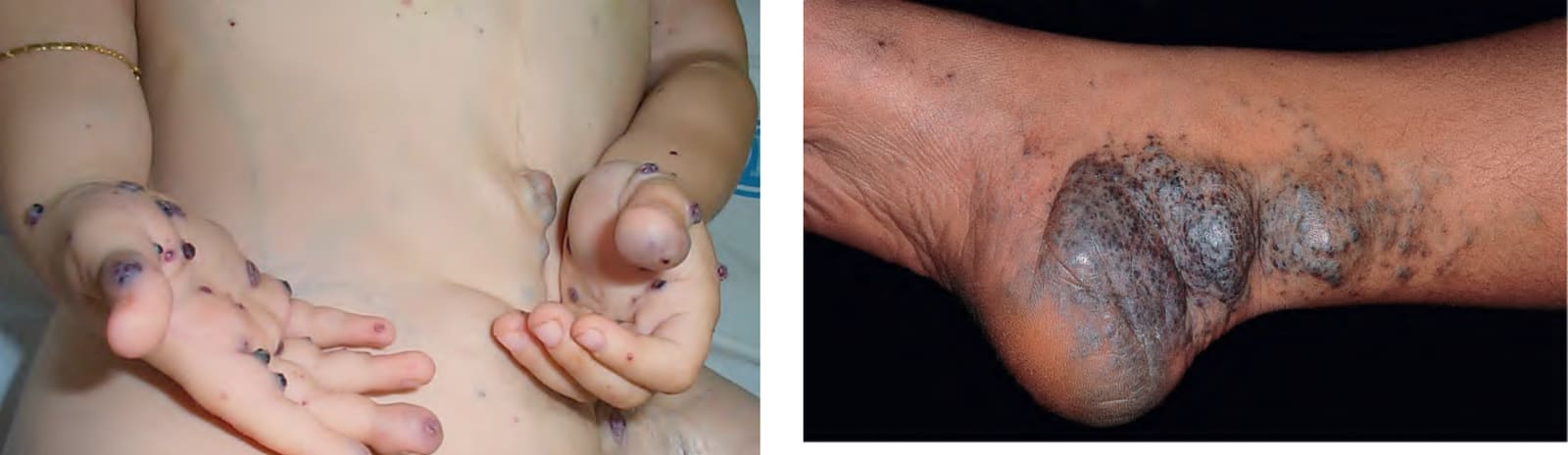
Fig. 104.19 Plaque-type glomuvenous malformation on the distal lower extremity.
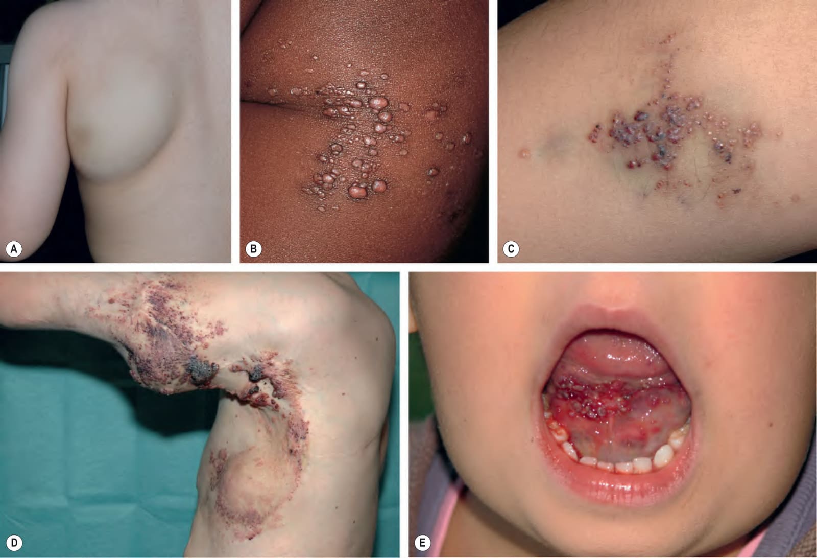
Fig. 104.20 Lymphatic malformations (LMs).A Macrocystic LM on the lateral trunk. The mass was soft except for a focal firm area of hemorrhage associated with bruise-like discoloration of the overlying skin. B–E Microcystic LMs presenting as clusters of clear or hemorrhagic vesicles, with active bleeding from lesions in the mouth (E). The crops of vesicles in (D) recurred years after surgical resection (utilizing grafting and linear closure) of a large microcystic LM affecting the skin and deeper structures of the arm and thorax. A, C, Courtesy Julie V. Schaffer, MD.
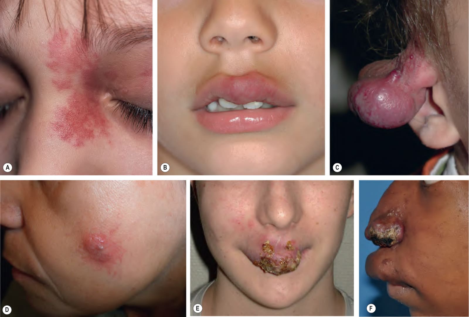
Fig. 104.21 Arteriovenous malformations (AVMs).A,B Dormant (stage 1) AVMs mimicking a port-wine birthmark (A) and infantile hemangioma (B). C, D Expanding (stage 2) AVMs on the external ear and cheek. E, F Destructive (stage 3) AVMs that have led to cutaneous necrosis. D, Courtesy, Fernando Alfageme, MD; E, Courtesy Juan Carlos López, MD.
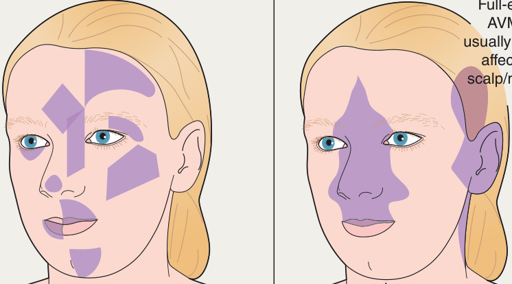
Fig. 104.22 Arteriovenous malformations – size, locations, and patterns.
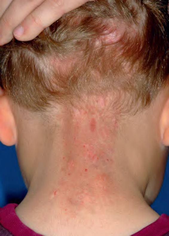
Fig. 104.23 Cobb syndrome. This birthmark on the nape has an appearance reminiscent of a salmon patch, but it was warm and superimposed darker red macules progressively appeared. Doppler ultrasonography, MRI, and arteriography confirmed the presence of a arteriovenous malformation of the skin and spine.
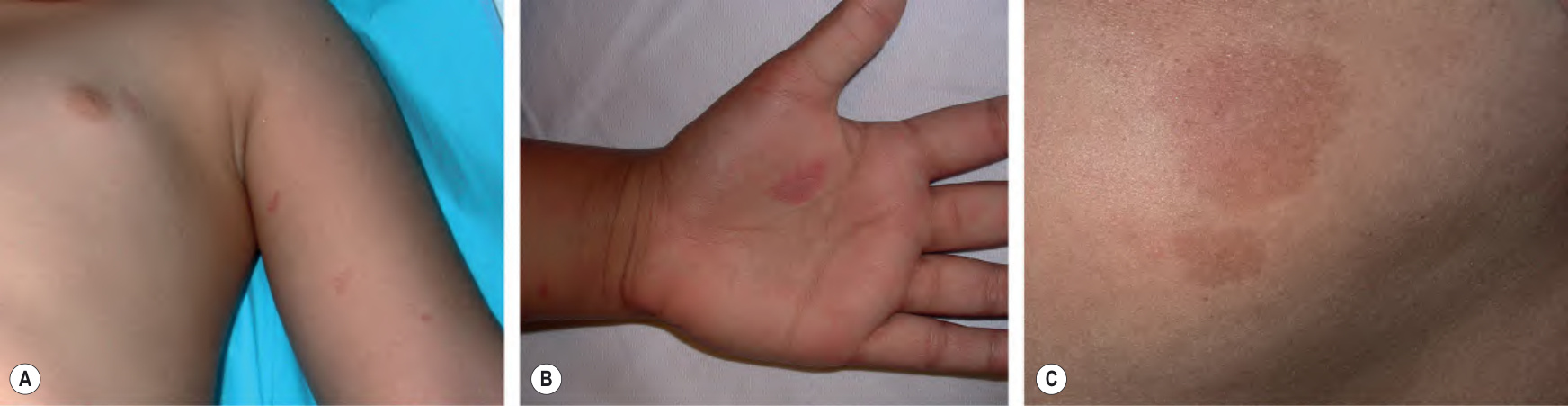
Fig. 104.24 Capillary malformation–arteriovenous malformation (CM-AVM) due to a RASA1 mutation. Multiple pink–red (A,B) to brownish (C) macules. A subtle blanched halo is often evident at the periphery of the lesions. B, Courtesy Margarita Larralde, MD.
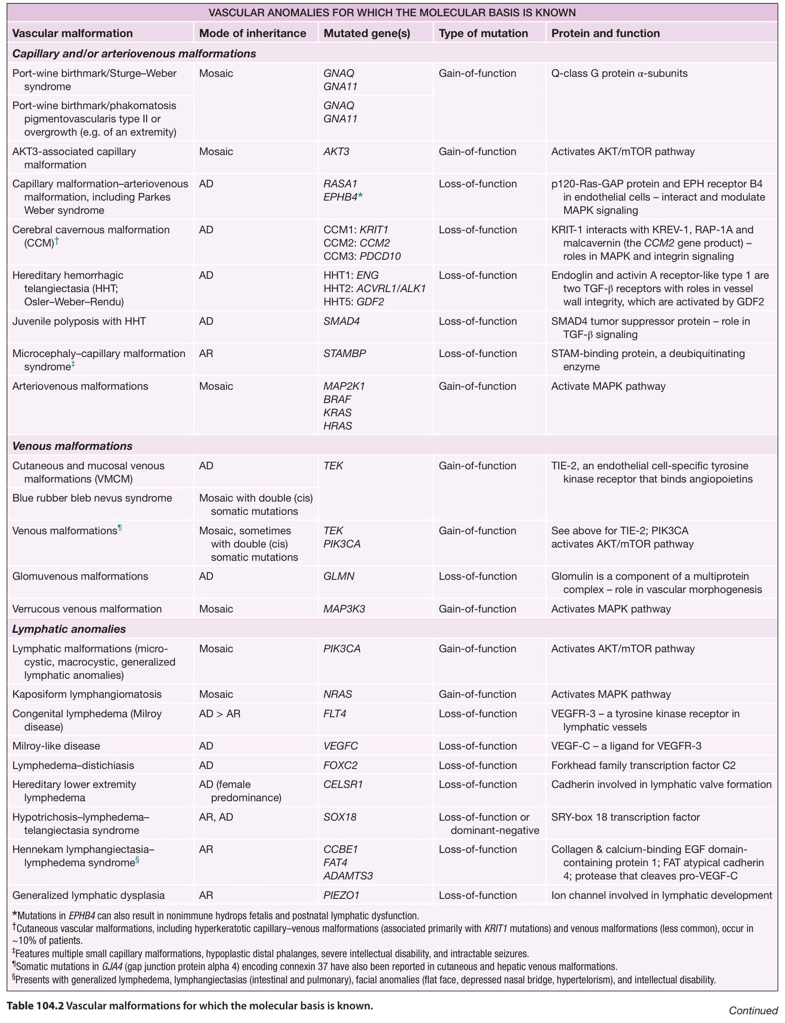
Table 104.2 Vascular malformations for which the molecular basis is known. Continued
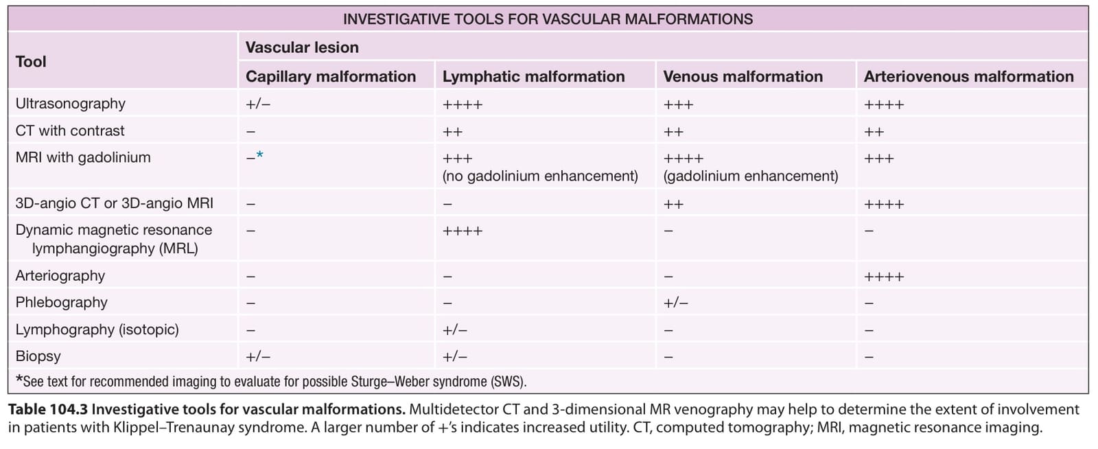
Table 104.3 Investigative tools for vascular malformations. Multidetector CT and 3-dimensional MR venography may help to determine the extent of involvement in patients with Klippel–Trenaunay syndrome. A larger number of +’s indicates increased utility. CT, computed tomography; MRI, magnetic resonance imaging.
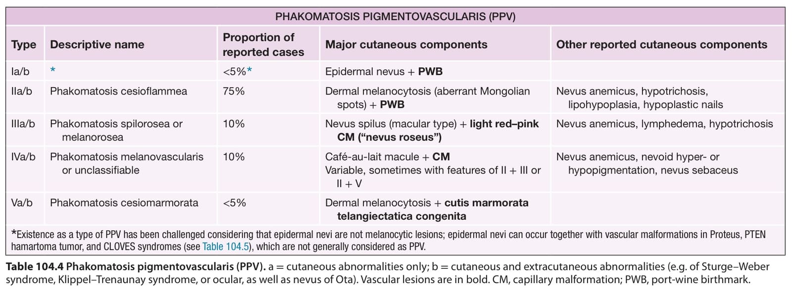
Table 104.4 Phakomatosis pigmentovascularis (PPV). a = cutaneous abnormalities only; b = cutaneous and extracutaneous abnormalities (e.g. of Sturge–Weber syndrome, Klippel–Trenaunay syndrome, or ocular, as well as nevus of Ota). Vascular lesions are in bold. CM, capillary malformation; PWB, port-wine birthmark.
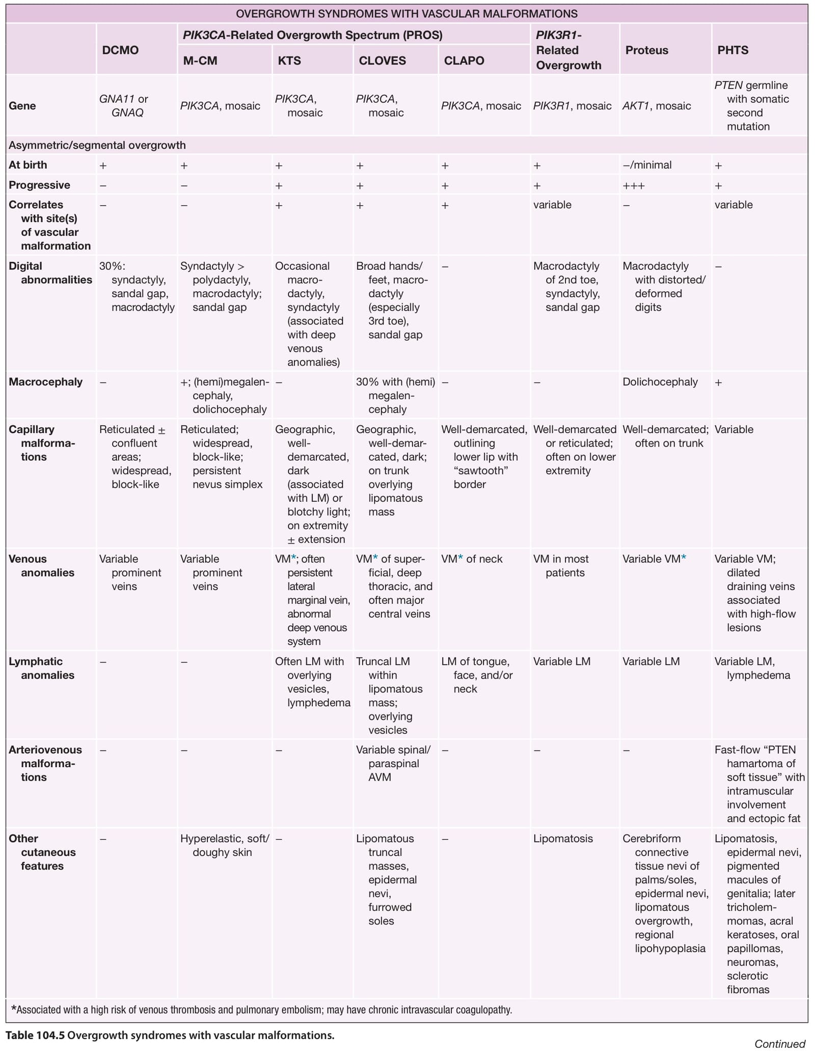
Table 104.5 Overgrowth syndromes with vascular malformations. Continued
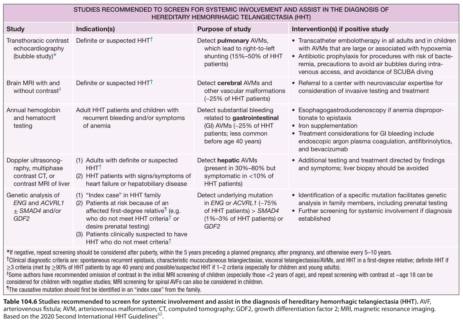
Table 104.6 Studies recommended to screen for systemic involvement and assist in the diagnosis of hereditary hemorrhagic telangiectasia (HHT). AVF, arteriovenous fistula; AVM, arteriovenous malformation; CT, computed tomography; GDF2, growth differentiation factor 2; MRI, magnetic resonance imaging. Based on the 2020 Second International HHT Guidelines.