PATHOLOGY
The histologic characteristics of the four major types of vascular malformations are illustrated in Fig. 104.25.
Capillary Malformations
A CM is composed of ectatic capillaries within the papillary and reticular dermis, lined by a flat continuous endothelium. Thickening of affected skin can occur over time due to a progressive increase in the diameter of the capillaries, involvement of the dermis and subcutis, fibrosis of the dermis, and, occasionally, venous channels in the subcutis.
Venous Malformations
Large convoluted and interconnected anomalous venous channels dissect the normal connective tissues and muscles. Organized thrombi, phleboliths (round calcifications), and intravascular papillary endothelial hyperplasia are frequently observed. Anomalous vessels of a VM have a continuous flat endothelium and a thin basement membrane. Their walls have discontinuous media due to foci with a relative lack or absence of smooth muscle cells.
Lymphatic Malformations
LMs of the microcystic type exhibit enlarged, distorted, irregular lymphatic channels with varying numbers of smooth muscle cells in their walls and a very thin endothelium, sometimes barely visible. Larger polyhedral lymphatic vessels may exhibit valves, and they are usually separated by fibrous septa. Lymphatic clefts dissect the host connective tissues. Hyperkeratotic epidermis may overlie some clusters of dermal lymphatic vesicles. LMs of the macrocystic type are composed of large lymphatic cisterns, often interconnected with thinner channels. Vesicles and large cysts in LMs usually contain watery fluid, lymphocytes, and macrophages; hemorrhage can also be seen. Lymphatic endothelium can be identified by immunohistochemical staining for podoplanin, LYVE-1, and Prox1 (see Ch. 102).
Arteriovenous Malformations
AVMs have irregularly thickened walls and are usually randomly distributed in the dermis and other involved tissues. Direct communications between vessels with different elastic components, typically arteries and veins, may be evident. A capillary component is often obvious in the overlying papillary dermis, and it may have a lobular architecture.
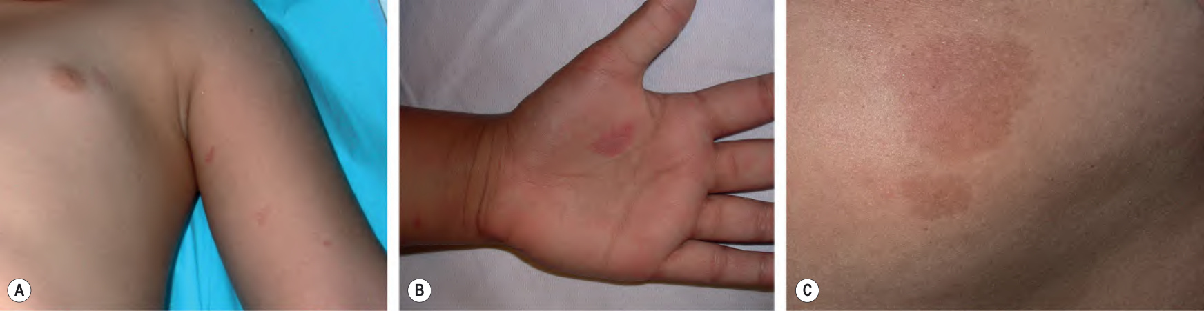
Fig. 104.24 Capillary malformation–arteriovenous malformation (CM-AVM) due to a RASA1 mutation. Multiple pink–red (A,B) to brownish (C) macules. A subtle blanched halo is often evident at the periphery of the lesions. B, Courtesy Margarita Larralde, MD.
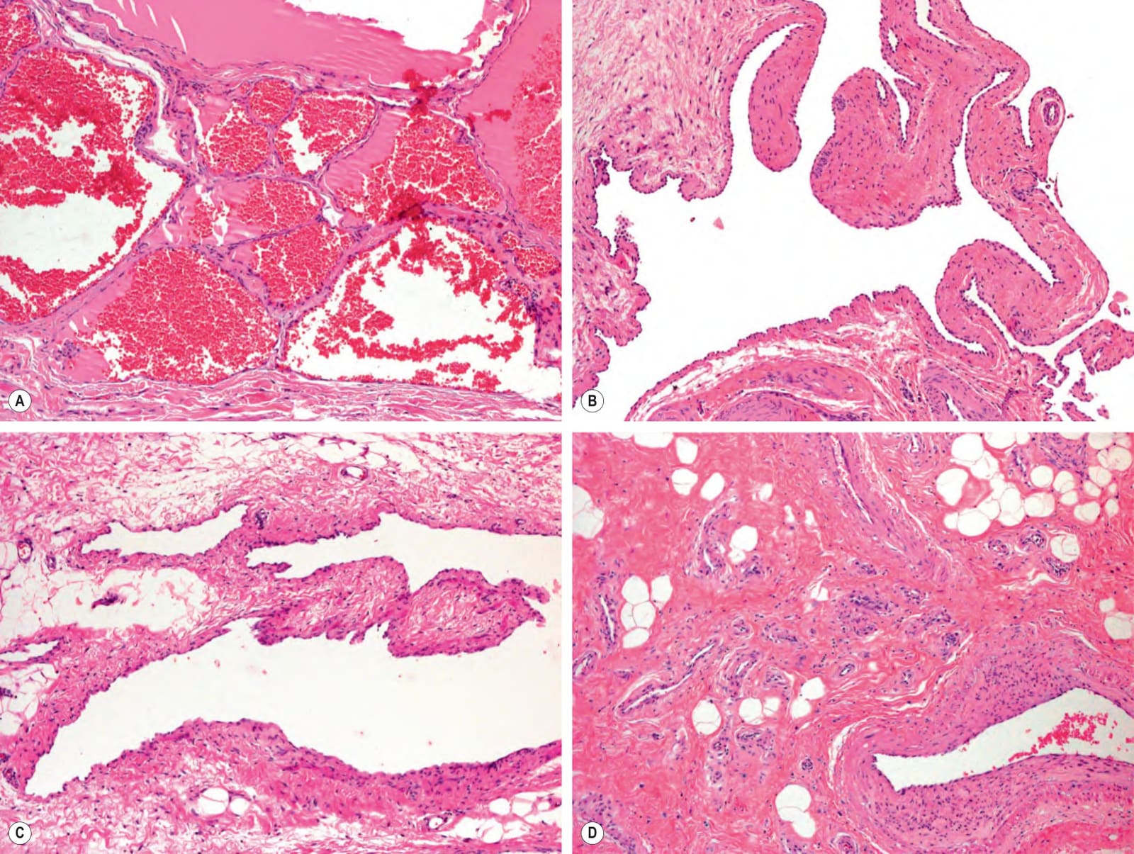
Fig. 104.25 Histologic features of vascular malformations.A Capillary malformation in an adult that was thickened and nodular. An increased number of dilated capillaries are present in the dermis. B Venous malformation composed of large, irregularly shaped, thin-walled channels. C Lymphatic malformation with large, irregularly shaped channels. D Arteriovenous malformation demonstrating an area of intense capillarogenesis near the AV nidus. This finding is sometimes misinterpreted as a “capillarovenous” malformation. A–D, Courtesy Lorenzo Cerroni, MD.