DIFFERENTIAL DIAGNOSIS
Vascular malformations must be distinguished from vascular neoplasms. Superficial infantile hemangiomas at the beginning of their proliferative phase or with minimal/arrested growth may resemble CMs, whereas deep infantile hemangiomas and other vascular tumors such as kaposiform hemangioendothelioma, infantile hemangiopericytoma, and benign lymphangioendothelioma (acquired progressive lymphangioma) can mimic VMs, LMs, and LVMs (see Chs. 103 & 114). Multifocal lymphangioendotheliomatosis with thrombocytopenia should be considered in patients with multiple vascular malformations, especially when associated with gastrointestinal bleeding (see Ch. 114).
Occasionally, other neoplasms and inflammatory disorders are misdiagnosed as vascular anomalies. A red or blue color and/or a mass underneath normal skin can represent potentially misleading clinical signs. For example, erythematous lesions of lupus erythematosus, plaque-type angiofibromas, telangiectasia macularis eruptiva perstans, and other telangiectatic conditions can mimic CMs (see Ch. 106). Dermal melanocytosis has a deep blue color that may lead to misdiagnosis as a VM. Masses beneath normal skin that may be erroneously diagnosed as a macrocystic LM in infants and children include a lipoblastoma, teratoma, bronchogenic cyst, branchial cleft cyst, thyroglossal duct cyst, myofibroma, rhabdomyosarcoma, and fibrosarcoma.
Acquired cutaneous lymphangiectasia imitating a LM can develop in several settings (Table 104.7).
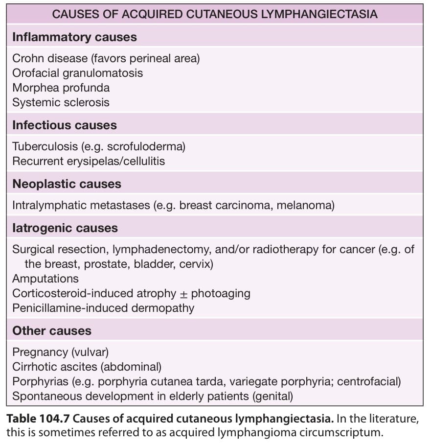
Table 104.7 Causes of acquired cutaneous lymphangiectasia. In the literature, this is sometimes referred to as acquired lymphangioma circumscriptum.