TREATMENT
No single treatment exists for this heterogeneous group of lesions.
Capillary Malformations
PWBs and telangiectasias are best treated with a pulsed dye laser (PDL; see Ch. 137). This treatment of PWBs, which is typically initiated early in life for facial lesions to decrease potential psychosocial impact and avoid lesional thickening, is often very effective. However, not all lesions respond well; PWBs on the central face and distal limbs tend to be more resistant to therapy than those in other sites. PDL treatment is safe, producing temporary purpura but a low risk of hypoor hyperpigmentation and scarring. Although the latter side effects
are more common in individuals with darkly pigmented skin (e.g. skin phototype V), these patients may have a good response and should not be excluded from PDL treatment.
Intervention during infancy with modified PDL therapy using a longer wavelength, increased pulse duration, higher fluence (11–12 J/cm), and dynamic cooling spray can maximize lightening or clearing of PWBs. Use of a topical anesthetic (see Ch. 143), together with the anesthetic effect of the cryogen spray, helps to reduce the need for general anesthesia in children. In a longitudinal study of 51 patients whose facial CM was treated with classic PDL, redarkening was evident in one-third of the lesions after a median follow-up period of 10 years. Relapses may be less common in patients treated at a younger age. If treatment is delayed, facial and gingival PWBs may develop hyperplastic changes that require additional dermatologic (e.g. dermabrasion prior to PDL treatment) as well as orthodontic and surgical interventions (e.g. to correct open-bite deformities and macrocheilia). For PWBs resistant to PDL, other lasers such as the alexandrite or Nd:YAG may be of utility. Topical application of sirolimus in conjunction with PDL may also provide additional benefit (see Ch. 129).
Multidisciplinary care is necessary for patients with SWS. Anticonvulsants are the mainstay of treatment for seizures, although selected patients with severe, uncontrolled seizures may benefit from surgical management. Low-dose aspirin (3–5 mg/kg) can also be considered in symptomatic and presymptomatic patients with brain involvement. Oral sirolimus also has potential benefit for uncontrolled seizures, cognitive impairment, and perhaps as a preventive treatment in SWS patients with extensive leptomeningeal involvement. Glaucoma usually responds to medical or surgical treatment. As discussed above, PDL therapy for PWBs can begin during infancy. Psychological counseling may be helpful for patients and families affected by SWS, and support is also available through the Sturge–Weber Foundation (www.sturge-weber.org).
Venous Malformations
Satisfactory treatment of a cephalic VM typically requires multiple sclerotherapy procedures over a period of years, often spanning from childhood to adulthood, combined with surgical excisions (see Fig. 104.16). The aims are to maintain facial symmetry, preserve muscular functions, and restore the dynamics of the smile. Limb VMs may also benefit from sclerotherapy and resection; however, many are too extensive and invasive for such interventions to be advisable. Use of elastic compression garments helps to reduce swelling and pain in the affected extremity. Treatment with low-molecular-weight heparin can control chronic coagulopathy associated with VMs, especially before surgery and during painful thrombotic events. Low-dose aspirin (3–5 mg/kg/day, up to 81 mg/day) may be of some benefit in patients with a mild coagulopathy.Treatment of VMs with oral sirolimus or the PI3K inhibitor alpelisib has also been reported to improve the associated coagulopathy as well as reduce lesion size and pain, although sirolimus appears to have greater benefit for combined lesions with a lymphatic component.
Lymphatic Malformations
The choice and outcome of treatment depend on the type and extent of the LM. Macrocystic LMs are best treated with percutaneous sclerotherapy utilizing agents such as killed bacteria (picibanil/OK-432), alcoholic solution of zein (Ethibloc®), sodium tetradecyl sulfate, pure ethanol, doxycycline, or bleomycin. This creates an inflammatory reaction with subsequent fibrosis and shrinkage of the treated cyst; repeated procedures are usually necessary. Surgical excision can be employed if sclerotherapy fails or gives incomplete results.
Surgical excision is the treatment of choice for microcystic LMs, either with direct closure or (for large lesions) use of skin grafts or tissue expanders. However, incomplete excisions occur in virtually all cases, whether because of concern regarding functional consequences of extensive surgeries or underestimation of the extent of the LM (see Fig. 104.21D). Other techniques that are often used in combination include laser therapy (e.g. CO, surface application or intralesional bare fiber Nd:YAG, diode), radiofrequency ablation, cryoablation, and sclerotherapy. Topical application of sirolimus may have benefit for the superficial component of microcystic LMs, with case series noting reductions in vesicle volume, lymphatic leakage, and bleeding. Microcystic LMs of the tongue and floor of the mouth are particularly difficult to treat, and careful dental hygiene is important. Transient inflammatory swelling of LMs usually responds to therapy with NSAIDs, corticosteroids, and/or antibiotics.
Patients with complex lymphatic anomalies may need replacement of albumin and immunoglobulins. A low-fat diet and substitution of long-chain fatty acids with medium-chain fatty acids may benefit those with intestinal lymphangiectasia. Oral sirolimus has been used with success in patients with LMs, GLA, and Gorham–Stout disease, and PI3K inhibition with alpelisib represents another promising therapeutic strategy. Sildenafil treatment has led to variable decreases in volume and softening of large LMs in some patients, but no responses were seen in other series. Zoledronate may be of benefit for osteolysis secondary to LMs in Gorham–Stout disease.
Arteriovenous Malformations
In general, stage 2 and 3 AVMs of the head and neck should be excised completely after careful preoperative embolization using arterial ± direct puncture approaches to prevent excessive intraoperative bleeding. Reconstruction often requires techniques such as skin grafting, preoperative tissue expansion, and free tissue transfer. Subtotal excision frequently results in subsequent progression of the AVM, with recruitment of adjacent vessels and apparent angiogenesis. Stage 1 and early stage 2 AVMs are often managed conservatively if postsurgical cosmetic results might be worse than the lesion itself. However, resection of early-stage AVMs can prevent further progression.
Embolization alone usually cannot cure a superficial AVM. The most effective embolic agent, pure ethanol, poses substantial risks (especially at higher doses) that include renal or pulmonary toxicity and cardiac arrest. For AVMs that begin to hemorrhage, embolization may be lifesaving. Distal hand or foot AVMs are rarely controlled long-term by embolotherapy, and ischemic complications resulting in extreme pain, necrosis, and bleeding may require amputation. In one report, treatment of a diffuse AVM in the arm of a young girl using a metalloproteinase inhibitor (Marimastat®) with antiangiogenic effects produced sustained improvement over a 12-year period, including reconstruction of affected bones and without adverse effects. Treatment with trametinib, a
MEK inhibitor, was beneficial in a child with a large, progressive truncal MAP2K1-related AVM and a young woman with cardiac compromise due to CM-AVM syndrome.
Combined Malformations and the PIK3CA-Related Overgrowth Spectrum (PROS)
A combined vascular malformation of a limb requires an individualized management plan. Longitudinal orthopedic evaluation is essential for lower extremity lesions in growing children. Carefully timed epiphysiodesis before the end of the growth period can potentially correct a leg-length discrepancy in KTS, but it may worsen vascular disease in Parkes Weber syndrome. Treatment of a component of the vascular malformation (e.g. surgery for varicose veins in KTS or embolization of the AV nidus in Parkes Weber syndrome) often has suboptimal results. Elastic support garments and a compensatory shoe-lift are indispensable conservative interventions.
The PI3K inhibitor alpelisib is FDA-approved for patients ≥2 years of age with severe manifestations of PROS. In a retrospective study in 32 patients with PROS, alpelisib therapy for 24 weeks led to improvement in vascular malformations and overgrowth in 79% and 74% of patients, respectively, with 38% achieving a ≥20% reduction in the volume of PROS lesions. Treatment of Proteus syndrome patients with miransertib, an oral AKT inhibitor, has led to stabilization of overgrowth and pain improvement. Additional clinical trials and long-term follow-up studies are needed to provide further data on the benefits of targeted therapies in these overgrowth syndromes.
The author wishes to acknowledge the late Odile Enjolras, MD for her valuable contribution to this chapter in the previous editions.
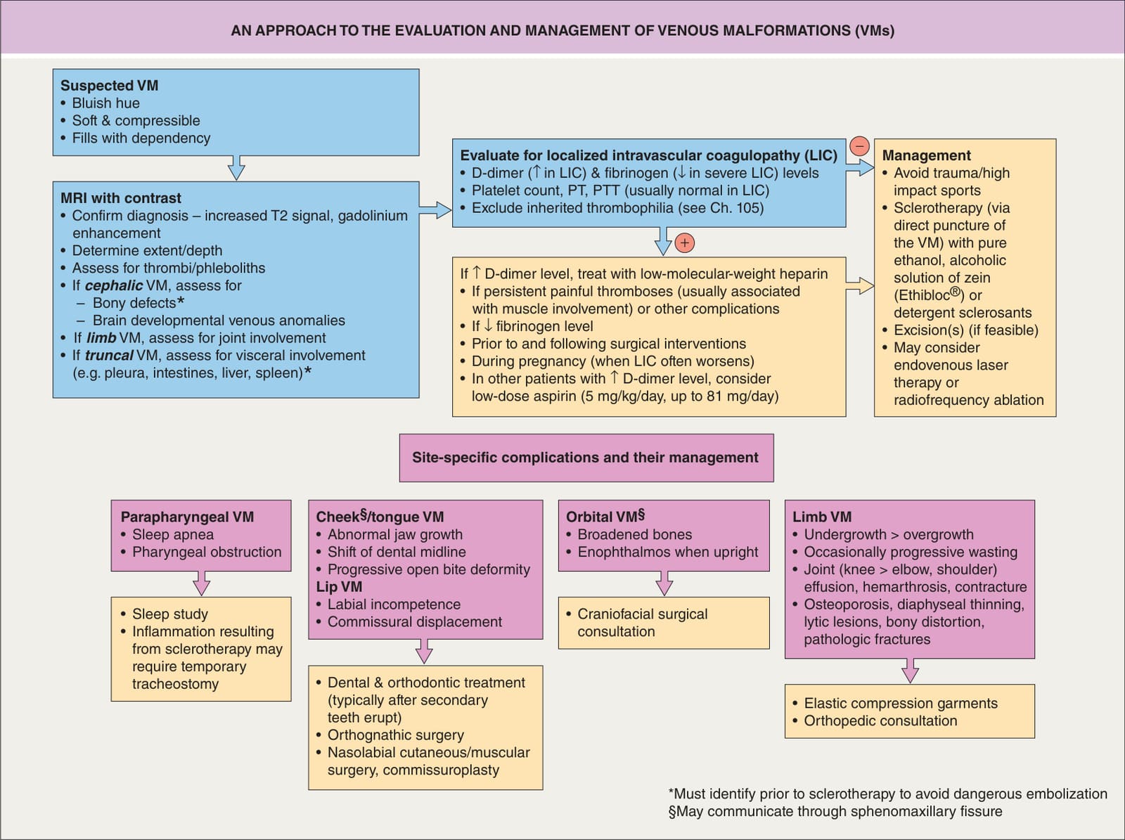
Fig. 104.16 An approach to the evaluation and management of venous malformations (VMs).
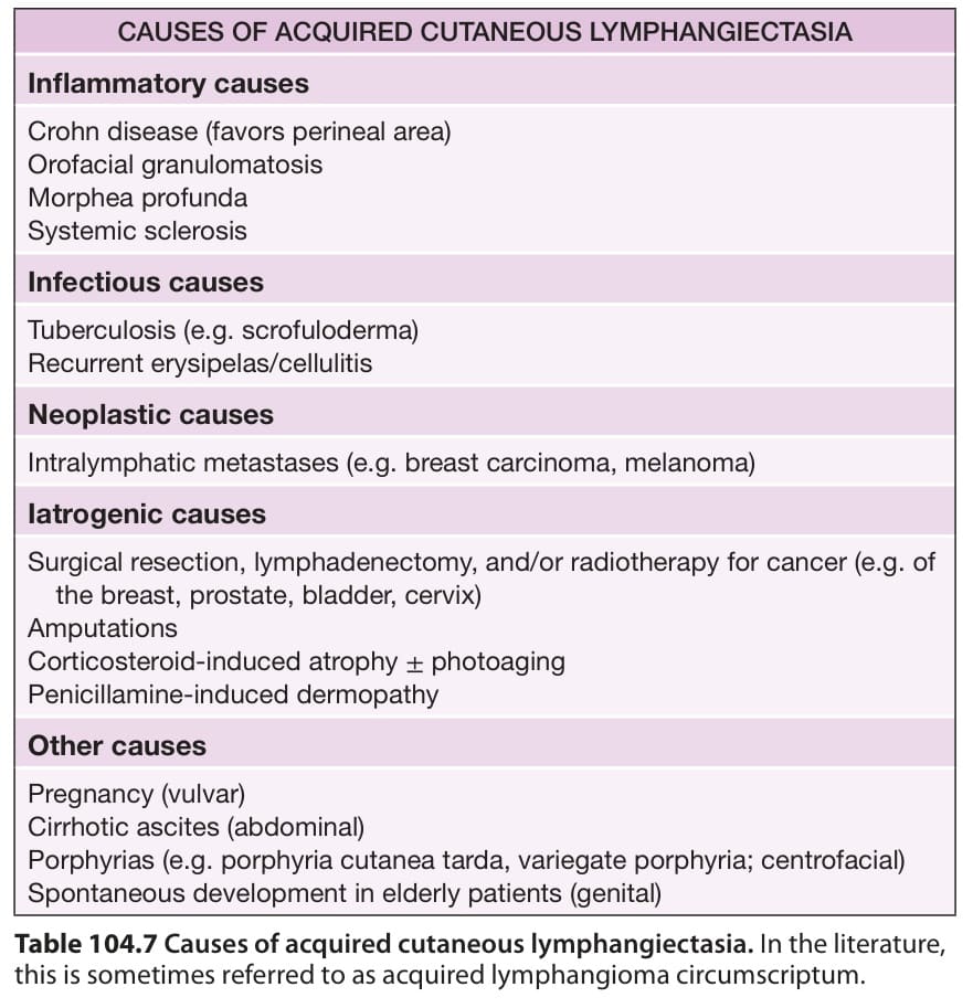
Table 104.7 Causes of acquired cutaneous lymphangiectasia. In the literature, this is sometimes referred to as acquired lymphangioma circumscriptum.