INTRODUCTION
A chronic wound is defined as one that fails to progress through a normal orderly and timely sequence of repair or a wound that passes through the repair process without restoring a functional result. A cutaneous ulcer is a wound that is associated with loss of both epidermal and dermal tissues. Chronic ulcers, which are due to a variety of systemic and/or local factors, can represent both a diagnostic and therapeutic challenge to the dermatologist. The majority of ulcers occur on the lower extremities, and most are related to venous insufficiency/venous hypertension, peripheral artery disease, or peripheral neuropathy (Table 105.1). Less common causes are outlined in Figure 105.1 and sites other than the legs may be involved (e.g. pressure-induced ulcers).
Wound therapy has advanced considerably over the past few decades with the advent of innovative dressings and technologies, but the key factor in the management of chronic ulcers remains proper patient evaluation and correction of the underlying cause, when identified.
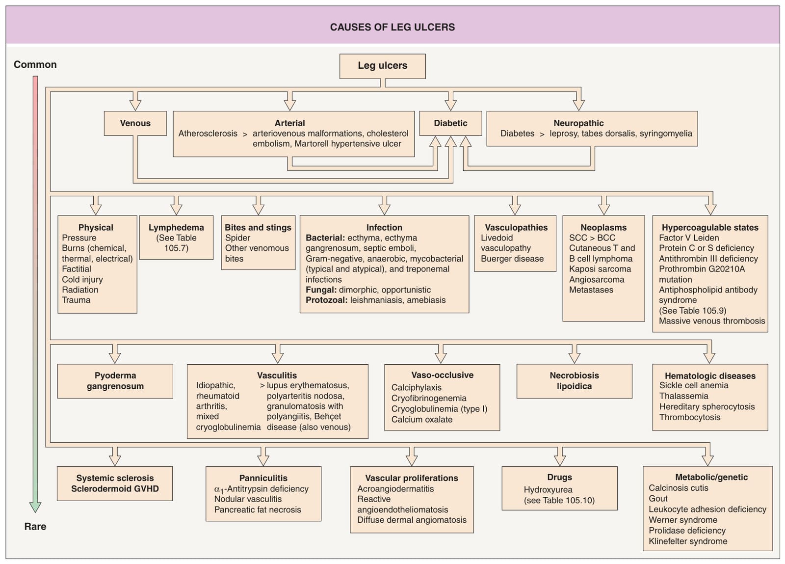
Fig. 105.1 Causes of leg ulcers. Patients with Behçet disease develop lower extremity ulcers due to vasculitis and/or venous insufficiency related to deep vein thromboses, and, occasionally, erosive pustular dermatosis is a cause of leg ulcers. Hydroxyurea-induced leg ulcers are often on the malleolus or tibial crest, exceedingly painful, and surrounded by atrophic skin. Additional genetic syndromes associated with ulceration are listed in Table 105.10. GVHD, graft-versus-host disease.
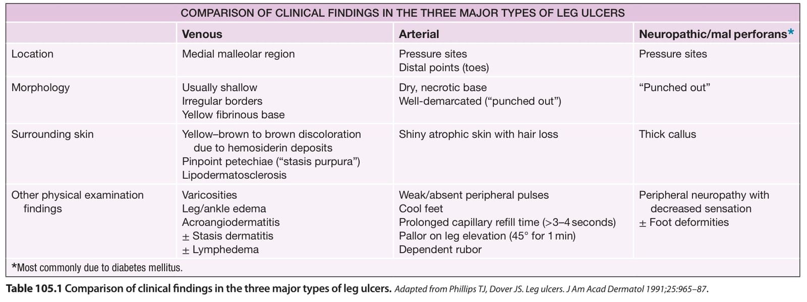
Table 105.1 Comparison of clinical findings in the three major types of leg ulcers.