VENOUS ULCERS
Chronic venous disease is a common problem in adults, with its prevalence increasing with age. Due in large part to a lack of standardized diagnostic criteria, the prevalence varies considerably, from <1% to 17% in men and from <1% to 40% in women.
Risk factors for chronic venous disease include family history, age, female sex, obesity, pregnancy, prolonged standing, and height. Venous hypertension and venous insufficiency represent the most common cause of chronic leg ulcerations. Venous ulcers tend to recur, are sometimes painful, and clearly have an impact on quality of life. Because of their prevalence and chronicity, there are significant healthcare costs, estimated to be ~$3 billion per year in the US.
Pathogenesis
The venous system of the lower extremities consists of an interconnected network of superficial veins, deep veins, and perforator or communicating veins which connect the superficial and deep systems (see Ch. 155). Throughout the superficial, communicating and deep veins, one-way bicuspid valves ensure unidirectional flow toward the deep system, thus allowing blood to flow in a cephalad direction and preventing reflux (Fig. 105.2A). It is primarily the contraction of calf muscles that drives blood from the leg toward the heart.
During standing, as leg muscles relax, venous pressure in the legs may reach up to 80 to 90 mmHg. Calf muscle contractions, e.g. when walking, transiently increase pressure within the deep leg veins, propelling blood towards the heart. When pressure rises within the deep system, the valves close, preventing retrograde flow and transmission of high pressure to the superficial system. When the deep system empties, the deep vein pressure abruptly falls to <30 mmHg and the valves open, enabling flow from the superficial into the deep system.
In the absence of competent valves, however, the decrease in venous pressure with leg movements is attenuated, and high pressures generated in the deep veins by calf muscle contraction are transmitted to the superficial venous system and to the microcirculation within the skin. A sustained elevation in venous pressure is responsible for the typical clinical picture seen in chronic venous disease and is termed venous hypertension or venous insufficiency.
In most patients, venous hypertension is caused by reflux through incompetent valves (Fig. 105.2B), whether primary (congenital) or secondary to deep vein thromboses or recurrent trauma. Additional causes of venous hypertension include venous outflow obstruction and failure of the calf muscle pump due to obesity or leg immobility.
Although venous hypertension appears to be central to the skin changes seen in chronic venous disease, the exact pathogenic cascade from valvular incompetence to frank ulceration remains obscure. A few pathomechanisms have been suggested, but it is still unclear whether they represent causative factors or epiphenomena. One proposal was that cuffs of fibrin around dermal capillaries (due to leakage of fibrinogen) could impede the diffusion of oxygen and nutrients to
Adapted from Phillips TJ, Dover JS. Leg ulcers. J Am Acad Dermatol 1991;25:965–87.
surrounding tissues and lead to degenerative skin changes. Other investigators suggested that venous pooling induced inter-endothelial space widening and deposition of fibrin and other macromolecules that then “trapped” growth factors, rendering them unavailable for wound repair.
Nowadays, however, the predominant theory is that chronic inflammation plays a key role in chronic venous ulcers (Fig. 105.3). With elevated venous pressure, shear stress on the endothelial wall is altered, leading to glycocalyx disruption and endothelial cell activation. As a result, there is release of vasoactive mediators, proinflammatory molecules (e.g. chemokines), and prothrombotic precursor proteins. Increased expression of adhesion molecules such as ICAM-1, VCAM-1, and platelet-endothelial leukocyte adhesion molecule-1 (PECAM-1) on endothelial cells leads to leukocyte recruitment followed by diapedesis
into the dermis (see Fig. 102.11). This inflammatory response then leads to further venous wall damage.
Increased expression and activity of matrix metalloproteinases (MMPs) also contribute to the breakdown of the extracellular matrix, which promotes the formation of ulcers and impairs healing. Elevated levels of cytokines such as transforming growth factor-beta (TGF-β) and vascular endothelial growth factor (VEGF) translate into increased dermal fibrosis and proliferation of dermal capillaries, respectively (see Fig. 141.3). Capillary hyperpermeability and extravasation of red blood cells lead to deposition of ferric iron within affected skin, resulting in brown discoloration.
While the persistent inflammatory response gives rise to ulcer formation (see above), continued production of MMPs and cytokines (e.g. interleukin-1, tumor necrosis factor [TNF]) leads to impaired fibroblast function and increased degradation of growth factors necessary for wound healing. In addition, oxidative stress-induced premature aging and dysfunction of wound fibroblasts contribute to the chronicity of venous ulcers.
Clinical Features
Chronic venous disease is associated with a wide spectrum of clinical findings, ranging from uncomplicated telangiectasias and varicose veins to lipodermatosclerosis and large chronic ulcerations. Patients with venous ulcers often complain of swelling and aching of the legs, typically worsened by dependency and prolonged standing but improved by leg elevation, use of gradient support stockings or walking, all of which decrease venous pressure. Additional symptoms sometimes ascribed to chronic venous disease include muscle cramps, pruritus, and restless legs.
Most commonly, venous ulcers are located above the medial malleolus (Fig. 105.4). The ulcer is usually shallow with a border that is typically irregular; the wound bed is often covered by a yellow, fibrinous exudate. If the latter is appropriately debrided, a base of healthy, non-ischemic granulation tissue is usually seen. When untreated, venous ulcers tend
Note the erythema, crusting, and scaling of the skin surrounding the ulcer of the medial malleolus. There is a yellowish fibrinous exudate and granulation tissue is seen in 15%–20% of the ulcer bed. In a chronic ulcer, the thickened rolled border is referred to as epibole.
to become larger than most other chronic wounds and can even become circumferential on the lower extremity. Pedal pulses are usually palpable. If they are not palpable because of induration or edema, an ankle–brachial index (see below) should be obtained to exclude concomitant peripheral artery disease.
Characteristic associated features include (Fig. 105.5): (1) varicose veins of varying severity (see Ch. 155); (2) pinpoint petechiae superimposed on a yellow–brown discoloration due to extravasation of red blood cells and deposition of hemosiderin within macrophages (see Fig. 22.7), which is sometimes accompanied by hyperpigmentation; (3) edema; and (4) stasis dermatitis with pruritus, erythema, scaling, and weeping (see Ch. 13). Of note, stasis dermatitis is sometimes aggravated by allergic contact dermatitis to topically applied medications, e.g. neomycin. The smooth, ivory-white, sclerotic plaques of atrophie blanche that are often surrounded by punctate telangiectasias and/or brown discoloration are observed in up to 40% of patients with chronic venous insufficiency.
Patients with longstanding venous hypertension and insufficiency may develop chronic lipodermatosclerosis (also known as sclerosing panniculitis) with induration and fibrosis that begins on the lower medial aspect of the leg above the malleolus (see Fig. 105.5C, D). Eventually, the entire lower third of the leg may become involved, leading to an appearance that has been likened to an “inverted champagne bottle”. Some authors believe the degree of lipodermatosclerosis correlates with delayed wound healing. Chronic lipodermatosclerosis may be preceded or accompanied by an acute inflammatory stage characterized by warm, erythematous, firm plaques that are tender and painful and are often misdiagnosed as cellulitis or another type of panniculitis, especially erythema nodosum.
Lipodermatosclerosis, along with ulcerations, is not limited to the lower extremities, as these changes can be seen in other sites such as the most dependent portion of the pannus (Fig. 105.6).
Laboratory Evaluation
Standardized evaluation of patients with chronic venous disease is based on the CEAP classification system which generates scores derived from objective clinical signs (C), etiology (E), anatomic (A) location of the affected veins, and pathophysiology (P) (reflux, obstruction, or both). Table 155.1 outlines the details of the CEAP classification system.
Color duplex ultrasonography, preferably performed with the patient in a standing position, is useful for assessing both reflux and obstruction within the deep, superficial, and perforating veins, from the inferior vena cava to the calf veins. It provides information that is necessary when determining the CEAP score. For most indications, color duplex ultrasonography has replaced venography. The latter is an invasive test that can provide a detailed view of the venous system in preparation for reconstructive surgery and stenting procedures.
Pathology
In the skin surrounding the ulceration, nonspecific features are seen including reactive epithelial hyperplasia, dermal fibrosis, and an inflammatory infiltrate, along with a proliferation of capillaries, extravasation of red blood cells, and deposits of hemosiderin within macrophages. Biopsies are occasionally performed to exclude a neoplastic, vasculitic, vasculopathic, or infectious process.
Differential Diagnosis
A significant number of patients may have a combination of venous and arterial insufficiency, so before treatments such as compression stockings are prescribed, the latter should be excluded (see below). In patients with livedoid vasculopathy (Fig. 105.7) or recurrent deep vein thromboses, the possibility of thrombophilia (hypercoagulability) as a contributing factor needs to be considered (see Table 105.9 & Ch. 23). Additional clues from the history, e.g. rate of expansion, and physical examination aid in narrowing the differential diagnosis (Table 105.2). Histologic examination of a longstanding or recalcitrant ulcer is recommended to exclude other diagnoses, such as squamous cell carcinoma or atypical mycobacterial infection (see Fig. 105.1), and the biopsy specimen should include surrounding non-ulcerated skin.
Treatment
Most of the therapeutic principles described in the following sections can be applied to the care of other types of cutaneous ulcers, unless otherwise specified. In general, normal wound healing requires adequate perfusion, good nutritional and immune status, and avoidance of deleterious mechanical forces. The initial step in the approach to an ulcer should be to address all systemic and local factors that may impair healing.
Moisture and occlusion
In the past, the mainstay of wound management was to dry the wound bed with absorptive gauzes. A major breakthrough in the management of chronic wounds was the realization that a moist environment is critical for wound healing and occlusive dressings do not increase the risk of infection. Moist retentive wound dressings stimulate collagen synthesis, promote angiogenesis by creating a hypoxic environment, encourage re-epithelialization, and decrease pain. By serving as a barrier against external contamination and by lowering wound pH, occlusive dressings actually decrease wound infection rates.
Dressings should be changed based on their potential for absorbency and the amount of exudate, with attention to the delicate balance between overhydration and excessive dryness. For example, occlusive dressings applied to highly exudative wounds may cause maceration and erosion of the surrounding skin, possibly leading to further deterioration of the wound. In addition, chronic wound fluid can perpetuate cytokine and MMP production (see above), thereby affecting the function of fibroblasts and growth factors and contributing to chronicity. It is therefore important to protect the skin surrounding the wound bed with an ointment in order to preserve barrier integrity.
Debridement
Debridement consists of the removal of necrotic, non-viable, or infected tissue from the wound bed in order to promote healing. In a sense, debridement is an attempt to shift the wound from a chronic state (with a milieu characterized by an abnormal extracellular matrix, an excess of proteolytic enzymes, and senescent cells) into an acute state. Debridement can be surgical (sharp debridement), mechanical (wet-todry), autolytic, chemical (enzymatic), and biologic (maggot therapy).
Sharp debridement with surgical instruments represents the most rapid method for debriding a wound and is most appropriate for large necrotic and/or infected wounds. It also enables the clinician to estimate the extent and severity of a wound. Although damage to viable tissue is possible, sharp debridement performed by a skilled physician is usually highly selective. Caution is advised in the case of arterial ulcers because of the potential risk for tissue desiccation and ulcer enlargement. Sharp debridement may be associated with bleeding and pain and may necessitate analgesia.
Wet-to-dry (mechanical) debridement consists of placing gauze moistened with saline over the wound, allowing the gauze to dry, and then removing the dry gauze along with adherent non-viable tissue. It is relatively fast and easy to perform but is painful and does not discriminate between viable and non-viable tissue. It is indicated for wounds with large amounts of necrotic tissue.
Autolytic debridement is the process by which the body uses its own proteolytic enzymes and phagocytic cells to clear necrotic debris. The process is enhanced by moist retentive wound dressings and may require weeks to accomplish. Autolytic debridement is selective, painless, and suited primarily for wounds with minimal debris. It is contraindicated if the wound is infected.
A Venulectasias of the instep referred to as corona phlebectasia. B Brown discoloration of the foot and ankle due to hemosiderin deposits within dermal macrophages in addition to hyperpigmentation. Note the cut-off at Wallace’s line. C Lipodermatosclerosis with violet– brown discoloration, tenderness, and induration that typically begins above the medial malleolus. D Stasis dermatitis and chronic lipodermatosclerosis with serous crusts and the “inverted champagne bottle” or “bowling pin” configuration. E Acroangiodermatitis (pseudo-Kaposi sarcoma). Violaceous plaque in a patient with venous hypertension. Histologically, these lesions can resemble Kaposi sarcoma. A, B, Courtesy Jean L. Bolognia, MD; C, Courtesy Kalman Watsky, MD.
Enzymatic (chemical) debridement utilizes topically applied proteolytic enzyme preparations such as papain–urea or collagenase to digest necrotic tissue, collagen, fibrin, and wound exudate. Chemical debridement is a gradual process. The papain–urea preparation contains papain, which is derived from the plant Carica papaya and is a non-selective cysteine protease. It is active over a broad pH range and is associated with an intense inflammatory response and digestion of viable tissue; local pain or a burning sensation is often described. Therefore, the product should be applied only to the wound bed and not to the surrounding intact skin. Collagenase preparations are derived from Clostridium histolyticum; they specifically target native collagen and are active within a narrow pH range of 6 to 8. These preparations are highly selective and do not harm viable cells. Prior to application, it is advisable to cross-hatch the eschar, if present, in order to increase penetration.
Multiple hemorrhagic crusts and small, very painful ulcers associated with brown discoloration due to hemosiderin deposits. B Stellate, porcelainwhite atrophic scars with peripheral telangiectatic papules, referred to as atrophie blanche. B, Courtesy Julie V. Schaffer, MD.
For therapy-resistant, chronic ulcers with marked necrosis and slough, maggot debridement therapy can be considered. Essentially, it exploits the ability of maggots to decompose necrotic tissue (benign myasis), but the debridement is done in a controlled manner so as to avoid undesirable effects on healthy tissues. Degradation via proteinases that are in larval excretory/secretory products is thought to contribute to wound debridement. It is important to select larval strains that feed preferentially on necrotic tissue and to avoid applying too many larvae to a wound. The flies most often employed are facultative calliphorids, in particular the green bottle blowfly, Lucilia sericata.
Maggots are used to treat various types of chronic ulcers, including pressure ulcers, venous ulcers, and neurovascular diabetic ulcers, as well as traumatic and postsurgical wounds. In a multicenter, randomized, open study that compared the clinical effectiveness of larval therapy to a standard debridement technique (hydrogel) for “sloughy” or necrotic leg ulcers, larval therapy did not improve the rate of healing or reduce bacterial load, but it did significantly reduce the time to debridement and increase ulcer pain.
Novel approaches to wound debridement are currently under investigation, including both hydrosurgery and ultrasound- and radio frequency-based techniques.
Wound dressings
The ideal wound dressing should: (1) provide a moist retentive environment; (2) absorb excess exudate without causing maceration of surrounding skin; (3) protect against bacteria; (4) leave no residual debris but cause no trauma when removed; (5) decrease odor; and (6) relieve pain. In addition, the ideal wound dressing should be cost-effective, easy to handle, and be neither irritating nor allergenic. While there is no single dressing that can fulfill all these criteria throughout the entire healing process, a vast array of dressings exists, and the choice of a dressing depends upon the wound type and its characteristics at a given time (Table 105.3).
Management of wound infection
The presence of a microbial infection is always detrimental to wound healing. However, all chronic open wounds are colonized with bacteria and it is important, although often difficult, to distinguish true infection from bacterial colonization. Of note, colonization is defined by the presence of replicating bacteria and adherent microorganisms, but without evidence of tissue damage.
Critical colonization is a relatively new concept in which the bacterial burden within a chronic wound does not elicit the typical symptoms or signs of an infection but does delay healing. The presence of >10 colonyforming units of bacteria/gram of tissue predicts delayed wound healing and a high risk for developing an infection. More recent studies have demonstrated that in many chronic wounds, bacteria persist in the form of biofilms. The latter represent communities of bacteria, protected by an adhesive polymeric matrix cover, that communicate with each other
via water channels. Through these communication channels, bacteria are able to regulate the transcription of genes and protein products in a manner that is beneficial to them, but which can delay healing (quorum sensing). Biofilms are particularly resistant to antimicrobial therapy.
Wounds become clinically infected when host defenses are overwhelmed. Signs and symptoms of infection include an increased exudate, purulent discharge, pain, and surrounding erythema and swelling. However, sometimes an increase in ulcer size and malodorous or friable granulation tissue are the only clues to the presence of an active infection within a chronic wound. Systemic signs of infection such as fever, chills, tachycardia, and leukocytosis may suggest that the infection has progressed to a bacteremia or septicemia, although the former may be subtle in elderly patients, necessitating a high index of suspicion.
Most chronic wounds have a polymicrobial flora. Bacterial cultures usually grow aerobic Gram-positive cocci, which are often mixed with Gram-negative bacilli and sometimes anaerobes. Clinically infected wounds should be cultured and treated initially with broad-spectrum systemic antibiotics. The antibiotic regimen is then adjusted based on the results of sensitivity testing.
In properly treated, non-healing wounds that demonstrate critical colonization, topical antimicrobials should be considered. Topical antimicrobial agents are divided into two major categories: antiseptics and antibiotics. Antiseptics have a broad antimicrobial spectrum as well as multiple microbial targets, but they are often toxic to host tissues. Topical antibiotics have specific cellular targets and a narrower spectrum of activity; they are not toxic to human cells but are more likely to induce bacterial resistance. Commonly used topical antibiotics include bacitracin, neomycin, mupirocin, retapamulin, ozenoxacin, gentamicin, and fusidic acid. However, topical antibiotics can cause allergic contact dermatitis, in particular neomycin and bacitracin. It is also preferable to avoid topical antimicrobials that have a systemic counterpart.
Commonly used antiseptics include hydrogen peroxide, chlorhexidine, iodine-based preparations (e.g. povidone-iodine, cadexomer iodine), and silver-releasing agents. Hydrogen peroxide and povidone-iodine have a broad antimicrobial spectrum with minimal resistance but may be cytotoxic. Cadexomer iodine is a complex of 0.9% elemental iodine plus cadexomer microbeads which are composed of a modified starch-based polymer. Upon absorption of exudate, the cadexomer beads swell and slowly release iodine into the wound bed, thus maintaining non-toxic iodine concentrations.
Silver is an effective antiseptic agent with broad-spectrum activity, including against methicillin-resistant Staphylococcus aureus (MRSA), vancomycin-resistant enterococci (VRE), and extended-spectrum β-lactamase producers. Resistance has rarely been reported, primarily with Gram-negative species. Only the ionized form of silver (Ag+) has antimicrobial properties, so exposure to wound fluid or exudate is required if the source is metallic silver. Silver ions kill bacteria by damaging bacterial cell walls and intracellular and nuclear membranes as well as denaturing bacterial DNA, RNA, and key enzymes (e.g. respiratory enzymes). Silver may also promote cellular proliferation and re-epithelialization by inducing the production of metallothionein by epidermal cells. Metallothionein increases zinc- and copper-dependent enzymes required for cellular proliferation and matrix remodeling. Silver has been incorporated into various dressing products (e.g. gauzes, hydrocolloids, alginates, foams) in addition to creams, gels, and barrier protectants which differ in their solubility and the rate at which silver ions are released into the wound bed. Disadvantages of silver products include potential irritation, permanent blue–gray discoloration (localized argyria), and cost. Topical silver sulfadiazine has induced neutropenia in children which was reversible once the cream was discontinued.
Lastly, as chronic wounds may serve as a portal of entry for Clostridium tetani, it is recommended that the tetanus immune status of patients with chronic leg ulcers be assessed.
Adjuncts to wound care
Compression therapy is a mainstay in the treatment of venous insufficiency. Compression stockings improve venous return, reduce edema, stimulate healthier granulation tissue within venous ulcers, and improve quality of life. A Cochrane meta-analysis of 39 randomized controlled trials concluded that compression therapy increased ulcer healing rates when compared to no compression.
Elastic stockings, preferably with graduated compression, are available in a wide range of compressive pressures (15 to 60 mmHg), lengths, and materials (Table 105.4). Lower levels of compression (20 to 30 mmHg) are sufficient for preventing mild edema, whereas higher levels of compression (30 to 40 mmHg) are required to control stasis dermatitis or lead to the healing of ulcers. Compression stockings also prevent the recurrence of venous ulcers once healed. Therefore, lifelong use of compression is recommended for patients with a history of venous ulcers. In addition to stockings, there are other forms of compression such as bandages and wraps, as outlined in Tables 105.5 and 105.6. Of note, early endovascular ablation of superficial venous reflux as an adjunct to compression therapy led to faster healing of venous ulcers than compression therapy alone.
It is important to remember that compression therapy in patients with undiagnosed arterial insufficiency can lead to ulcer worsening, gangrene, or even limb amputation. As many patients with venous disease have concomitant arterial insufficiency, clinically significant arterial disease must be excluded before prescribing compression therapy. The latter is also relatively contraindicated in patients with uncompensated congestive heart failure.
Edema is a significant cause of delayed healing of venous ulcers. Identifying the cause of the edema is instrumental in choosing the appropriate therapy, which in turn is critical for ulcer resolution.
However, even with identification and treatment of contributing factors (e.g. afterload reduction, diuretics), edema may be persistent.
Manual lymph drainage is a massage technique in which lymph conduits are contracted more frequently and lymphatic fluid is moved away from affected lymphedematous regions toward more centrally located, functioning lymph basins, thereby reducing edema volume. This manual drainage is employed during the early, intensive phase of lymphedema therapy and is combined with short-stretch compression bandaging applied immediately after treatment. The regimen also includes a series of exercises to increase lymphatic flow through functioning collateral pathways, skin care, and sometimes the use of intermittent pneumatic compression. This intensive, short-term
Adapted from Phillips TJ. Current approaches to venous ulcers and compression. Dermatol Surg 2001;27:611–21.
Adapted from Choucair M, Phillips TJ. Compression therapy. Dermatol Surg 1998;24:141–8.
therapy is referred to as “complete decongestive physiotherapy” and is followed by a maintenance phase that consists of skin care, elastic compression, and exercise.
In topical negative pressure therapy, ideally performed using a vacuumassisted closure (VAC) system, negative pressure is applied to hasten wound healing. It is used primarily for ulcers that are due to venous disease or diabetes. The system includes an open pore foam conduit (cut to the wound size) sealed with a semi-occlusive drape which is then attached to a vacuum source via a tube (see Fig. 145.13). When subatmospheric pressure (100–125 mmHg) is applied to the foam– wound interface (continuously or intermittently), accumulated fluid is transferred to a canister. Topical negative pressure therapy promotes wound healing by creating mechanical forces that stimulate a biological response while maintaining a moist environment. It also removes exudate, reduces edema, increases local perfusion, decreases the bacterial count, and enhances granulation tissue formation.
Before application, the wound must be debrided of necrotic tissue in order to prevent infection. Topical negative pressure therapy is contraindicated for ischemic wounds and ulcerated malignancies because it may cause necrosis of the wound edges and tumor growth, respectively. The foam should be changed every 48 to 72 hours. Sometimes, new granulation tissue grows into the foam, which can result in pain during foam changing. If this occurs, then the foam should be changed more frequently or a denser foam employed. In up to 25% of patients, there are complications such as pain, odor, periwound irritation or maceration, infection, bleeding, and necrosis; most of these problems can be avoided with careful patient selection and skilled management.
Patients with recalcitrant chronic ulcers can sometimes benefit from autologous skin grafting, especially when the ulcers are due to venous disease (see Ch. 148). However, there has been an increase in the use of biologic dressings and synthetic skin substitutes in such patients. Living skin substitutes are tissue-engineered products that contain a cellular component of autologous or allogeneic origin plus a biodegradable scaffold, and they serve as a replacement for the epidermis, dermis or both. These skin mimetics provide temporary wound coverage, serving primarily as a source for growth factors and extracellular matrix. Although effective in wound repair, their clinical use is still limited due to their high cost and complex technology (see Ch. 145). In general, these living skin substitutes are indicated for venous and diabetic ulcers.
There are also biologic dressings that consist of just an extracellular matrix (i.e. they have no cellular component). Structural extracellular matrix components, such as collagen, fibronectin, and proteoglycans, provide a temporary scaffold that supports cellular proliferation and migration. In addition, they contain cytokines, e.g. TGF-β, bFGF, which promote wound healing. These dressings are also used for diabetic and venous ulcers.
Another approach is a bio-inspired artificial hydrogel matrix that utilizes natural enzymatic reactions to become bioactive. It is able to incorporate and then deliver biomolecules (e.g. MMPs, growth factors) and at the same time be degraded and remodeled by them, ultimately being replaced by newly formed tissue. Emerging therapies include application of bone marrow-derived mesenchymal cells, mesenchymal stem cell-derived extracellular vesicles, and spraying of autologous fibroblasts and keratinocytes onto the wound.
Daily, short-contact application of topical tretinoin solution (0.05%; currently requires compounding) was reported to stimulate granulation tissue formation and healing of chronic leg ulcers. Application of small amounts of 0.025% tretinoin cream was also observed to induce neovascularization in ischemic wound beds. Daily application of 0.5% timolol maleate solution (one drop per 2 cm of wound perimeter) can also improve healing of chronic diabetic and venous leg ulcers via promotion of keratinocyte migration. Topical growth factors, in particular PDGF, have been utilized to enhance wound healing (see below).
Pentoxifylline, a drug that targets inflammatory cytokine release, leukocyte activation and platelet aggregation at the microcirculatory level, is occasionally a component of the treatment regimen for chronic venous insufficiency. Although evidence of a beneficial effect when the drug is used as monotherapy is largely lacking, pentoxifylline has been shown (at a dose of 1200 mg/day), in conjunction with compression therapy, to modestly improve ulcer healing as compared to compression plus placebo. This effect has been attributed to the drug’s fibrinolytic properties, antithrombotic effects, and/or ability to inhibit the production of proinflammatory cytokines.
The use of veno-active drugs, especially flavonoids such as micronized purified flavonoid fraction (MPFF), either alone or in combination with compression, may lead to improved healing of venous ulcers and a reduction in edema. The proposed mechanism is inhibition of leukocyte trapping and as a consequence, a decrease in the release of proinflammatory molecules that damage the endothelium and increase capillary permeability.
Ulcers secondary to livedoid vasculopathy may benefit from antiplatelet and anticoagulant agents including aspirin, direct oral anticoagulants (DOACs), and low-molecular-weight heparin, as well as fibrinolytics, vasodilators, and immunosuppressants or immunomodulators (e.g. IVIg) (see Ch. 23).
Figure 105.8 outlines an approach to the patient with a presumed venous ulcer.
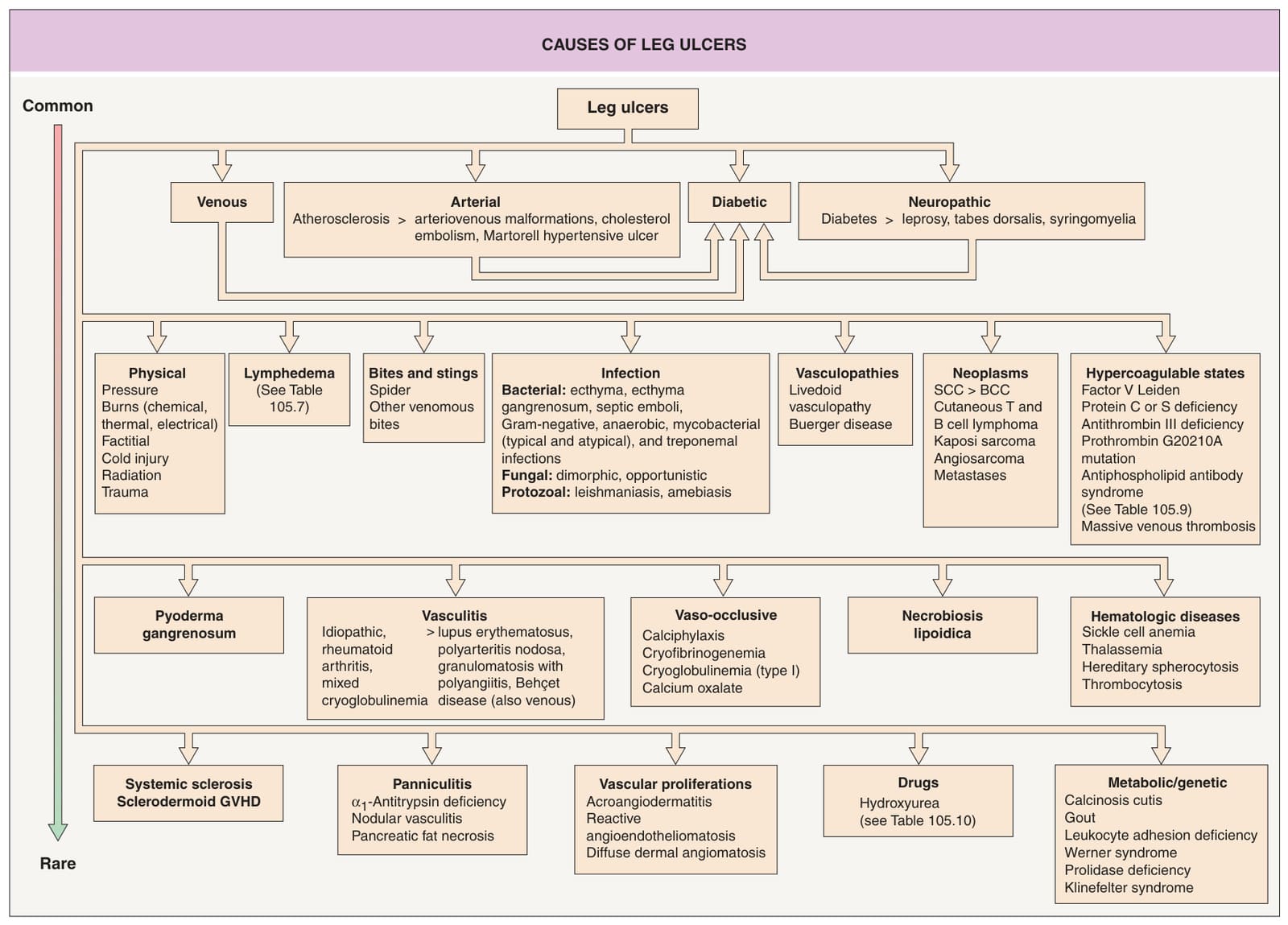
Fig. 105.1 Causes of leg ulcers. Patients with Behçet disease develop lower extremity ulcers due to vasculitis and/or venous insufficiency related to deep vein thromboses, and, occasionally, erosive pustular dermatosis is a cause of leg ulcers. Hydroxyurea-induced leg ulcers are often on the malleolus or tibial crest, exceedingly painful, and surrounded by atrophic skin. Additional genetic syndromes associated with ulceration are listed in Table 105.10. GVHD, graft-versus-host disease.
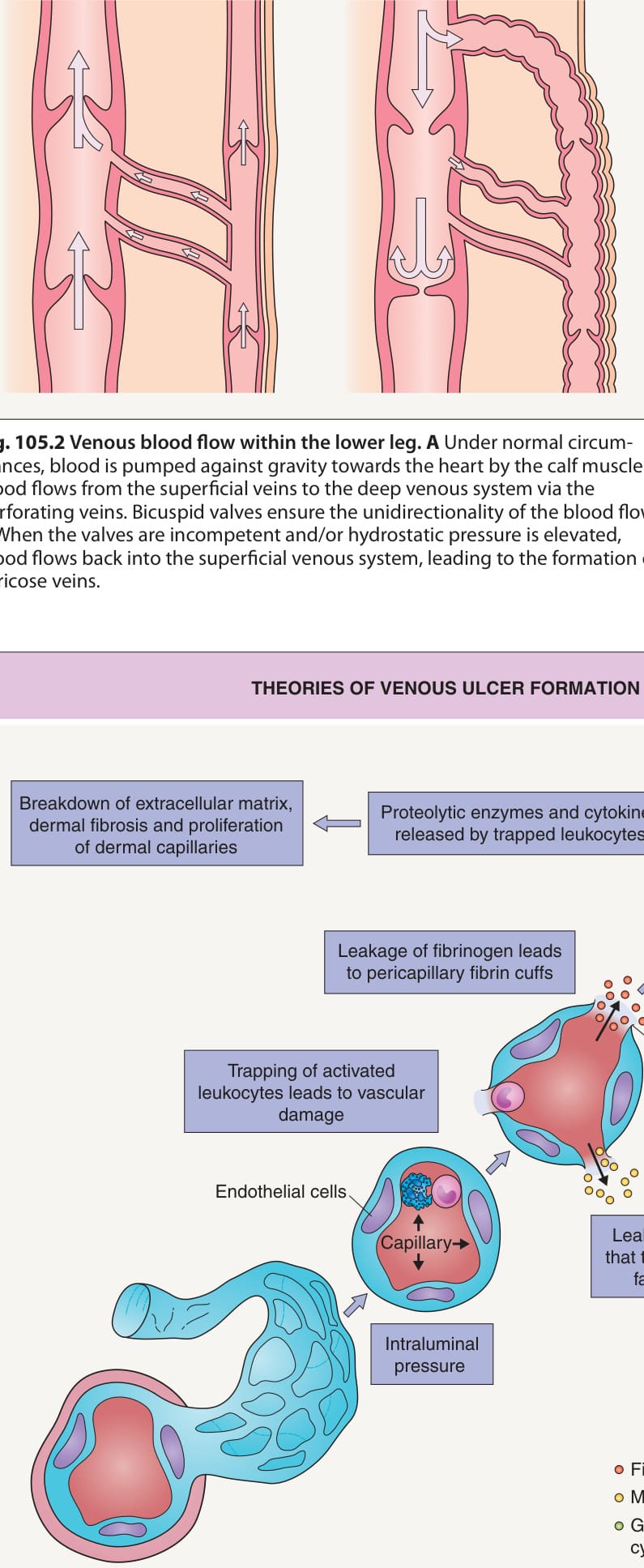
Fig. 105.2 Venous blood flow within the lower leg.A Under normal circumstances, blood is pumped against gravity towards the heart by the calf muscles. Blood flows from the superficial veins to the deep venous system via the perforating veins. Bicuspid valves ensure the unidirectionality of the blood flow. B When the valves are incompetent and/or hydrostatic pressure is elevated, blood flows back into the superficial venous system, leading to the formation of varicose veins.
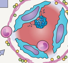
Fig. 105.3 Theories of venous ulcer formation. The theories include the presence of pericapillary cuffs due to leakage of fibrinogen, binding of growth factors by leaked molecules, and the trapping of leukocytes, with subsequent chronic inflammation, dermal changes, and ulcer formation. TGF, transforming growth factor; VEGF, vascular endothelial growth factor.
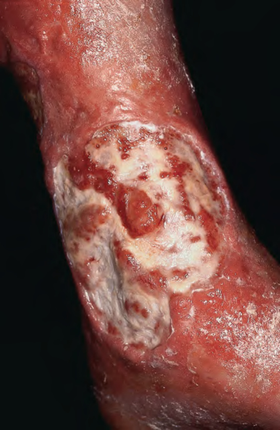
Fig. 105.4 Venous ulcer and stasis dermatitis.
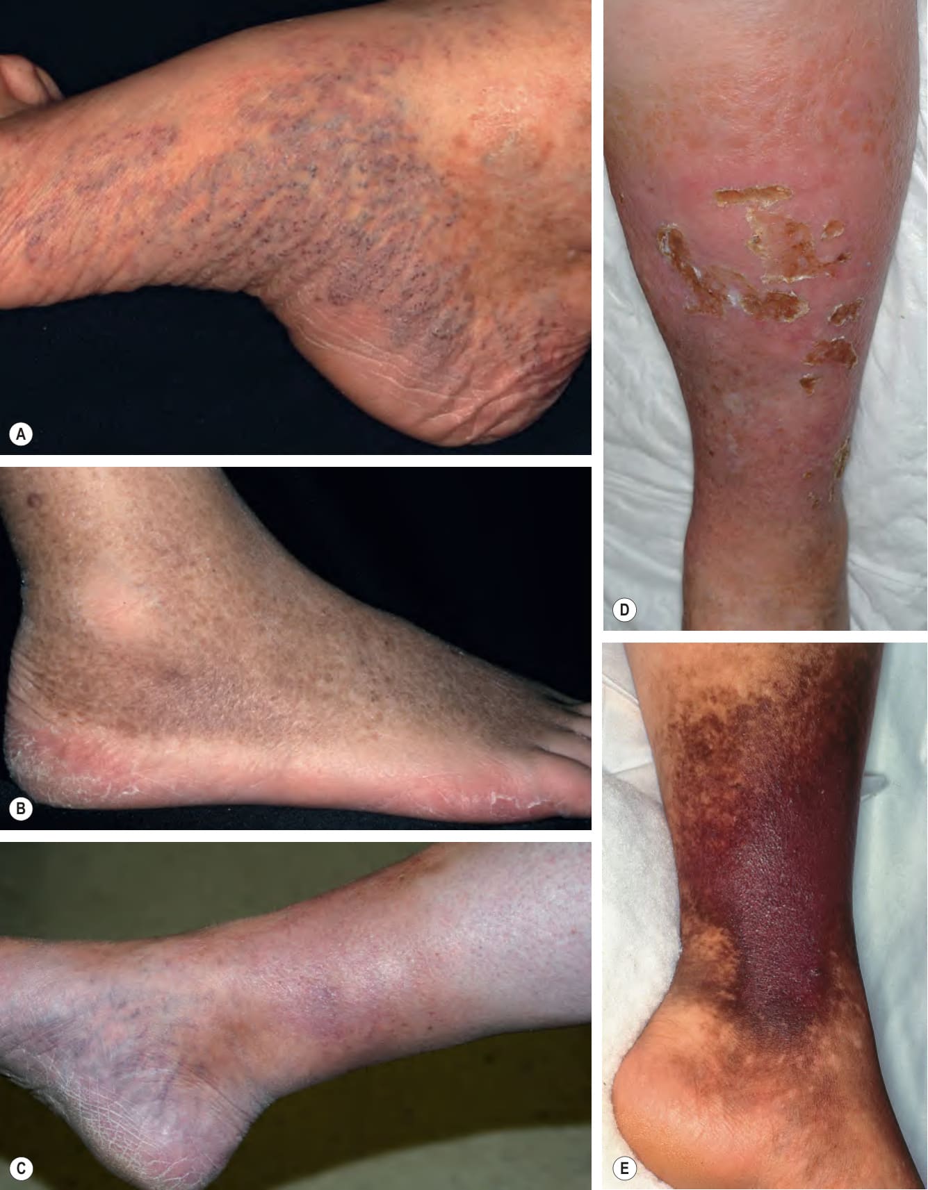
Fig. 105.5 Associated findings in patients with venous hypertension and venous insufficiency.
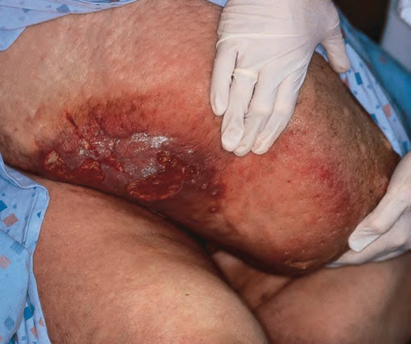
Fig. 105.6 Lipodermatosclerosis, chronic ulceration and lymphedema of the dependent portion of the pannus. The changes are similar to those seen on the distal lower extremities. Courtesy Jean L. Bolognia, MD.
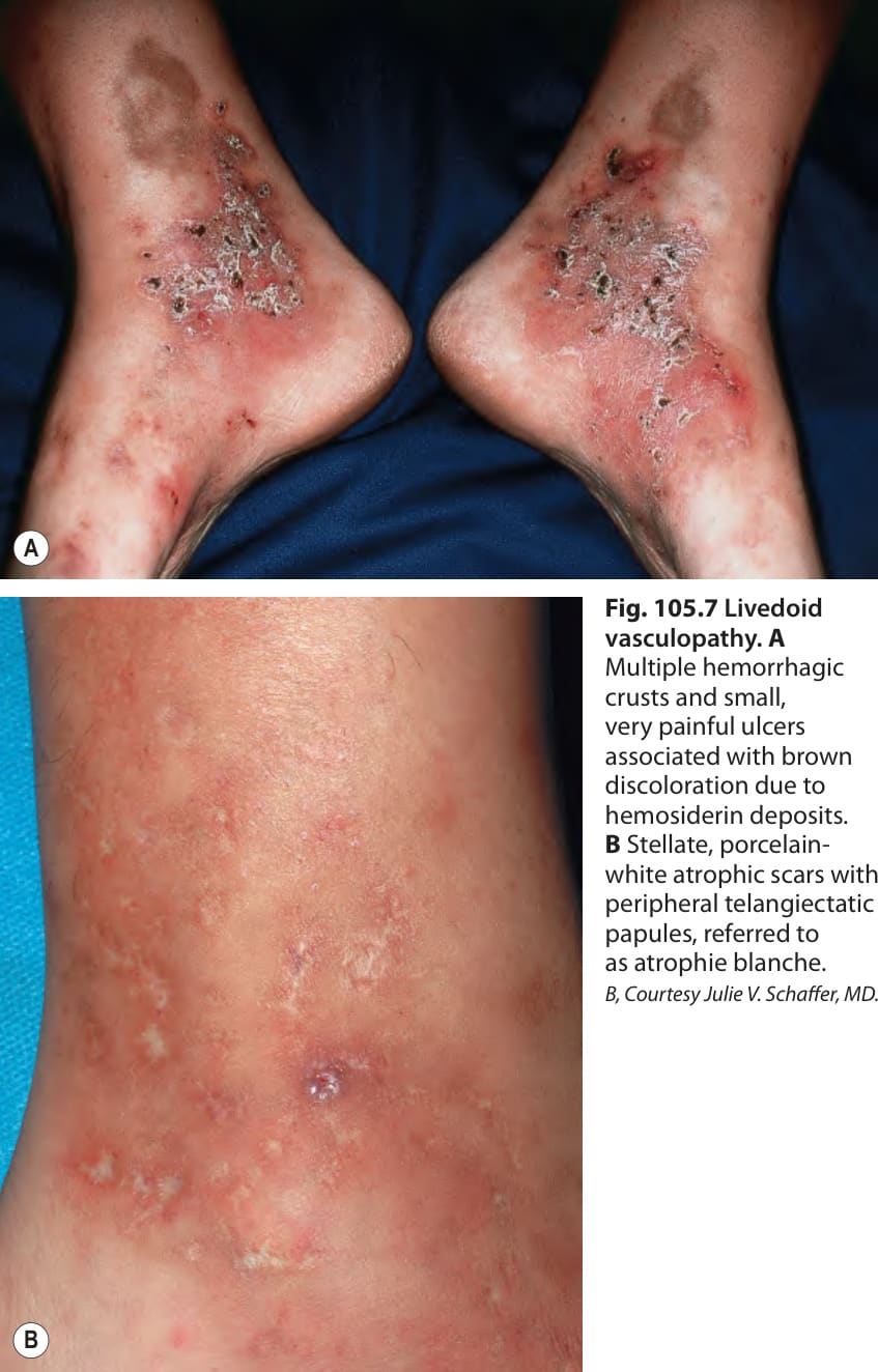
Fig. 105.7 Livedoid vasculopathy.A
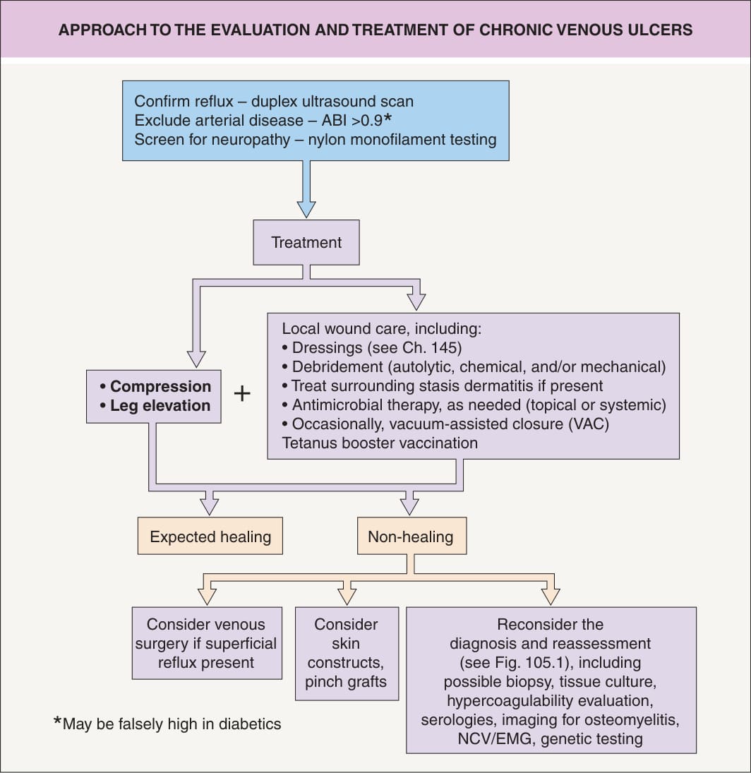
Fig. 105.8 Approach to the evaluation and treatment of chronic venous ulcers. See Table 105.8 for determination and interpretation of the ankle–brachial index (ABI). EMG, electromyography; NCV, nerve conduction velocity test.
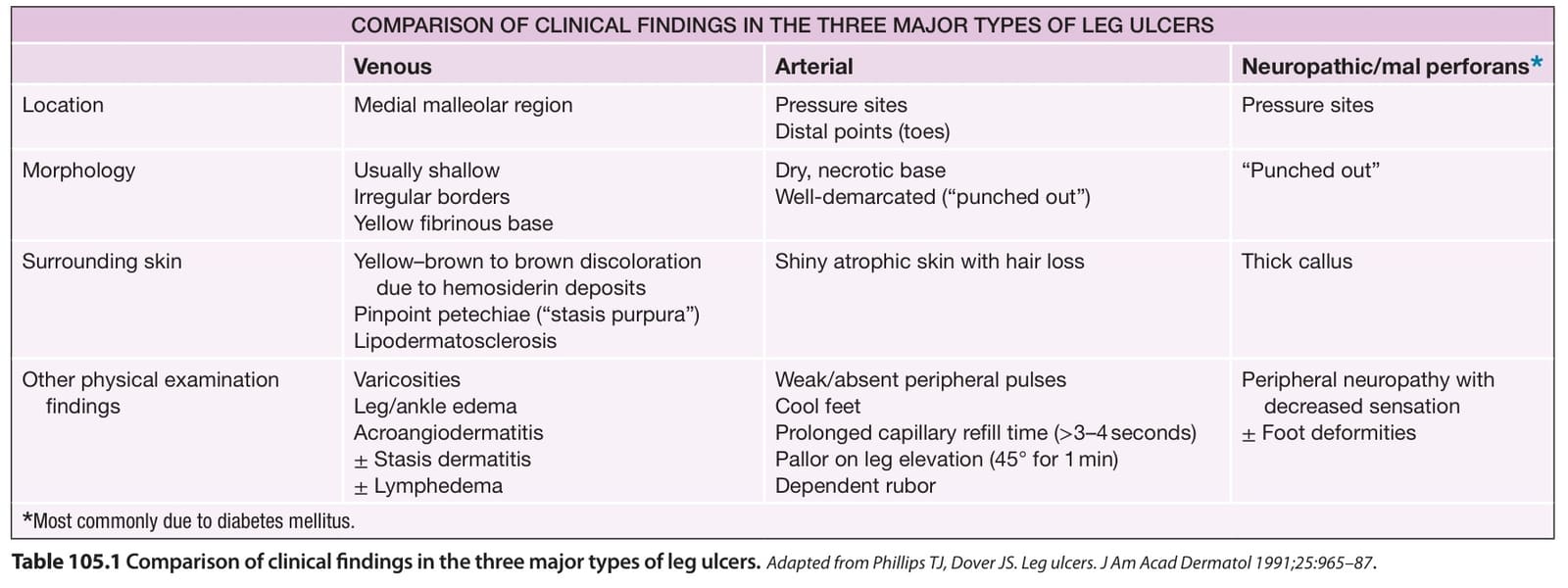
Table 105.1 Comparison of clinical findings in the three major types of leg ulcers.
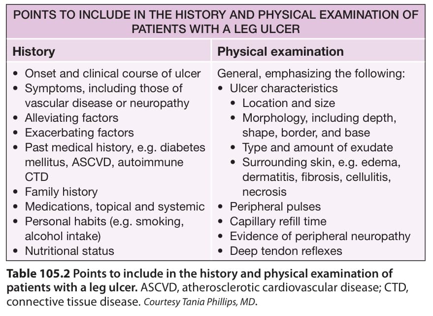
Table 105.2 Points to include in the history and physical examination of patients with a leg ulcer. ASCVD, atherosclerotic cardiovascular disease; CTD, connective tissue disease. Courtesy Tania Phillips, MD.
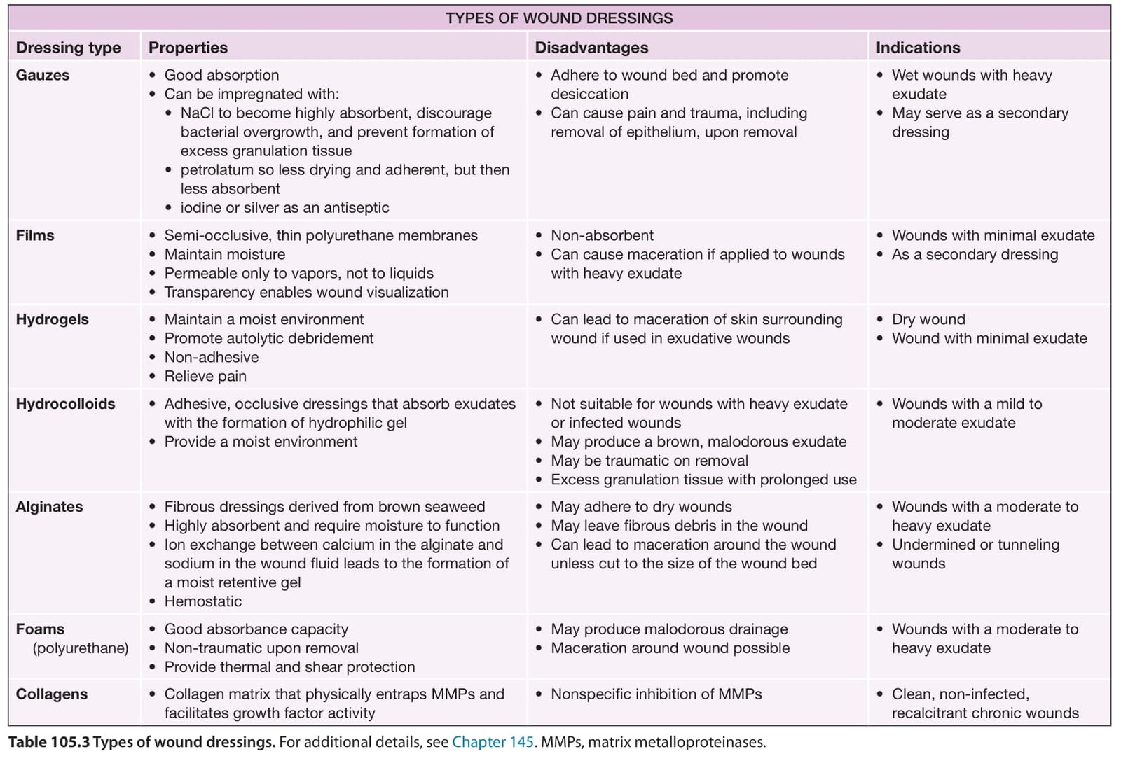
Table 105.3 Types of wound dressings. For additional details, see Chapter 145. MMPs, matrix metalloproteinases.
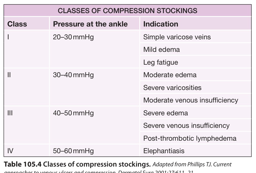
Table 105.4 Classes of compression stockings.
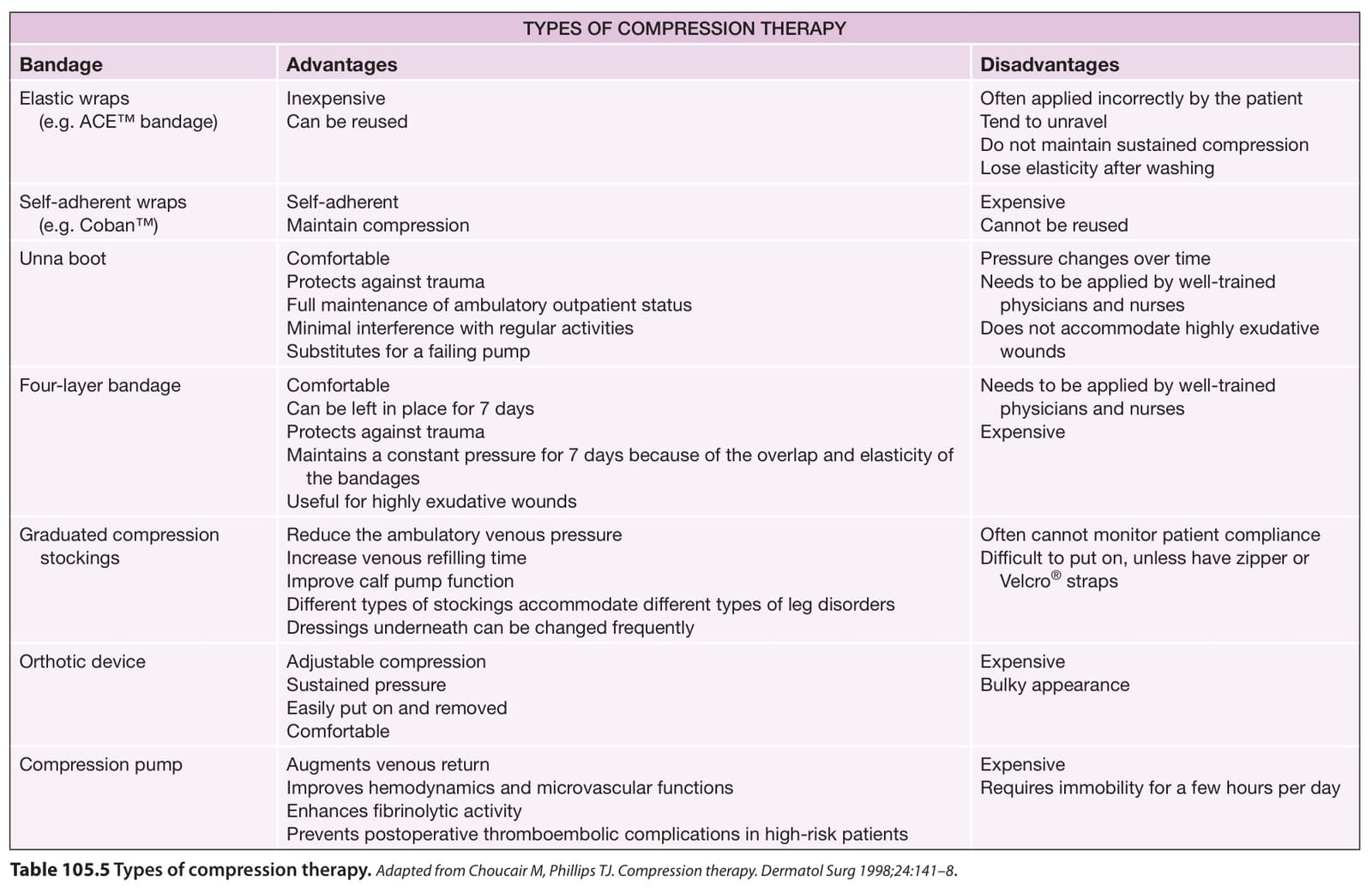
Table 105.5 Types of compression therapy.
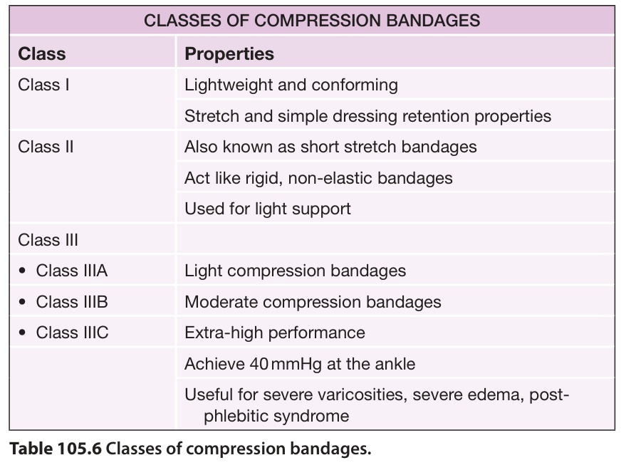
Table 105.6 Classes of compression bandages.
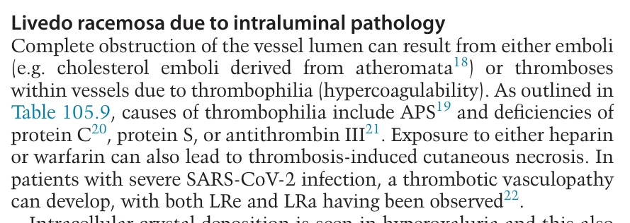
Table 105.9 Evaluation for thrombophilia/hypercoagulability. Shaded areas represent first-tier screening tests. The initial laboratory evaluation should also include a complete blood count with differential and platelet count, examination of a peripheral blood smear, ESR, activated partial thromboplastin time (PTT), and hepatic and renal function panels. Testing for anti-neutrophil cytoplasmic antibodies (ANCA) can be considered for patients with retiform purpura, as ANCA-positive vasculitides occasionally present with minimally inflammatory lesions; patients with levamisole-induced vasculopathy may have anti-phospholipid antibodies (as well as ANCA). Infections can also lead to coagulopathy (e.g. COVID-19-associated) in which abnormalities such as elevated fibrinogen or antiphospholipid antibodies are detected. Additional inherited causes of hypercoagulability include polymorphisms in the genes encoding the endothelial protein C receptor, the protein Z-dependent protease inhibitor, and E-selectin. AD, autosomal dominant; CBS, cystathionine β-synthase; DOACs, direct-acting oral anticoagulants; MTHFR, methylenetetrahydrofolate reductase; ND, not determined; OCP, oral contraceptive pills; RVVT, Russell viper-venom time.