LYMPHEDEMA
Lymphedema is the accumulation of lymph fluid within the inter-stitium that is due to a reduction in lymph drainage in the presence of normal capillary filtration. Lymphedema should be differentiated from edema which results from an imbalance between capillary filtration and lymph drainage.
Lymphedema can be primary or secondary (Table 105.7). Primary lymphedema can be further subdivided based on age at time of clinical presentation, but because the time of onset may vary, even within the same family, genetic bases when known are currently used for classification. Congenital lymphedema usually appears before 1 year of age and is sporadic or familial (e.g. lymphatic malformation 1 [Milroy disease]; Fig. 105.9). Meige disease (lymphatic malformation 5), classically a type of lymphedema praecox, is the most common form of primary lymphedema and it usually presents around puberty and more often affects girls. Lymphedema tarda usually presents after 35 years of age and is probably the result of an environmental trigger such as trauma or infection superimposed on inherently weakened lymphatics.
Common causes for secondary or acquired lymphedema are malignancy, radiation, and surgical lymph node dissection, as well as recurrent cellulitis, trauma, filariasis, morbid obesity, and chronic venous insufficiency (Fig. 105.10). Filariasis, due to infection of the lymphatics by the nematode Wuchereria bancrofti, is the most common cause of lymphedema in low-income tropical and subtropical countries. Podoconiosis, due to pedal exposure to volcanic soils, occurs most commonly in eastern Africa (Fig. 105.11A).
Clinically, lymphedema begins as painless pitting edema of the dorsal aspect of the foot which then progresses proximally. Over time, the skin becomes indurated due to fibrosis, and the affected areas are prone to ulceration and secondary infection. Recurrent infections lead to worsening of the lymphedema and a vicious cycle is established.
Elephantiasis nostras verrucosa is a complication of chronic lymphedema and most commonly affects the feet and distal lower extremities. In addition to profound non-pitting edema and progressive fibrosis of the dermis and subcutaneous tissue, there are verrucous changes with papillomatosis and hyperkeratosis (Fig. 105.11B). The skin acquires a mossy or cobblestone appearance and discrete firm fibrous papules or nodules may develop. Colonization with bacteria and fungi often results in ulcers becoming crusted and malodorous.
Elephantiasis nostras verrucosa is difficult to treat. Compression stockings, pneumatic pumps, and manual lymph drainage (see above)
are used to decrease lymph accumulation. Topical keratolytics or humectants, such as salicylic acid and urea, may improve the hyperkeratosis. Oral retinoids have been reported to be of benefit for both the verrucous changes and the lymphedema. Surgical management of lymphedema
This middle-aged woman has bilateral “stovepipe” enlargement of the legs and minimal involvement of the feet. Note the sharp demarcation between normal and abnormal tissue at the ankle, referred to as the “cuff sign”. Courtesy Jean L. Bolognia, MD.
includes physiologic procedures such as lymphaticovenous anastomosis for relatively early (stage I) lymphedema. Debulking and reduction surgery with preservation of perforators are reserved for patients with advanced (stage III) disease.
The differential diagnosis includes lipedema, in which there is an abnormal deposition of subcutaneous fat and lymphatic vessel dysfunction with sparing of the feet (Fig. 105.12), thyroid dermopathy (pretibial myexedema), and obesity-associated lymphedematous mucinosis, where there can be dermal mucin deposition in the absence of thyroid disease.
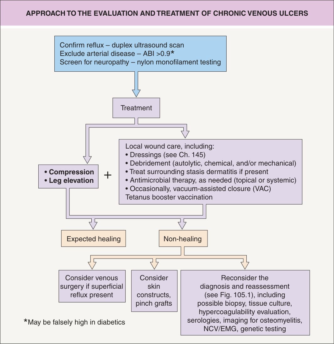
Fig. 105.8 Approach to the evaluation and treatment of chronic venous ulcers. See Table 105.8 for determination and interpretation of the ankle–brachial index (ABI). EMG, electromyography; NCV, nerve conduction velocity test.
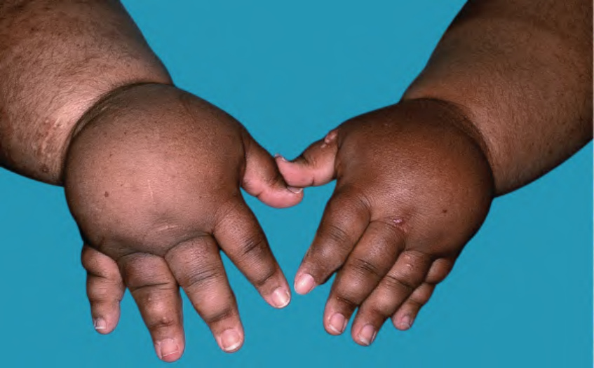
Fig. 105.9 Bilateral primary lymphedema due to lymphatic malformation 1 (also referred to as Milroy disease).
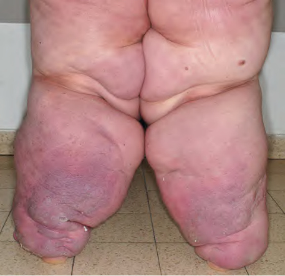
Fig. 105.10 Lymphedema secondary to morbid obesity associated with myxedema.
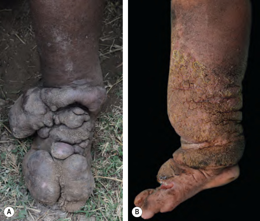
Fig. 105.11 Elephantiasis nostras verrucosa.A Podoconiosis (endemic, nonfilarial elephantiasis) due to pedal exposure to volcanic soils. B The patient had both lymphedema and venous insufficiency. Note the involvement of the foot. A, Courtesy Claire Fuller, MD.
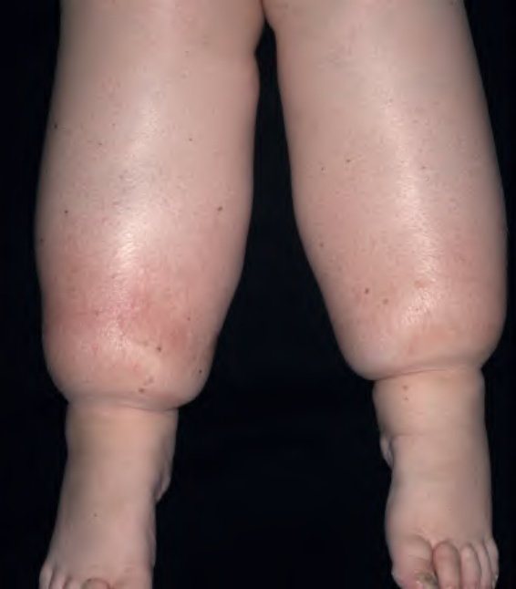
Fig. 105.12 Lipedema.
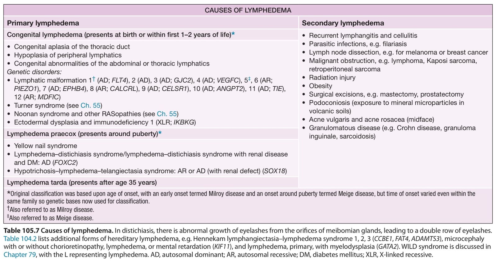
Table 105.7 Causes of lymphedema. In distichiasis, there is abnormal growth of eyelashes from the orifices of meibomian glands, leading to a double row of eyelashes. Table 104.2 lists additional forms of hereditary lymphedema, e.g. Hennekam lymphangiectasia–lymphedema syndrome 1, 2, 3 (CCBE1, FAT4, ADAMTS3), microcephaly with or without chorioretinopathy, lymphedema, or mental retardation (KIF11), and lymphedema, primary, with myelodysplasia (GATA2). WILD syndrome is discussed in Chapter 79, with the L representing lymphedema. AD, autosomal dominant; AR, autosomal recessive; DM, diabetes mellitus; XLR, X-linked recessive.