ARTERIAL ULCERS
Peripheral arterial disease (PAD) is a common manifestation of atherosclerosis. Cigarette smoking and diabetes mellitus are the strongest risk factors, with hypertension, dyslipidemia, and hyperhomocysteinemia representing additional risk factors. Up to 25% of patients with leg ulcers have PAD, and a significant number of patients have a combination of arterial and venous insufficiencies.
Pathogenesis
A lack of blood perfusion decreases tissue resilience and leads to tissue necrosis; it also impedes wound healing by reducing the supply of oxygen, nutrients, and soluble mediators involved in the repair process. Although PAD alone infrequently precipitates ulceration, arterial insufficiency plays a major role in delayed wound healing and complications including gangrene. Ischemic foot ulcers are often precipitated by trauma in a patient with relatively mild symptoms of arterial insufficiency. In such an individual, the degree of skin perfusion is sufficient to maintain cutaneous integrity but is not adequate enough to support proper wound healing. In such a setting, the ulcer will inevitably progress into a chronic wound or gangrene unless perfusion is restored.
Clinical Manifestations
Most patients with significant PAD are symptom-free. Intermittent claudication, defined as leg pain induced by ambulation and relieved by resting, represents the earliest and the most common presenting symptom of PAD of the lower extremities. With disease progression, patients begin to complain of pain at rest, especially when the legs are elevated in bed at night, which is ameliorated by dependency. While symptoms of claudication are usually localized to the calf or the thigh, pain at rest is often felt in the feet. In advanced stages of PAD, the reduction in cutaneous blood supply may lead to ischemic ulceration and gangrene, and this may necessitate amputation.
Arterial ulcers are usually round with a sharply demarcated border and are characterized by an absence of bleeding (Fig. 105.13). These wounds typically occur on the distal lower extremities, often over bony prominences. The surrounding skin may be hairless, shiny, and atrophic. Wound pain is often significant and is exacerbated by limb elevation.
Additional manifestations of impaired arterial perfusion to the foot include diminished or absent pedal pulses, cool feet, pallor of the foot with elevation of the leg and then subsequent redness with lowering (dependent rubor), sluggish refilling of toe capillaries, thickened nails, and absence of toe hair (see Table 105.1).
Laboratory Evaluation
The ankle–brachial pressure index, more commonly referred to as the ankle–brachial index (ABI), is a simple, non-invasive test that is widely used for diagnosis of PAD and to assess its severity. The ABI is calculated by dividing the systolic blood pressure in the ankle by the systolic blood pressure in the arm while the patient is in a resting supine position (Table 105.8). A decrease in arterial blood pressure at the ankle (relative to the central blood pressure) implicates arterial stenosis at any point between the aorta and the ankle. The normal range for the ABI is 0.91 to 1.40, with a reading <0.9 being indicative of PAD.
Depending on the patient’s presentation and symptoms, further non-invasive imaging techniques may be employed, including duplex ultrasound, computed tomography angiography (CTA), and magnetic resonance angiography (MRA). Both CTA and MRA produce images of vascular structures in cross-sectional slices that can be reconfigured into three-dimensional angiographic images. CTA is less expensive than MRA, with higher spatial resolution. However, CTA involves ionizing radiation and the use of iodinated contrast material. The gold standard for diagnosis and evaluation of PAD is invasive digital subtraction angiography, but amongst radiologists, it is being replaced by CTA and MRA.
Measurement of transcutaneous oxygen pressure (TcPO2) quantifies skin oxygenation and indirectly assesses blood flow. The reduction in TcPO2 correlates with the severity of lower extremity ischemia, and when the TcPO2 is <40 mmHg, wound healing is impaired.
Differential Diagnosis
Patients with PAD can also develop cutaneous ulcers due to cholesterol emboli (Fig. 105.14) and risk factors include arterial or coronary catheterization and anticoagulation (see Ch. 23). In Buerger disease, ulcers occur distally on both the upper and lower extremities, with smoking as a risk factor. Martorell hypertensive ischemic ulcers result from arteriolosclerosis and in contrast to arterial ulcers, they are typically located on the posterolateral leg. These ulcers are characterized by excruciating pain and patients have treatment-resistant hypertension. Histologically, subcutaneous stenotic arteriolosclerosis and medial calcinosis are prominent. Treatment includes surgical debridement, skin grafting, and pain and blood pressure control.
Additional disorders to consider are shown in Figure 105.1, including diffuse dermal angiomatosis (see below).
Treatment
Arterial ulcers can benefit from restoration of peripheral arterial blood flow, either by endovascular interventions (e.g. percutaneous angioplasty, stent placement, cryoplasty) or by more invasive surgical reconstruction (e.g. femoral popliteal bypass). In principle, local treatment of arterial ulcers is similar to that of other types of skin ulcers, with two major exceptions: (1) sharp debridement should be performed very cautiously or avoided in order to prevent further necrosis and ulcer enlargement; and (2) VAC therapy should be avoided as it can lead to worsening of the ulcer. Additional interventions include smoking cessation, exercise training, antiplatelet drugs (e.g. aspirin, clopidogrel), cilostazol, and treatment of diabetes, hyperlipidemia, and hypertension.
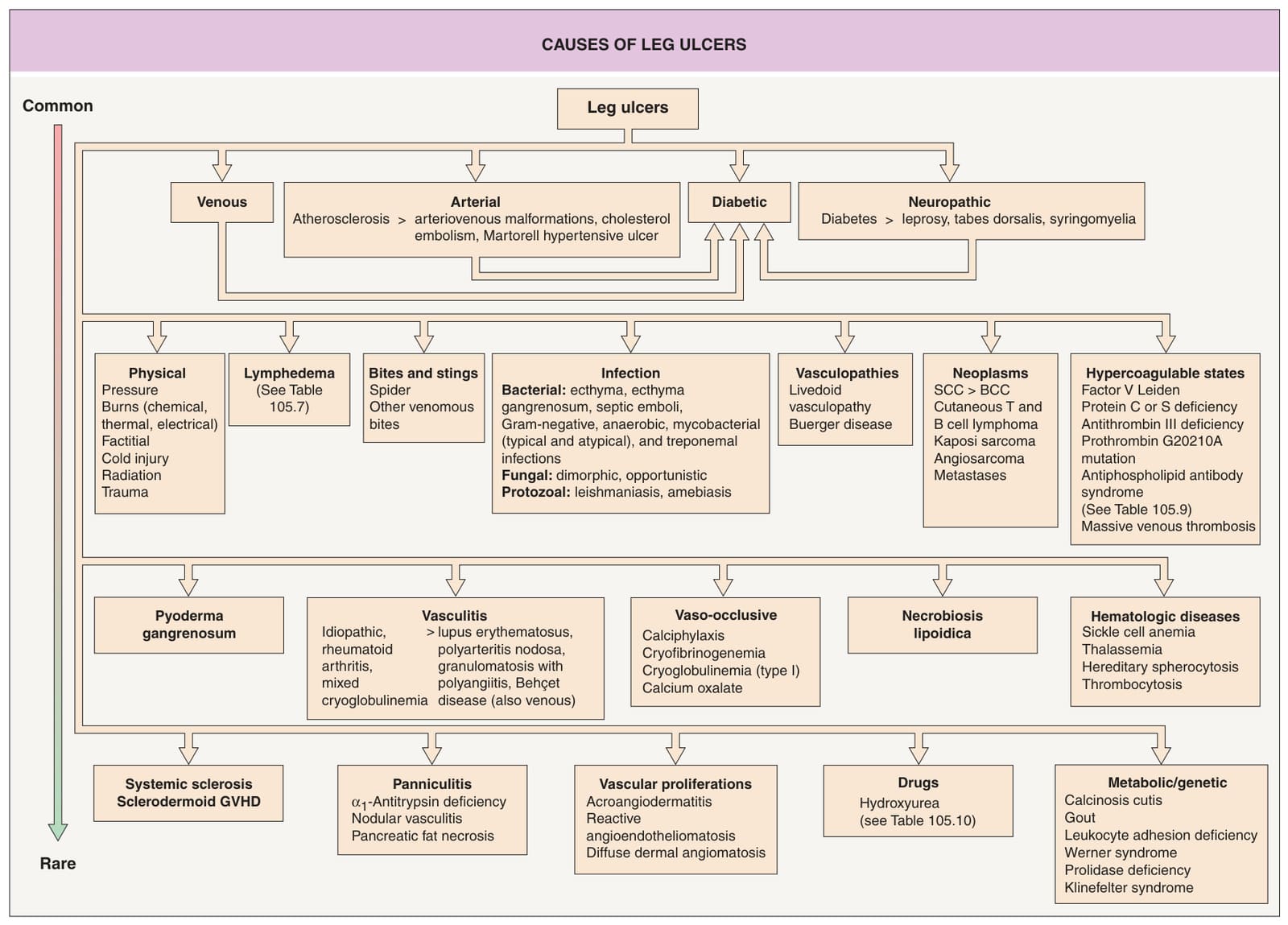
Fig. 105.1 Causes of leg ulcers. Patients with Behçet disease develop lower extremity ulcers due to vasculitis and/or venous insufficiency related to deep vein thromboses, and, occasionally, erosive pustular dermatosis is a cause of leg ulcers. Hydroxyurea-induced leg ulcers are often on the malleolus or tibial crest, exceedingly painful, and surrounded by atrophic skin. Additional genetic syndromes associated with ulceration are listed in Table 105.10. GVHD, graft-versus-host disease.
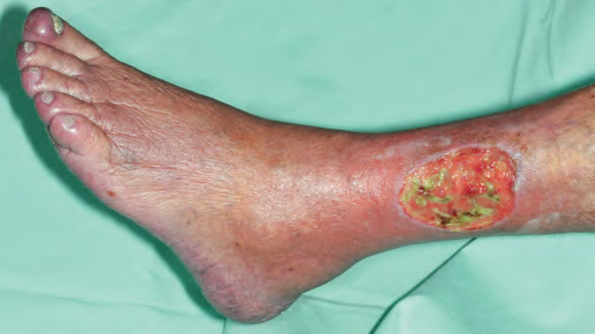
Fig. 105.13 Arterial ulcer. The punched-out appearance and surrounding smooth shiny skin are common features.
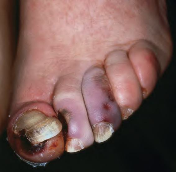
Fig. 105.14 Cholesterol emboli. Ischemia of the toes and necrosis with early ulcer formation.
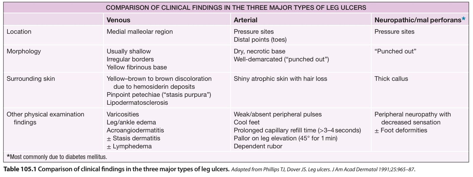
Table 105.1 Comparison of clinical findings in the three major types of leg ulcers.
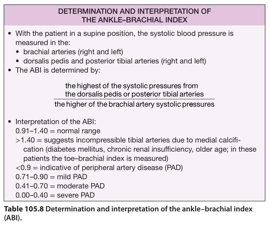
Table 105.8 Determination and interpretation of the ankle–brachial index (ABI).