DIABETIC (AND NEUROPATHIC) ULCERS
Patients with diabetes mellitus have a 10%–25% lifetime risk of developing a foot ulcer. These foot ulcers constitute a major medical and economical burden, with a substantial morbidity and impaired quality of life. Approximately 15% of diabetic patients with foot ulcers will eventually undergo a lower extremity amputation. The rising incidence of both type 1 and type 2 diabetes has made diabetic foot ulcers the most important risk factor for lower extremity amputation in high-income countries.
Pathogenesis
The etiology of diabetic ulcer formation is multifactorial. Although ischemia, trauma, and infection have traditionally been incriminated as underlying pathogenic factors, nowadays diabetic neuropathy is considered to be the major cause of foot ulcers in diabetic patients.
Diabetic neuropathy typically involves the sensory, motor and autonomic nerves, with sensory neuropathy substantially impairing the patient’s perception of touch, deep pressure, temperature, and joint position. With the loss of protective sensation, foot trauma is unrecognized and this can lead to ulceration. Peripheral motor neuropathy results in weakness and atrophy of the intrinsic muscles of the feet, producing altered biomechanics and structural deformities that, in the presence of sensory neuropathy, increase the risk for skin injury and ulceration from repetitive mechanical stress. Continuous pressure to the skin overlying bony prominences typically results in the formation of a thick, keratotic plaque (callus), which eventually breaks down and ulcerates. Autonomic neuropathy also plays a role because it can lead to a loss of sweating, with the resultant dry skin being susceptible to cracks and fissures that serve as a portal of entry for bacteria. In addition, a potential consequence of autonomic neuropathy is arteriovenous shunting within the skin microcirculation which results in decreased cutaneous perfusion and oxygen saturation, thus contributing to ulceration and an impaired response to infection. Moreover, this abnormal vascular autoregulation, coupled with sensory neuropathy, contributes to the development of Charcot joints (degenerative arthropathy), further adding to the risk of ulceration from an abnormal distribution of pressure forces and foot deformities.
Diabetes is also associated with an increased risk of developing PAD, which characteristically affects vessels between the knee and the ankle. Although microcirculatory dysfunction occurs early on in the course of diabetes, it is primarily atherosclerotic macrovascular disease that accounts for ischemic complications in these patients. In general, diabetic foot ulcers are not purely related to vascular occlusion; rather, mechanical damage that typically occurs to the insensate foot, in the presence of a decreased blood supply, rapidly results in neuroischemic ulcers. PAD also contributes to an altered response to foot infections and poor wound healing. The combination of a depressed immune response plus a decrease in cutaneous blood supply plays a key role in the development of cellulitis, abscesses, and osteomyelitis, all of which increase the risk of limb amputation.
Occasionally, patients with diabetes develop ulcerations within lesions of necrobiosis lipoidica as well as ulcers due to streptococcal infections.
Clinical Features
Diabetic ulcers are usually located on pressure points and bony prominences such as the metatarsal heads, great toes, and heels (Fig. 105.15). They often arise within exuberant, moist, malodorous calluses (mal perforans). In the absence of peripheral atherosclerotic disease, the diabetic foot is warm, with good color and palpable pulses, but with decreased sensation. Foot deformities are frequently present, e.g. hammer toes, claw toes, collapse of the longitudinal arch (Charcot foot).
Laboratory Evaluation
Because diabetic ulcers are heterogeneous in nature, a thorough evaluation of the patient is crucial in determining the underlying causes. More specifically, peripheral neuropathy can be detected via a neurologic examination of the lower extremities that includes the use of a 10 g monofilament to test for sensation. More invasive testing, i.e. nerve conduction studies/electromyography, is sometimes required. The degree to which vascular perfusion is compromised can be assessed via determination of the ABI (see above).
A complete blood count, ESR, C-reactive protein level, and blood glucose level are obtained when assessing for a possible associated infection. Of note, sometimes an elevated blood glucose level is the only sign of infection. Microbial cultures (bacterial, mycobacterial, and fungal) should be obtained from the wound bed. Although swabs are often used, deep tissue specimens, obtained aseptically, are preferable.
Diagnostic imaging, including plain radiographs of both feet, bone scintigraphy, MRI, CT and sometimes ultrasound, is used to better define the presence or absence of a bone or deep soft tissue infection. Of these, MRI is the most accurate test for diagnosing osteomyelitis. If osteomyelitis is suspected, a bone biopsy processed for culture and histology is the gold standard.
Treatment
Conservative treatment of diabetic foot ulcers consists of providing a proper wound healing environment, eradication of infection, and avoidance of repetitive trauma by alleviating the mechanical load on ulcers (off-loading). Methods of off-loading include bed rest and the use of a wheelchair, crutches, total contact casting, cast walkers, felted foam, half-shoes, or therapeutic shoes. Total contact casting is considered to be the best approach as it enables pressure redistribution and continuous off-loading of the ulcer area. The inability of the patient to remove the cast ensures compliance and reduces the level of activity. However, total contact casting is somewhat impractical because it is expensive and requires skill and experience. In addition, as the edema subsides, cast changing and size adjustments are often required (usually twice a week or more often). Total contact casting is contraindicated if the wound is infected or has a significant exudate and it can lead to new ulcerations over bony prominences.
Another approach to off-loading is the use of specialized shoes that reduce pressure about the forefoot and heel and multilayered insoles that enhance shock absorption. Half-shoes assist in off-loading forefoot plantar ulcers as they support only the rear and mid foot, while the forefoot is left suspended in the air.
Initial wound management consists of cleansing the wound and proper debridement of any necrotic material, including the callus; the latter can contribute to increased pressure on the insensate foot. In addition, debridement promotes wound healing at the cellular level by removing excess extracellular matrix, thus liberating trapped growth factors and facilitating cell migration. Moist wound healing is recommended as with other types of chronic ulcers but avoidance of maceration of surrounding skin is of particular importance.
Apart from standard therapies (see above), hyperbaric oxygen has been shown to be of specific benefit to patients with diabetic foot ulcers. While it is unclear to what extent it benefits other types of ulcers, hyperbaric oxygen therapy has been shown to be effective in the treatment of diabetic foot ulcers, with a decrease in healing time and amputation rate. Oxygen (100%) at supra-atmospheric pressures is intermittently inhaled, either in a full-body (monoplace) chamber or in a multiplace chamber via a gas mask. In theory, tissue hypoxia impairs wound healing and hyperbaric oxygen therapy leads to an increase in the blood oxygen level within the wound. In addition, there is reallocation of blood flow to hypoxic areas due to hyperoxic vasoconstriction of surrounding normal tissue. Side effects include oxygen toxicity to the brain (e.g. seizures) and lungs, and barotrauma to the lungs, ears, sinuses, and eyes (transient myopia).
As endogenous growth factors are often degraded or trapped within chronic wounds, genetically engineered exogenous growth factors have been used to try to enhance wound healing. Becaplermin gel (Regranex®) is recombinant platelet-derived growth factor whose biologic activity is similar to its endogenous counterpart, with stimulation of granulation tissue production and release of other growth factors important to wound repair. It is the only growth factor currently approved for use in chronic neuropathic diabetic ulcers. In the setting of best wound care, becap lermin gel has been shown to increase healing rates and shorten healing time of diabetic ulcers. However, in routine clinical practice, becap lermin has not been as successful and it has an FDA black box warning of an increased risk of cancer mortality (when exposed to three or more tubes). More recently, strategies have focused on improving growth factor activity and protecting these factors from degradation.
Living skin equivalents (e.g. Apligraf®, Dermagraft®) are bioengineered products that act as delivery systems for growth factors and extracellular matrix (see Ch. 145). Both are indicated for recalcitrant diabetic foot ulcers that have no exposed muscle, bone or tendon, are not infected, and have had aggressive debridement.
Prevention
Preventing wound recurrence is of critical importance. The risk of ulceration and/or amputation is increased in patients who have had diabetes for >10 years, are male, have poor glucose control, or have target organ complications. Foot-related conditions that are associated with an increased risk of amputation are peripheral neuropathy, altered biomechanics, evidence of increased pressure, foot deformity, PAD, history of an ulcer or previous amputation, and severe nail pathology. Tight control of glucose levels is required in order to prevent diabetic complications that are due to microvascular changes, as the latter predispose the patient to neuropathy, ischemia, and infection. Lower HbA1c values are associated with a decrease in healing times for leg and foot ulcers.
Smoking cessation and control of low-density lipoprotein (LDL) and total cholesterol levels are also important in the prevention of atherosclerotic vascular disease.
All diabetic patients should receive an annual foot examination to assess neurologic and vascular status, biomechanics, skin integrity, and the presence and types of foot deformities. Patients with significant risk factors for ulcer formation should be evaluated more frequently. Education regarding daily foot care that includes inspection for any scratch, blister or erythema, daily washing in warm water, appropriate footwear selection (e.g. avoiding pointed shoes), skin moisturizing, and treatment of nail and foot fungal infection should be provided.
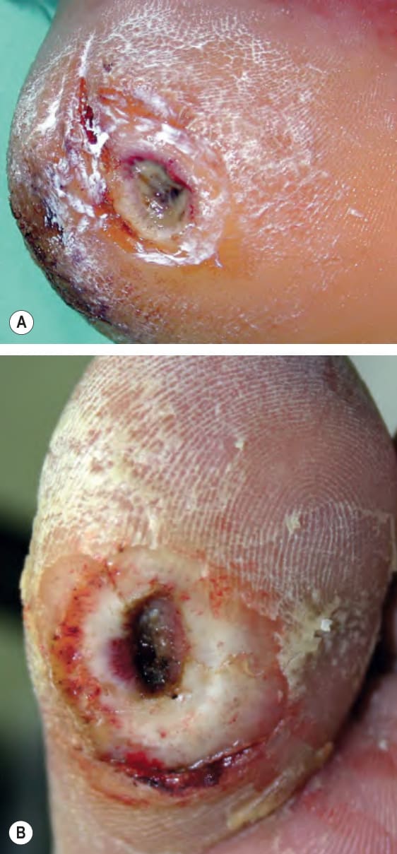
Fig. 105.15 Neuropathic ulcers (mal perforans) in patients with diabetes mellitus and peripheral neuropathy. Common locations are the plantar surface of the heel (A) and the great toe (B). Note the thick rim of callus.
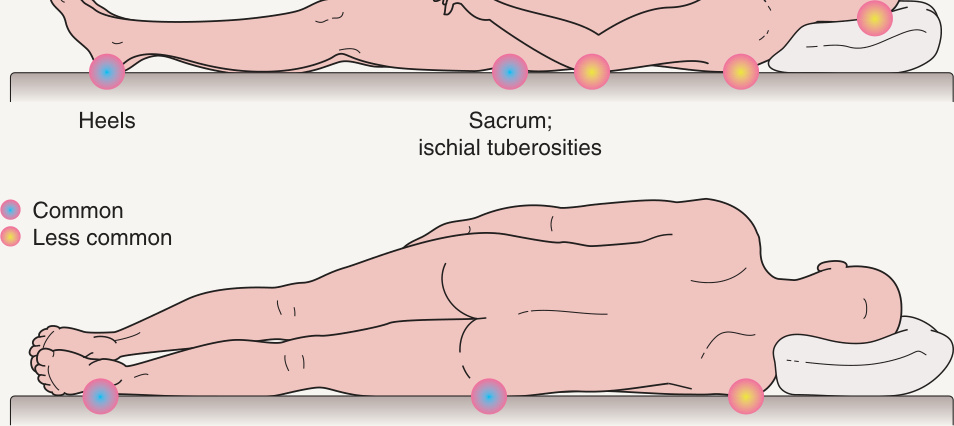
Fig. 105.16 Most common sites for pressure ulcers.