ULCERS DUE TO PHYSICAL FACTORS
Any form of mechanical trauma to the skin can result in the formation of an ulcer if factors that compromise the skin’s ability to recover from physical insults are present. The most common form of physical injury which leads to ulceration is prolonged pressure due to immobilization. The resultant ulcer is commonly referred to as a pressure ulcer or decubitus ulcer (decubitus means lying down in Latin). These ulcers are caused by unrelieved pressure to soft tissues compressed between a bony prominence and an external surface. The most common bony prominences involved are the sacrum, ischial tuberosities, greater trochanters, heels, and lateral malleoli (Fig. 105.16). A special type of pressure ulcer, often presenting as a pear- or butterfly-shaped ulcer in the sacral area, is termed a Kennedy terminal ulcer because it often precedes death.
Approximately 1.5–3 million people in the US have pressure ulcers, with an annual cost of treatment of up to $5 billion. It has also been estimated that 10% of hospitalized patients and 25% of nursing home residents have pressure ulcers, with most developing during the first few weeks of hospitalization. An estimated 70% of pressure ulcers occur in patients over 70 years of age; 95% are located on the lower portion of the body: 65% in the pelvic girdle area (Fig. 105.17) and 30% on the lower limbs (Fig. 105.18). Risk factors that predispose to the development of pressure ulcers include prolonged immobility, sensory deficit, circulatory disturbance, and poor nutrition.
Pathogenesis
Four major etiologic factors have been identified as playing an important role in the development of pressure ulcers: external pressure, shearing forces, friction, and moisture.
Interstitial pressures >32 mmHg (normal capillary pressure range = 12–32 mmHg) usually compromise oxygenation and microcirculation. There is an inverse time–pressure curve, with slow ulcer
formation at low pressures and rapid ulcer formation at high pressures (>70 mmHg). The length of continuous exposure of the skin to pressure is also of critical importance, thus explaining why inter-mittent relief of the pressure can prevent ulcer formation. Because subcutaneous tissues are most susceptible to the damaging effects of prolonged exposure to pressure, deep tissue trauma can occur with relatively little superficial damage that would alert caregivers to the extent of tissue injury.
Shearing forces result from the sliding and relative displacement of two apposing surfaces. Although externally applied pressure is more effective than shearing forces alone in reducing skin arteriolar blood flow, these two factors can combine to enhance vascular occlusion. When the head of a supine patient is raised more than 30°, shearing forces occur in the sacral and coccygeal areas. Sliding of the torso transmits pressure to the sacrum and deep fascia, as the outer sacral skin is fixed because of friction with the bed. Vessels within the deeper portion of the superficial fascia angulate and thrombose, which manifests clinically as undermining of the ulcer.
Friction is often associated with shearing and it reflects the degree of resistance generated when two surfaces move across each other. When a bedridden patient is dragged across the bed sheets, friction is produced.
Damage to the protective stratum corneum enhances ulcer formation by compromising the skin barrier.
Moisture results from perspiration due to fevers and from urine and feces. It has been shown to increase the risk of pressure ulcer formation fivefold.
Clinical Features
Pressure ulcers are usually classified according to a four-stage system designed by the National Pressure Ulcer Advisory Panel (NPUAP) (Fig. 105.19). Of note, the ulcers do not necessarily progress sequentially from stage I to stage IV, nor do they necessarily heal from stage IV to stage I.
●Stage I: non-blanchable erythema of intact skin which heralds skin ulceration. For individuals with more darkly pigmented skin, warmth, edema, discoloration of the skin, and induration may serve as indicators of impending ulceration.
●Stage II: partial-thickness skin loss involving the epidermis, dermis, or both. This superficial lesion presents as an erosion, blister, or shallow ulcer.
●Stage III: full-thickness skin loss with damage to the subcutaneous tissue, extending down to (but not including) the underlying fascia. This deep lesion presents as a crater-like ulcer and sometimes involves adjacent tissue.
●Stage IV: full-thickness skin loss and extensive tissue necrosis with destruction extending to muscle, bone, or supporting structures such as tendons or joint capsules. Undermining or sinus tracts can be present. As mentioned previously, a major limitation in the clinical classification of pressure ulcers stems from the fact that extensive deep tissue damage may be accompanied initially by minimal superficial manifestations. Adequate evaluation may also be hampered by the presence of an eschar, which should be debrided for full assessment.
Pathology
When the diagnosis is in doubt or when a secondary malignancy is suspected in the setting of a longstanding lesion, histologic examination is indicated. In all four stages, the histopathologic findings are rather nonspecific. When there is blanchable erythema clinically, the super-ficial dermal capillaries and venules are dilated. Mild to moderate edema within the papillary dermis is seen in association with a mild perivascular lymphocytic infiltrate. The epidermis, pilosebaceous structures, and reticular dermis remain normal. At the stage of non-blanchable erythema, engorgement of capillaries and venules with red blood cells, platelet thrombi, and hemorrhage are observed in the papillary dermis. Although the epidermis still appears normal, sweat gland and subcutaneous fat degeneration is often present. Subepidermal separation can occur with formation of a subepidermal bulla. In early ulcers, the epidermis is lost and acute inflammation of the papillary and reticular dermis is seen. Chronic ulcers have a diffusely fibrotic dermis with a loss of adnexa. The surface may have a hemorrhagic crust containing acute inflammatory cells or a thin zone of coagulation necrosis. In the black eschar stage, full-thickness destruction of the skin occurs. General dermal architecture is preserved, but there is obliteration of cellular details.
Treatment
Pressure ulcers are preventable, primarily by relief of pressure on the skin. This can be accomplished by frequent position changes in addition to use of a variety of support surfaces to relieve pressure; the latter include air- or liquid-filled flotation devices, foam products, and pillows or foam wedges as positioning devices. Adequate nutrition, education, pain management, and psychosocial support are also important inter-ventions. Causes of immobility and systemic conditions that interfere with wound healing or decrease tissue perfusion must be addressed, including congestive heart failure, diabetes, and/or spastic paresis.
The general principles of ulcer and chronic wound care discussed previously also apply to pressure ulcer management. Briefly, debridement may be accomplished via mechanical, enzymatic, and/or autolytic means (see above). Wounds should be cleansed as non-traumatically as possible, and normal saline for irrigation is preferred rather than cytotoxic agents such as hydrogen peroxide or povidone-iodine.
As always, bacterial colonization and infection must be controlled. Dressings should provide a moist, but not macerated, environment and occlusive dressings are often utilized.
In general, stage I, II, and III pressure ulcers are more likely to heal with local therapy, whereas stage IV ulcers, particularly those over the ischial tuberosities, often require surgical intervention. Adjuvant therapies such as laser, ultrasound, hyperbaric oxygen, and UV irradiation are investigational and to date cannot be considered as standard of care. The application of growth factors, cultured keratinocyte grafts, and skin substitutes is promising, but these are also still in the investigational stage.
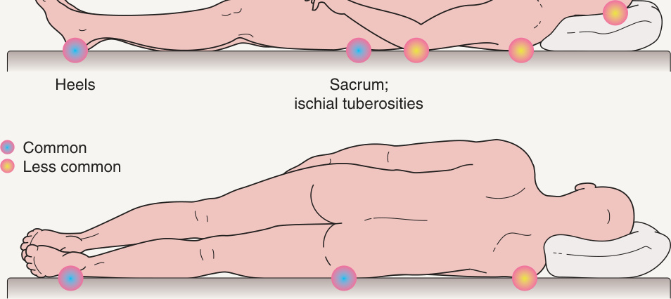
Fig. 105.16 Most common sites for pressure ulcers.
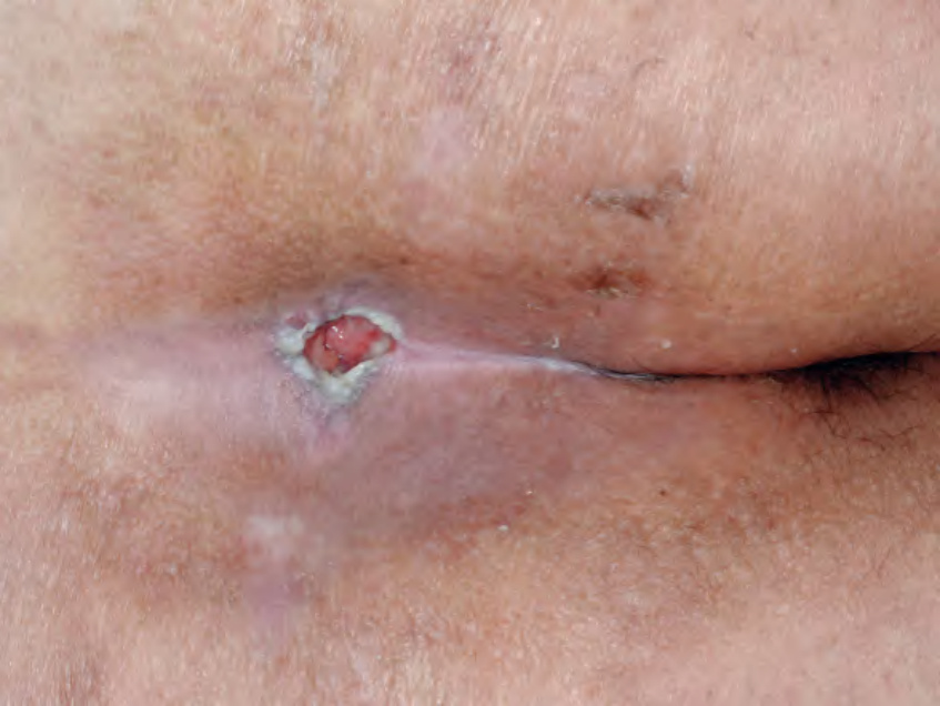
Fig. 105.17 Pressure ulcer (stage III) over the sacral area.
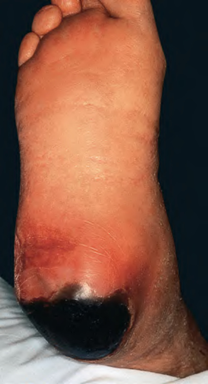
Fig. 105.18 Black eschar of the heel at site of pressure necrosis.
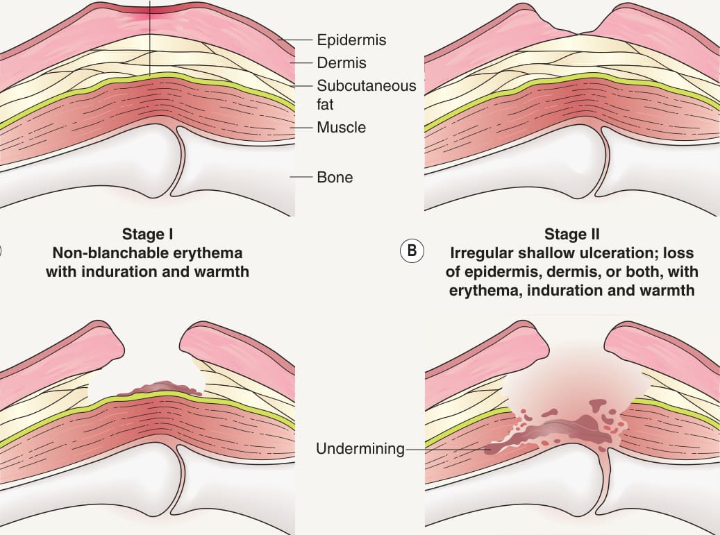
Fig. 105.19 National Pressure Ulcer Advisory