OTHER CAUSES OF SKIN ULCERATION
Ulcers that are apparently recalcitrant to prescribed treatments should be reevaluated regarding compliance with the program of care, and when non-compliance is excluded, then rare or unusual causes should be considered (Fig. 105.20; see Fig. 105.1).
Diffuse Dermal Angiomatosis
Diffuse dermal angiomatosis is an unusual manifestation of vascular atherosclerosis. There is rather rapid development of one or several violaceous plaques in a reticulated pattern, often with central ulceration. The lesions are typically painful and are usually located on the lower extremities, but they may appear elsewhere (e.g. breast, forearm) (see Fig. 105.20A). Histologically, a diffuse interstitial proliferation of CD31-positive endothelial cells is present within the papillary and reticular dermis, with focal formation of small vascular channels. Cytologic atypia and atypical mitoses are absent.
Interventions to correct the underlying PAD, such as femoral artery angioplasty or femoral popliteal bypass surgery, lead to rapid resolution. Less often, lesions resolve without therapy.
Panel classification of pressure ulcers.A Stage I: non-blanchable erythema of intact skin. This lesion is the heralding sign of impending skin ulceration. For darker-skinned individuals, other signs may be indicators and include warmth, edema, discoloration of the skin, and induration. B Stage II: partial-thickness skin loss involving the epidermis, dermis or both. This superficial lesion presents as an abrasion, blister, or shallow crater. C Stage III: fullthickness skin loss, in which subcutaneous tissue is damaged or necrotic and may extend down into, but not including, the underlying fascia. This deep lesion presents as a crater and sometimes involves adjacent tissue. D Stage IV: full-thickness skin loss and extensive tissue necrosis, destruction to muscle, bone, or supporting structures such as a tendon or joint capsule. Undermining or sinus tracts can be present.
Hematologic Disorders
Anemia is a major and often underrecognized cause for delayed wound healing. It prevents proper tissue oxygenation. In addition, some forms of anemia are associated with a marked tendency for ulcer formation, such as the hemoglobinopathies (see Fig. 105.20C). Hematologic malignancies are also associated with ulcerative processes including pyoderma gangrenosum, vasculitis, and cryoglobulinemia (particularly type I). Clotting abnormalities can play a direct role in the formation of ulcers (e.g. antiphospholipid antibody syndrome), as well as serving as a predisposing factor for more common types of cutaneous ulcerations such as venous ulcers. Acute massive venous thrombosis, also known as phlegmasia cerulea dolens, can lead to massive edema in the legs, with subsequent ischemia and ulceration due to venous limb gangrene. Table 105.9 summarizes the recommended evaluation when thrombophilia is suspected.
Tropical Ulcers
These ulcers are found in children and adults residing in the tropics who typically live in rural areas and are malnourished or otherwise debilitated. They may also occur in travelers returning from those areas. Although many tropical diseases manifest with chronic ulceration, the term “tropical ulcer” is often used to describe a phagedenic ulcer, usually located on the leg, which often occurs secondarily to minor trauma. The ulcer is invariably associated with a polymicrobial infection including Fusobacterium spp., other anaerobic bacteria, and spirochetes.
Tropical ulcers are always painful and progress rapidly to often involve deep tissues. The edges are undermined and violaceous, and malignant degeneration can occur. Assessment includes a smear and cultures plus soft tissue and bone imaging when deep involvement is suspected. These ulcers are best managed with systemic antibiotics
(e.g. tetracycline, metronidazole) and non-adherent dressings; they occasionally require surgical debridement.
The differential diagnosis of tropical ulcers includes other common causes of ulceration in tropical areas such as bacterial (including anthrax), mycobacterial, deep fungal, and parasitic (leishmaniasis) infections as well as non-infectious causes such as sickle cell anemia, venous insufficiency, neuropathy, and trauma.
Additional Causes
A variety of physical, inflammatory, infectious, metabolic, and inherited disorders are also associated with cutaneous ulceration (see Fig. 105.20B, D–G) and they are outlined in Fig. 105.1 and Table 105.10.
Additional figures available in our eBook (see inside front cover for access code).
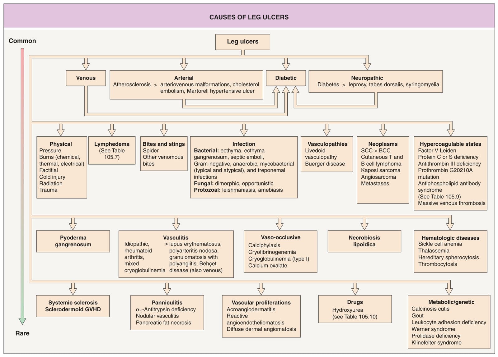
Fig. 105.1 Causes of leg ulcers. Patients with Behçet disease develop lower extremity ulcers due to vasculitis and/or venous insufficiency related to deep vein thromboses, and, occasionally, erosive pustular dermatosis is a cause of leg ulcers. Hydroxyurea-induced leg ulcers are often on the malleolus or tibial crest, exceedingly painful, and surrounded by atrophic skin. Additional genetic syndromes associated with ulceration are listed in Table 105.10. GVHD, graft-versus-host disease.
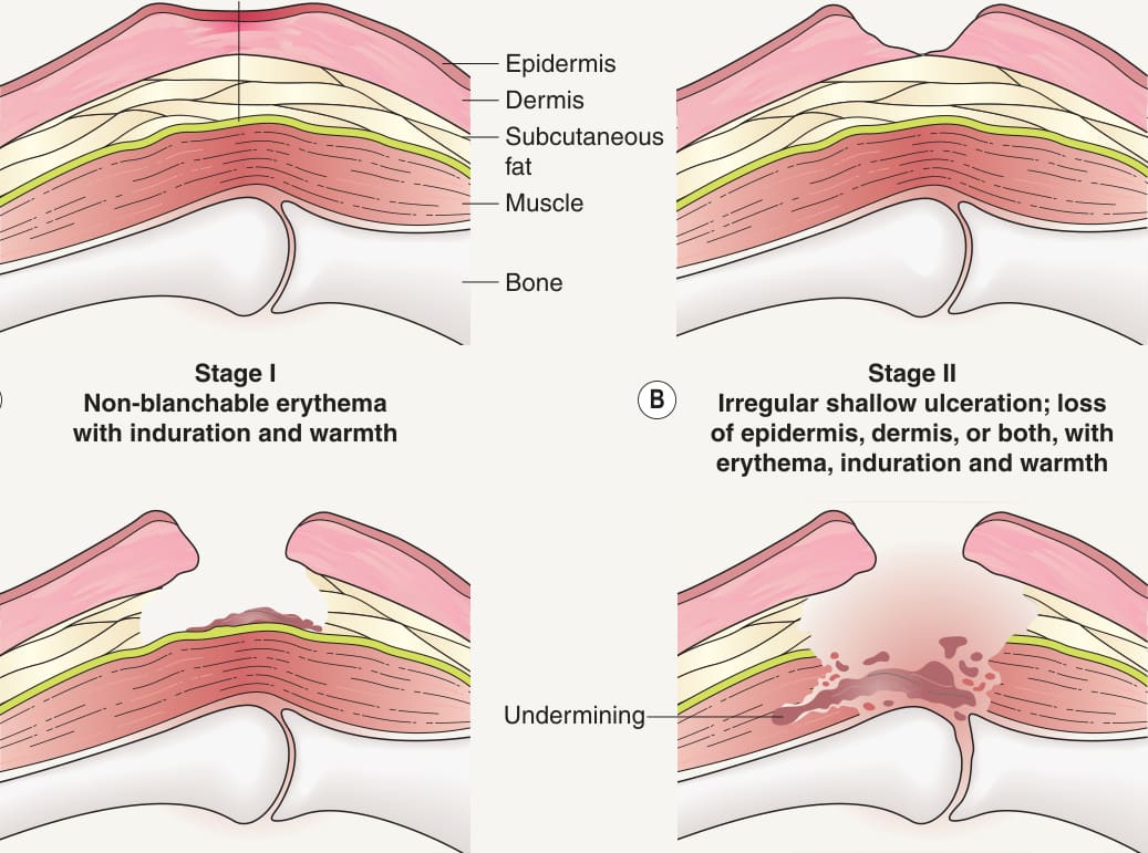
Fig. 105.19 National Pressure Ulcer Advisory
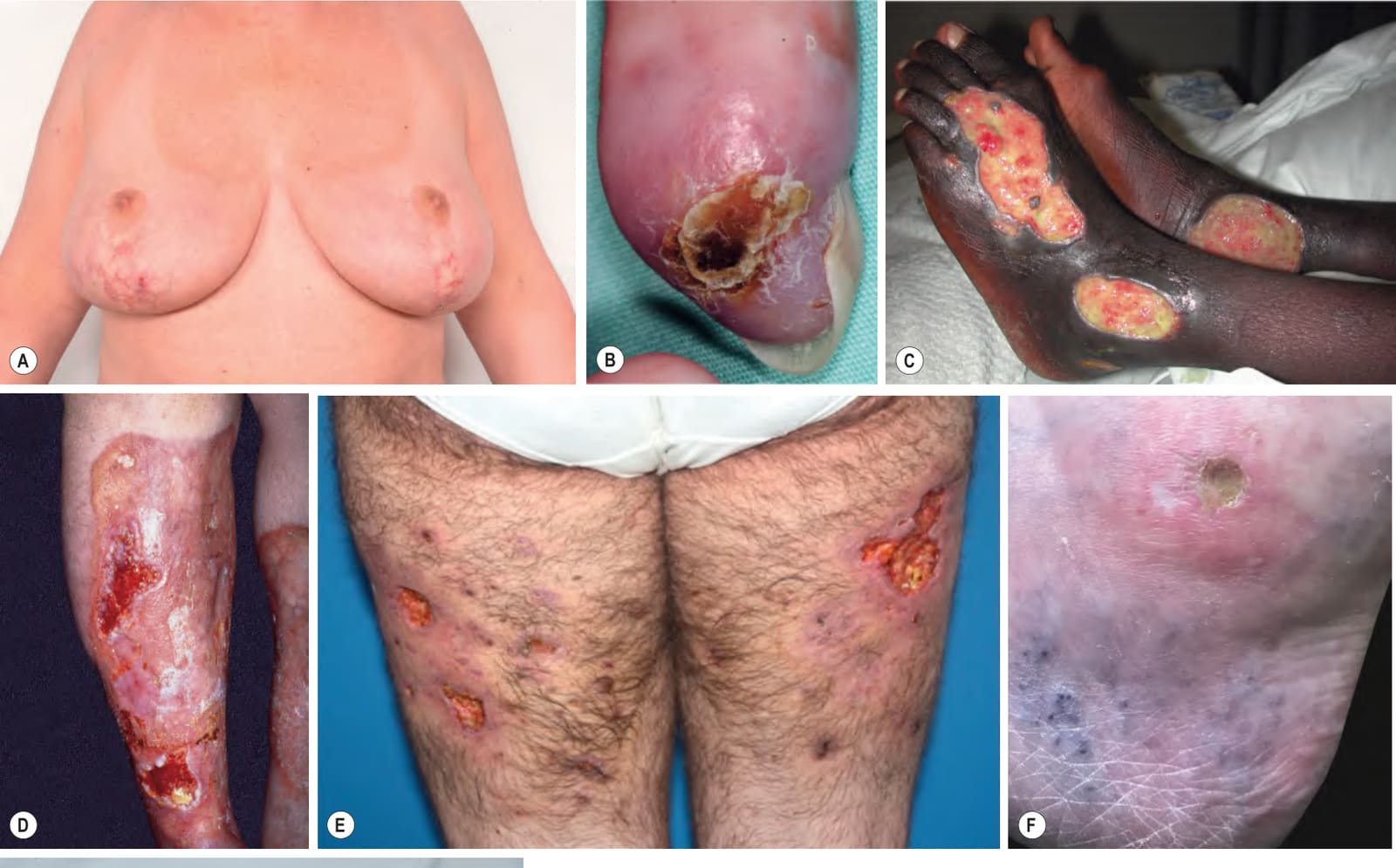
Fig. 105.20 Additional causes of ulcers.A Diffuse dermal angiomatosis develops most often in pendulous breasts (note the scars from previous breast reduction surgery) and the lower extremities in association with atherosclerosis. B Systemic sclerosis with an ulcer of the fingertip. C Multiple ulcers in a patient with sickle cell anemia. D Necrobiosis lipoidica. Controversy exists about the exact risk of diabetes mellitus in such patients, but it is more strongly associated with diabetes than is granuloma annulare. E Prolidase deficiency with spontaneous ulcers and scarring from previous ulcerations. F Ulceration of the lateral malleolus in a patient receiving hydroxyurea. G Henoch–Schönlein purpura in an adolescent. A, Courtesy Margo Peters, MD; D, Courtesy Jeffrey P. Callen, MD; E, Courtesy Duygu Gülseren, MD; F, Courtesy Karynne O. Duncan, MD; G, Courtesy Julie V. Schaffer, MD.
Table 105.9 Evaluation for thrombophilia/hypercoagulability. Shaded areas represent first-tier screening tests. The initial laboratory evaluation should also include a complete blood count with differential and platelet count, examination of a peripheral blood smear, ESR, activated partial thromboplastin time (PTT), and hepatic and renal function panels. Testing for anti-neutrophil cytoplasmic antibodies (ANCA) can be considered for patients with retiform purpura, as ANCA-positive vasculitides occasionally present with minimally inflammatory lesions; patients with levamisole-induced vasculopathy may have anti-phospholipid antibodies (as well as ANCA). Infections can also lead to coagulopathy (e.g. COVID-19-associated) in which abnormalities such as elevated fibrinogen or antiphospholipid antibodies are detected. Additional inherited causes of hypercoagulability include polymorphisms in the genes encoding the endothelial protein C receptor, the protein Z-dependent protease inhibitor, and E-selectin. AD, autosomal dominant; CBS, cystathionine β-synthase; DOACs, direct-acting oral anticoagulants; MTHFR, methylenetetrahydrofolate reductase; ND, not determined; OCP, oral contraceptive pills; RVVT, Russell viper-venom time.
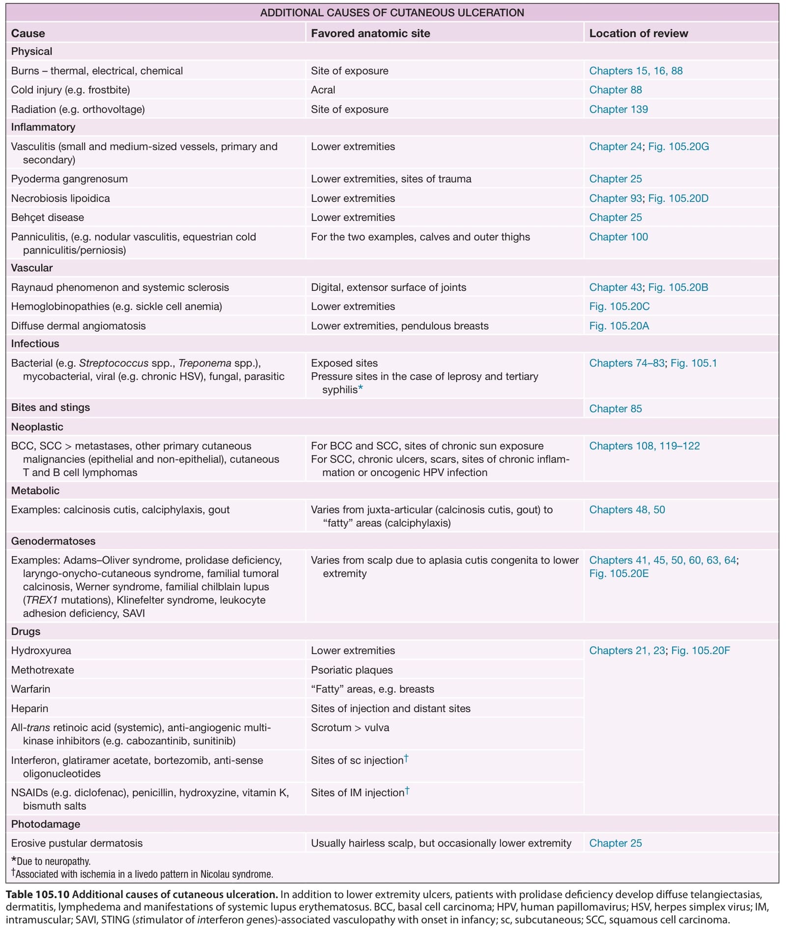
Table 105.10 Additional causes of cutaneous ulceration. In addition to lower extremity ulcers, patients with prolidase deficiency develop diffuse telangiectasias, dermatitis, lymphedema and manifestations of systemic lupus erythematosus. BCC, basal cell carcinoma; HPV, human papillomavirus; HSV, herpes simplex virus; IM, intramuscular; SAVI, STING (stimulator of interferon genes)-associated vasculopathy with onset in infancy; sc, subcutaneous; SCC, squamous cell carcinoma.