ERYTHROMELALGIA
Synonyms: Erythermalgia Erythralgia
Key features
Characterized by painful burning and erythema of the distal extremities (lower > upper)
Precipitated by heat and relieved by cooling
May be idiopathic, familial, or arise secondarily due to an under- lying condition (e. g. thrombocythemia)
Introduction
Erythromelalgia is an episodic condition characterized by a burning sensation, erythema, and increased skin temperature. It affects acral sites, particularly the lower extremities. Classically, there are three major forms, including:
●type 1 – associated with thrombocythemia
●type 2 – primary or idiopathic form
●type 3 – associated with underlying causes other than thrombocythemia.
History
The term “erythromelalgia” was first coined by Mitchell in 1878. It was used to describe redness (erythros), involvement of extremities (melos), and pain (algos). The diagnostic criteria were established by Thompson in 1979: (1) burning pain in the extremities; (2) pain aggravated by warming; (3) pain relieved by cooling; (4) erythema of affected skin; and (5) increased temperature of affected skin.
Epidemiology
In Norway, the estimated incidence of erythromelalgia is 0.25/100 000 with a prevalence of 2/100 000. While types 1 and 3 usually appear during adulthood, type 2 may appear in childhood and may be familial. The female : male incidence varies from 2 : 1 to almost 3 : 1. In one study, the mean age of onset was 56 years (range, 5–91 years) with 4% having their symptoms begin during childhood.
Pathogenesis
While the discovery of SCN9A mutations in patients with primary familial erythromelalgia has provided insights into this disorder, in general, the pathogenesis of erythromelalgia is not entirely understood. Firstly, it may vary depending on the underlying cause and between different patient subgroups. For example, in patients with thrombocythemia, the pathophysiology is likely to be related to both increased numbers of platelets as well as abnormalities in platelet function. Of note, one explanation for why type 1 erythromelalgia does not respond to heparin or warfarin is that the microthrombi that form do not require thrombin activation.
In other types of erythromelalgia, changes in vascular dynamics may be a factor. It has been proposed that hyperemia results from increased blood flow through arteriovenous shunts with a resultant reduction in blood flow within nutritional vessels, thus leading to cutaneous hypoxia. Another proposed mechanism is vasoconstriction (as occurs in Raynaud phenomenon), but in erythromelalgia this is followed by a prolonged phase of hyperemia. A temperature-triggered release of vasoactive substances and chemical pain mediators may also play a role.
In 2004, gain-of-function missense mutations in SCN9A, which encodes the voltage-gated sodium channel alpha subunit Nav1.7, were first described in patients with primary familial erythromelalgia. Nav1.7 produces threshold currents and is expressed primarily within dorsal root ganglia of sensory neurons (especially nociceptors) and sympathetic ganglion neurons. Mutations lead to lowered thresholds and “over-excitability” of pain-signaling sensory neurons while at the same time producing “under-excitability” of sympathetic neurons. Certain mutations that produce a lesser effect on sodium channel activation and subsequent neuron excitability are associated with a later onset of clinical signs and symptoms.
Another dominantly inherited pain syndrome, paroxysmal extreme pain disorder, has also been found to be due to gain-of-function mutations in SCN9A, whereas loss-of-function mutations in both copies of this gene lead to a recessively inherited insensitivity to pain. Lastly, there exist additional families that are not linked to chromosome 2q31–32 (SCN9A locus), suggesting genetic heterogeneity. Associations in secondary erythromelalgia include myelodysplastic syndromes, diabetes mellitus, peripheral arterial disease, vasculitis, systemic lupus erythematosus, and other autoimmune connective tissue diseases.
Clinical Features
Erythromelalgia is characterized by burning, erythema, and warmth of acral sites (Fig. 106.6). Attacks most commonly occur late in the day, last through the night, and frequently impair sleep. The symptoms are usually episodic, although occasionally they may be continuous. The feet are involved in 90% of patients, whereas the hands are affected in 25%. Less commonly, there is also involvement of the head and neck. Type 1 erythromelalgia may be unilateral and is more frequently associated with progression to ischemic necrosis. In contrast, the idiopathic form is more likely to be bilateral. The pain is precipitated by minor elevations in temperature (between 32°C and 36°C). Other exacerbating factors include exercise, standing, walking, fever, and limb dependency. Cooling and limb elevation generally reduce the symptoms.
The affected areas will often appear red and swollen. Other findings include acrocyanosis, livedo reticularis, facial flushing, cutaneous necrosis, and ulceration. In up to 40% of patients, the limb may appear normal between attacks. Prolonged immersion in water can lead to extensive maceration and contribute to ulcer formation.
Prognosis is variable. In one long-term study, ~30% of patients fell into each of three categories: worsening, no change, and improvement. A further 10% had complete resolution of their symptoms.
Pathology
In type 1 erythromelalgia, vessels may show intimal proliferation and occlusive thrombosis followed by complete fibrosis of the affected arterioles. In general, however, biopsies are not required and histology is nonspecific.
Differential Diagnosis
Complex regional pain syndrome (CRPS; reflex sympathetic dystrophy) may sometimes have features similar to erythromelalgia (see Ch. 6). Abnormal warmth, erythema, and burning pain may be seen in both, but CRPS does not have the characteristic close relationship to temperature and tends to be constant rather than episodic. Peripheral neuropathy may also cause tingling and burning and may require differentiation by nerve conduction studies. Calcium channel blockers, autonomic dysfunction, acrodynia, and mushroom poisoning, as well as occlusive vascular diseases such as thromboangiitis obliterans, may be associated with erythromelalgia-like symptoms. It is important to consider under-lying causes of erythromelalgia – in particular, chronic myeloproliferative disorders – as erythromelalgia may be the first sign of the latter.
Treatment
Although numerous treatments have been reported as possible therapies for erythromelalgia, no single therapy is consistently effective and many cases can prove difficult to control. The assistance of a specialist pain clinic can be valuable. Various methods of cooling the limbs during attacks, such as fans, wet dressings, and ice packs wrapped in towels, should be explored and have often already been tried by the time of presentation. Frequent periods of leg elevation can be helpful in reducing both discomfort and leg edema, whereas prolonged periods of leg dependency should be avoided. Simple oral analgesia is also important.
Aspirin can be effective for type 1 erythromelalgia and treatment for the thrombocythemia with medications such as hydroxyurea and (based upon mutational analysis) JAK inhibitors should also be considered. Topical therapies include capsaicin cream, 1%–2% amitriptyline/0.5% ketamine gel, and lidocaine patches, while possible oral medications include SSRIs (e.g. venlafaxine), tricyclic antidepressants (e.g. amitriptyline), anticonvulsants (e.g. gabapentin, carbamazepine), calcium channel blockers (e.g. diltiazem), the prostaglandin analogue misoprostol, and especially
in primary inherited erythromelalgia, sodium channel blocking agents (e.g. mexiletine, flecainide). With the report of functional expression studies (based upon a specific mutation) correlating with clinical responsiveness to carbamazepine or mexiletine, the possibility exists of utilizing such assays in the future to guide therapy. Rizatriptan, a selective agonist of serotonin 5-HT1B and 5-HT1D receptors, can be used for acute pain attacks in patients with primary erythromelalgia. In addition, there are ongoing trials of Nav1.7 channel blockers, both oral and topical.
Several intravenous therapies have been used for more severe cases, including nitroprusside, prostaglandin E1, and lidocaine (combined with oral mexiletine). More invasive approaches include epidural infusions of bupivacaine and opiates, lumbar sympathetic blocks, and bilateral lumbar sympathectomies. Oral medications should be tried initially, but more invasive procedures may be necessary for more severe cases.
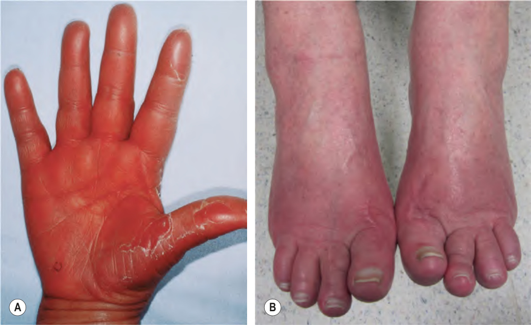
Fig. 106.6 Erythromelalgia. Red hot painful hand (A) and feet (B) in two patients with erythromelalgia. A, Courtesy Agustin Aloma, MD.