TELANGIECTASIAS
Key features
Telangiectasia is due to persistently dilated dermal vessels and not angiogenesis
Can occur as a primary process, a result of cutaneous damage, or secondary to systemic disease
Introduction
Telangiectasia refers to abnormal, small, persistently dilated blood vessels visible in the skin (Fig. 106.7). Individual vessels can be discerned
and range in color from light red to deep purple and will usually empty with pressure. They occur as a result of vascular dilation rather than new vessel growth and are thought to arise from capillaries, venules, or small arteriovenous malformations. Telangiectasias are seen in a range of clinical settings (Table 106.6).
Treatment is often not required; however, options include cosmetic camouflage, light or fine wire diathermy, injection sclerotherapy, and laser or intense pulsed light therapies.
Spider Telangiectasia
Synonyms: Nevus araneus Spider nevus Spider angioma
This localized lesion has a slightly raised central red papule (which often becomes more prominent with time) and multiple small, radiating, dilated vessels (“legs”) (Fig. 106.9). The lesion can vary from several millimeters to more than a centimeter in diameter. They are commonly located on the face, neck, upper trunk, and hands.
Spider telangiectasias represent a form of telangiectasia with a central feeding arterial vessel. They are usually seen in otherwise healthy individuals, especially women and children. Lesions, often multiple, commonly occur with pregnancy, liver disease, and oral contraceptive pills (OCPs). Spontaneous resolution may occur, especially after pregnancy. Treatment options include electrosurgery or vascular lasers.
Generalized Essential Telangiectasia
This is a primary disorder that typically affects adult women but may commence during childhood. Initially, the limbs are affected, especially the distal lower extremities, with sheets of asymptomatic blanchable telangiectasias (Fig. 106.10). The involvement is usually symmetric. Over time, there is often a progression proximally such that the telangiectasias become more widespread. Although the telangiectasias are persistent, patients do not have associated systemic illnesses.
The differential diagnosis includes cutaneous collagenous vasculopathy; typically it is not confused with telangiectasia macularis eruptiva perstans. While improvement with vascular lasers is possible, the progressive nature must be considered and the cost of treating extensive involvement may prove prohibitive.
Cutaneous Collagenous Vasculopathy
Cutaneous collagenous vasculopathy (CCV) was first described in
2000. Clinically, it can resemble generalized essential telangiectasia (GET). However, based upon the limited number of reported cases, there is not a female predominance, the telangiectasias may involve
just the trunk and/or proximal extremities, and history of an “upward march” of lesions is lacking (Fig. 106.11A). The etiology is unknown and there is no clear association with any underlying systemic illness or medication.
CCV does have unique histopathologic findings consisting of ectatic superficial blood vessels with hyalinized and laminated, concentric concretions surrounding the basement membrane of affected vessels. With special staining, this material is PAS-positive and diastaseresistant, and by immunohistochemistry, it reacts with antibodies against collagen type IV (Fig. 106.11B). Ultrastructurally, the deposits have been shown to consist of collagen with abnormal banding patterns (Luse bodies).
This condition, like generalized essential telangiectasia, is benign in nature. There are scattered case reports of improvement following pulsed dye laser therapy.
Unilateral Nevoid Telangiectasia
Telangiectasias in this condition are usually confined to the trigeminal or upper cervical dermatomes (Fig. 106.12) and may follow the lines of Blaschko. Congenital and acquired forms are recognized. It has been proposed that an increase in estrogen receptors on blood vessels in affected areas and/or an increase in estrogen levels is causative. Situations of relative estrogen excess such as pregnancy, puberty, and liver disease are associated with the acquired form.
Angioma Serpiginosum
Angioma serpiginosum is a rare vascular disorder with a characteristic appearance. It is usually sporadic, but familial cases have been reported. Typically, the condition affects female patients and
commences during the first two decades of life. Multiple, small, asymptomatic, non-palpable, deep-red to purple puncta occur in small clusters and sheets (Fig. 106.13). The arrangement and extension of the lesions may produce a serpiginous pattern. The extremities are most commonly affected, initially with a unilateral distribution; over months to years, the involvement may become more widespread. The palms, soles, and mucous membranes are not involved.
The puncta represent dilated, non-inflamed capillaries within the dermal papillae. These can be seen with dermoscopy. Incomplete blanching occurs with pressure, but lesions are not purpuric. The differential diagnosis includes pigmented purpuric eruptions, particularly the Majocchi variant (purpura annularis telangiectoides), which is more likely to be bilateral and on biopsy has perivascular lymphocytes and extravasated erythrocytes. Treatment is not necessary, but lesions can be improved with the pulsed dye laser.
Hereditary Hemorrhagic Telangiectasia
Synonyms: Osler–Weber–Rendu disease Osler–Rendu– Weber disease
Hereditary hemorrhagic telangiectasia (HHT) is an autosomal dominant condition in which there are multiple mucocutaneous and gastrointestinal telangiectasias (which are actually arteriovenous malformations)
as well as variable visceral involvement (lung, liver, and CNS). The diagnosis may first be suspected in children who have repeated episodes of epistaxis; however, the initial presentation can occur during the second or third decades of life. The characteristic mat-like and papular telangiectasias on the mucous membranes are first seen during adolescence. Cutaneous lesions usually appear after puberty or even later in life.
Lesions, which increase in size and number as the patient ages, are most commonly seen on the face, tongue, lips, nasal mucosa, hands, and fingertips (Fig. 106.14). They frequently occur throughout the gastrointestinal tract and may result in obvious hemorrhage or iron deficiency anemia. Hemorrhage from vascular lesions in the lung, liver, CNS, spleen, and urinary tract may occur, as well as paradoxical emboli due to pulmonary arteriovenous malformations.
HHT can result from mutations in several genes, most commonly HHT1 and HHT2 which encode endoglin and ALK-1, respectively (see Table 104.2). Both of these glycoproteins are TGF-β receptors expressed by vascular endothelium, and they are thought to play a role in angiogenesis and vessel wall integrity.
Treatment of the mucocutaneous vascular lesions may not be required. Destructive treatments such as diathermy, cautery, or laser may be used to treat individual lesions. Surgical management or embolization may be required for uncontrolled hemorrhage from mucosal lesions or complications arising from visceral lesions. Initial and longitudinal evaluation of patients with HHT is outlined in Table 104.6.
Ataxia–Telangiectasia
Synonym: Louis-Bar syndrome
Ataxia–telangiectasia was first described by Louis-Bar in 1941. It is an autosomal recessive disorder characterized by cerebellar ataxia, chromosomal instability (frequent translocations between chromosomes 7 and 14), growth retardation, oculocutaneous telangiectasias, pulmonary infections (including bronchiectasis), immunodeficiency, and the development of lymphomas.
It is covered in more detail in Chapter 60. Linear telangiectasias first appear on the bulbar conjunctivae between 4 and 6 years of age. Cutaneous telangiectasias favor the head and neck region, and they are most common on the malar prominences, ears, and eyelids in addition to the popliteal and antecubital fossae. Patients may have poikiloderma (hypopigmentation, hyperpigmentation, atrophy, telangiectasia), premature hair graying, and decreased subcutaneous fat.
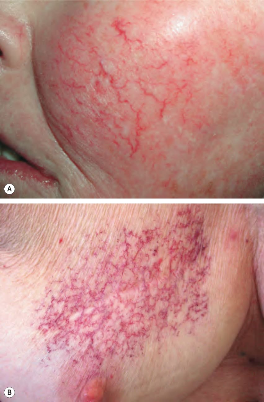
Fig. 106.7 Telangiectasias.A Sun-induced telangiectasias on the cheek. B Prominent telangiectasias on the breast following radiation therapy.
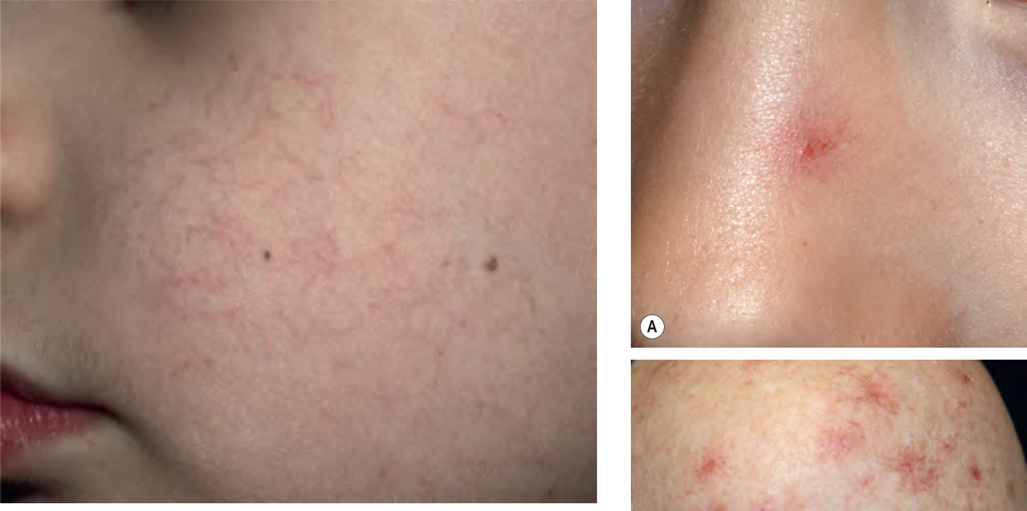
Fig. 106.8 Hereditary benign telangiectasia. Fine linear and branching telan giectasias are seen on the cheeks bilaterally in this five-year-old boy. There is no associated visceral involvement. Courtesy Julie V. Schaffer, MD.
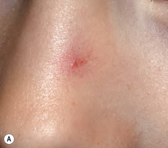
Fig. 106.9 Spider telangiectasia. They are characterized by a central arteriole with radiating telangiectatic “legs”. A Single lesion on the nose of a young child. B Multiple lesions on the shoulder of an adult with liver disease. A, Courtesy Julie V. Schaffer, MD; B, Courtesy Jeffrey P. Callen, MD.
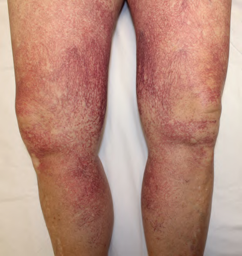
Fig. 106.10 Generalized essential telangiectasia. Bilateral sheets of asymptomatic blanchable telangiectasias. The initial site of involvement is usually the distal lower extremity. Courtesy Jean L. Bolognia, MD.
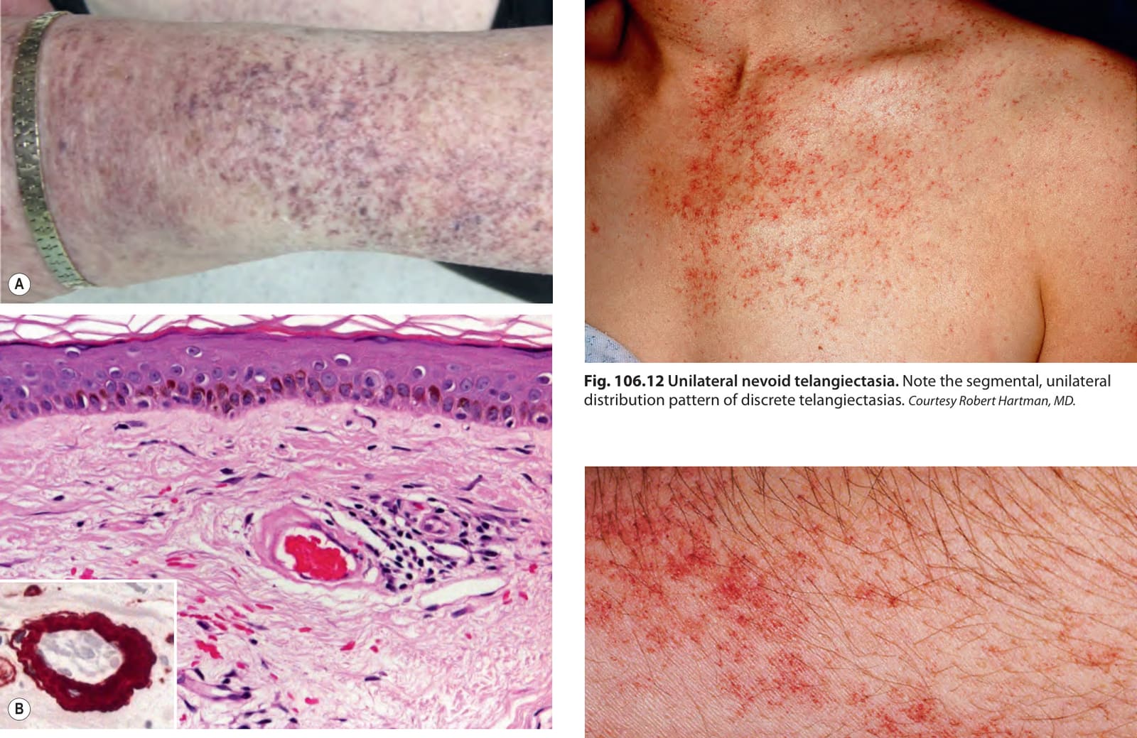
Fig. 106.11 Cutaneous collagenous vasculopathy – clinical and histologic features.A Multiple violaceous branching telangiectasias on the forearm; the patient also had involvement of the thighs, abdomen, and back. B In the upper dermis, there are some extravasated red blood cells and clumped RBCs within an ectatic blood vessel. Note the concentric hyaline deposits at the periphery of the affected blood vessel. Immunohistochemical staining with antibodies to collagen type IV highlight the deposits (inset). B, Courtesy Lorenzo Cerrroni, MD.
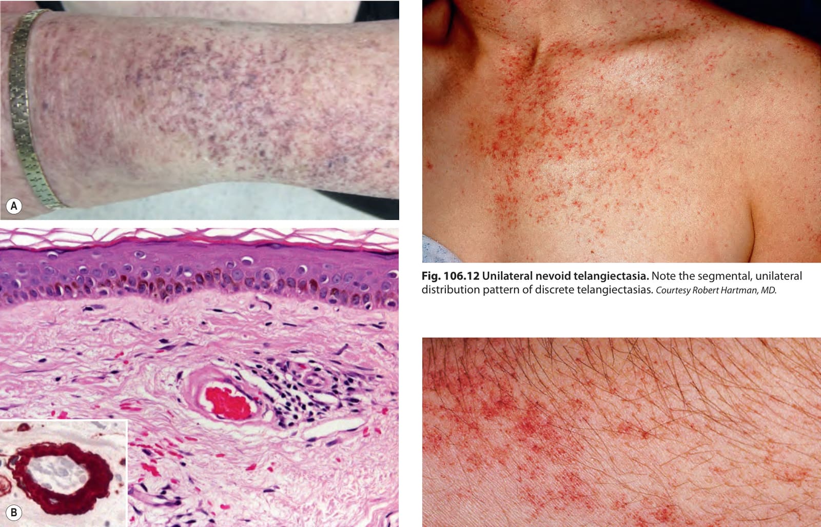
Fig. 106.12 Unilateral nevoid telangiectasia. Note the segmental, unilateral distribution pattern of discrete telangiectasias. Courtesy Robert Hartman, MD.
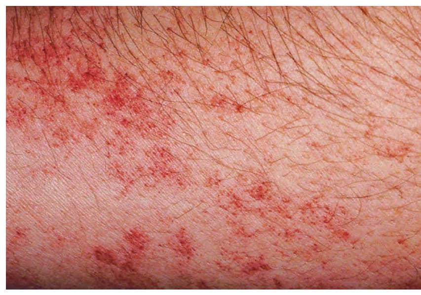
Fig. 106.13 Angioma serpiginosum. Grouped dark red puncta on the arm in a serpiginous pattern.
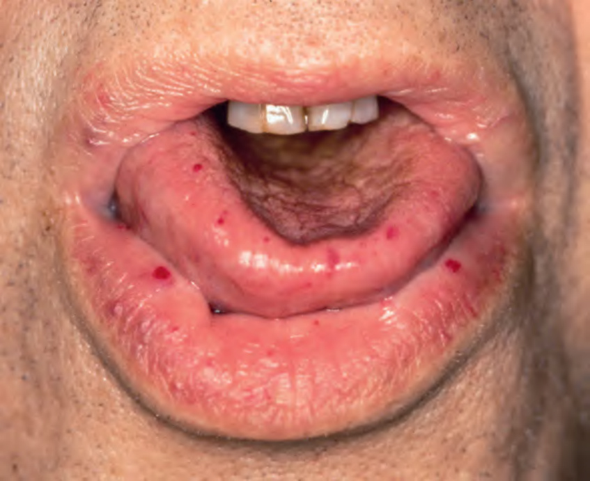
Fig. 106.14 Hereditary hemorrhagic telangiectasia. The tongue and lips are a common location for the mat-like and papular telangiectasias that actually represent small arteriovenous malformations.
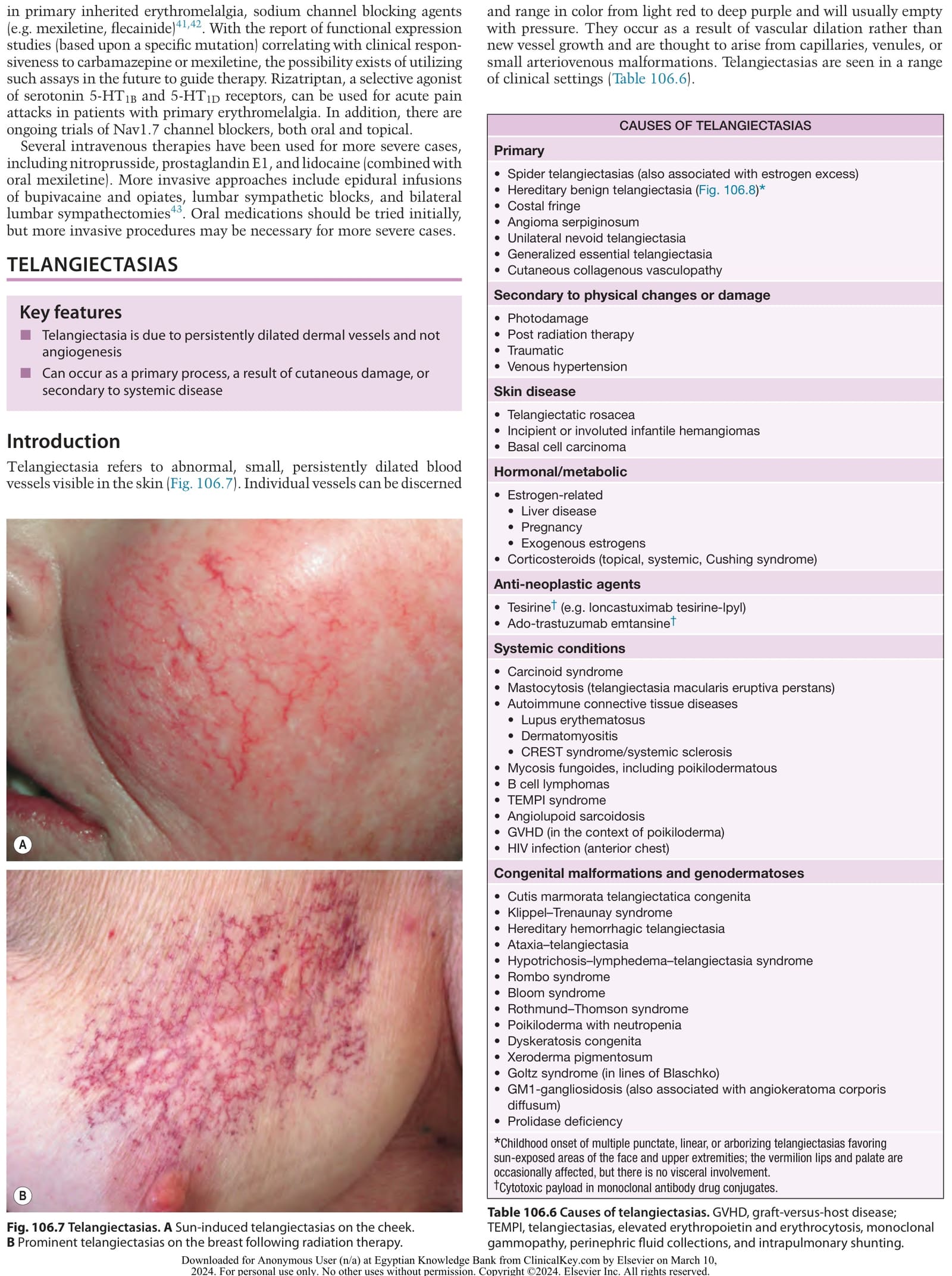
Table 106.6 Causes of telangiectasias. GVHD, graft-versus-host disease; TEMPI, telangiectasias, elevated erythropoietin and erythrocytosis, monoclonal gammopathy, perinephric fluid collections, and intrapulmonary shunting.