ANGIOSPASTIC MACULES (BIER SPOTS)
These were first described by Bier in 1898 and represent a pattern of vascular mottling consisting of white macular areas surrounded by a red to occasionally blue cyanotic background. Bier spots most commonly occur on the arms and legs of young adults (Fig. 106.17), but may also occur on the trunk. They are more noticeable in patients with lightly pigmented skin. Angiospastic macules may be induced by venous congestion and can be elicited by placing a limb in a dependent position or by placing a tourniquet around a limb. The spots disappear with limb elevation or release of the tourniquet. As with nevus anemicus, the border of the lesion becomes imperceptible with pressure (diascopy). The vascular pattern is thought to result from a benign physiological response consisting of vasoconstriction. Bier spots have also been associated with pregnancy, lymphedema, cryoglobulinemia, capillary malformationarteriovenous malformation syndrome (especially type 2 due to EPHB4 mutations), tuberous sclerosis complex, and aortic hypoplasia. BASCULE syndrome features Bier anemic spots, cyanosis (acral), and an urticarialike eruption in association with orthostatic intolerance.
Additional figures available in our eBook (see inside front cover for access code).
- Weir NU, Snowden JA, Greaves M, Davies-Jones GAB.
Clin. 1990;8:347–354.2. Gibbs MB, English JC, Zirwas MJ. Livedo reticularis: an
- Fischer TZ, Waxman SG. Familial pain syndromes from
Livedo reticularis associated with hereditary protein C deficiency and recurrent thromboembolism. Br J Dermatol. 1995;132:283–285.21. Donnet A, Khalil R, Terrier G, et al. Cerebral infarction, update. J Am Acad Dermatol. 2005;52:1009–1019.3. Bashir S, Chew A. Cutaneous reactions to cold and heat.
In: Griffiths C, Barker J, Bleiker T, et al. , editors. Textbook of Dermatology. 8th ed. Blackwell Science; 2017. p. 125. 6.4. Kelly RI, Wee E, Balta S, Williams RA. Lymphocytic throm- mutations of the NaV1. 7 sodium channel. Ann N Y Acad Sci. 2010;1184:196–207.39. Burns TM, Te Morsche RH, Jansen JB, Drenth JP. Genetic heterogeneity and exclusion of a modifying locus at 2q in a family with autosomal dominant primary erythermalgia. Br J Dermatol. 2005;153:174–177.40. Davis MDP, O’Fallon WM, Rogers III RS, Rooke TW.
livedo reticularis, and familial deficiency in antithrombin-III. Stroke. 1992;23:611–612.22. Freeman EF, Devon E, Lipoff JB, et al. The spectrum of bophilic arteritis and cutaneous polyarteritis nodosa: clinicopathologic comparison with blinded histologic assessment. J Am Acad Dermatol. 2020;83:501–508.5. Lee JS, Kossard S, McGrath MA. Lymphocytic thrombo-
COVID-19 associated dermatologic manifestations: an international registry of 716 patients from 31 countries. J Am Acad Dermatol. 2020;83:1118–1129.23. Spiers EM, Sanders DY, Omura EF. Clinical and histo- philic arteritis: a newly described medium-sized vessel arteritis of the skin. Arch Dermatol. 2008;144:1175–1182.6. Francés C, Piette JC. The mystery of Sneddon syndrome:
Natural history of erythromelalgia. Arch Dermatol. 2000;136:330–336.41. Iqbal J, Bhat MI, Charoo BA, et al. Experience with oral mexiletine in primary erythromelalgia in children. Ann Saudi Med. 2009;29:316–318.42. Natkunarajah J, Atherton D, Elmslie F, et al. Treatment logic features of primary oxalosis. J Am Acad Dermatol. 1990;22:952–956.24. Sladden MJ, Nicolaou N, Johnston GA, Hutchinson PE.
relationship with antiphospholipid syndrome and systemic lupus erythematosus. J Autoimmun. 2000;15:139–143.7. Francés C, Papo T, Wechsler B, et al. Sneddon syndrome with carbamazepine and gabapentin of a patient with primary erythermalgia (erythromelalgia) identified to have a mutation in the SCN9A gene, encoding a voltage-gated sodium channel. Clin Exp Dermatol. 2009;34:e640–e642.43. Cohen JS. Erythromelalgia: new theories and new
Livedo reticularis induced by amantadine. Br J Dermatol. 2003;149:655–680.25. Wilkin JK. Why is flushing limited to a mostly facial with or without antiphospholipid antibodies: a comparative study in 46 patients. Medicine (Baltimore). 1999;78:209–219.8. Devillers AC, de Waard-van der Spek FB, Oranje AP. Cutis cutaneous distribution? J Am Acad Dermatol. 1988;19:309–313.26. Freeman R, Waldorf HA, Dover JS. Autonomic neuroder- marmorata telangiectatica congenita: clinical features in 35 cases. Arch Dermatol. 1999;135:34–38.9. Gerritsen MJP, Steijlen PM, Brunner HG, Rieu P. Cutis therapies. J Am Acad Dermatol. 2000;43:841–847.44. Bean WB. Vascular Spiders and Related Lesions of the Skin.
matology (Part II): disorders of sweating and flushing. Semin Neurol. 1992;12:394–407.27. Wilkin JK. The red face: flushing disorders. Clin Dermatol.
marmorata telangiectatica congenita: report of 18 cases. Br J Dermatol. 2000;142:366–369.10. Speight EL, Lawrence CM. Reticulate purpura,
Oxford: Blackwell Scientific; 1958.45. Rothe MJ, Grant-Kels JM. Nomenclature of the primary
1993;11:211–223.28. Wilkin JK. Flushing reactions. Recent Adv Dermatol.
telangiectasias. Int J Dermatol. 1992;31:320.46. McGrae Jr JD, Winkelmann RK. Generalized essential
1983;6:157–187.29. Clement D, Ramage J, Srirajaskanthan R. Update on cryoglobulinaemia and livedo reticularis. Br J Dermatol. 1993;129:319–323.11. Filo V, Brezová D, Hlavcák P, Filová A. Livedo reticularis telangiectasia: report of a clinical and histochemical study of 13 patients with acquired cutaneous lesions. J Am Med Assoc. 1963;185:909–913.47. Salama S, Rosenthal DJ. Cutaneous collagenous vascu- pathophysiology, treatment, and complications of carcinoid syndrome. J Oncol. 2020;2020;8341426.30. Thompson GH, Hahn G, Rang M. Erythromelalgia. Clin as a presenting symptom of polycythemia vera. Clin Exp Dermatol. 1999;24:428.12. Bauzá A, España A, Idoate M. Cutaneous polyarteritis lopathy with generalized telangiectasia: an immunohistochemical and ultrastructural study. J Cutan Pathol. 2000;27:40–48.48. Davis TL, Mandal RV, Bevona C, et al. Collagenous
Orthop Relat Res. 1979;144:249–254.31. Kvernebo K. Erythromelalgia: a condition caused by nodosa. Br J Dermatol. 2002;146:694–699.13. Vakili S, Zampella JG, Kwatra SG, et al. Lymphocytic microvascular arteriovenous shunting. Vasa. 1998(Suppl. 51):1–40.32. van Genderen PJ, Lucas IS, van Strik R, et al. Erythromelalgia thrombophilic arteritis: a review. J Clin Rheumatol. 2019;25:147–152.14. Anavekar NS, Kelly R. Heterozygous prothrombin vasculopathy: a report of three cases. J Cutan Pathol. 2008;35:967–970.49. Wilken JK. Unilateral dermatomal superficial telangiec- in essential thrombocythemia is characterized by platelet activation and endothelial cell damage but not by thrombin generation. Thromb Haemost. 1996;76:333–338.33. Mork C, Asker CL, Salerud EG, Kvernebo K. Microvascular gene mutation associated with livedoid vasculopathy. Australas J Dermatol. 2007;48:120–123.15. Zhou Q, Yang D, Ombrello AK, et al. Early-onset stroke tasia. Arch Dermatol. 1984;120:579–580.50. Uhlin SR, McCarty Jr KS. Unilateral nevoid telangiec- tatic syndrome: the role of estrogen and progesterone receptors. Arch Dermatol. 1983;119:226–228.51. Hunt SJ, Santa Cruz DJ. Acquired benign and ‘borderline’
arteriovenous shunting is a probable pathogenetic mechanism in erythromelalgia. J Invest Dermatol. 2000;114:643–646.34. Berlin AL, Pehr K. Coexistence of erythromelalgia and vasculopathy associated with mutations in ADA2. N Engl J Med. 2014;370:911–920.16. Navon Elkan P, Pierce SB, Segel R, et al. Mutant adenosine deaminase 2 in a polyarteritis nodosa vasculopathy. N Engl J Med. 2014;370:921–931.17. Nigwekar SU, Kroshinsky D, Nazarian RM, et al.
vascular lesions. Dermatol Clin. 1992;10:97–115.52. Marriott PJ, Munro DD, Ryan T. Angioma serpiginosum:
and Raynaud’s phenomenon. J Am Acad Dermatol. 2004;50:456–460.35. Yang Y, Wang Y, Li S, et al. Mutations in SCN9A, encoding
Calciphylaxis: risk factors, diagnosis, and treatment. Am J Kidney Dis. 2015;66:133–146.18. Rosman HS, Davis TP, Reddy D, Goldstein S. Cholesterol familial incidence. Br J Dermatol. 1975;93:701–706.53. Abrahamian LM, Rothe MJ, Grant-Kels JM. Primary telan- giectasia of childhood. Int J Dermatol. 1992;31:307–313.54. Smith LL, Conerly SL. Ataxia-telangiectasia or Louis-Bar a sodium channel alpha subunit, in patients with primary erythermalgia. J Med Genet. 2004;41:171–174.36. Choi JS, Dib-Hajj SD, Waxman SG. Inherited embolization: clinical findings and implications. J Am Coll Cardiol. 1990;15:1296–1299.19. Gibson GE, Su WPD, Pittelkow MR. Antiphospholipid syndrome. J Am Acad Dermatol. 1985;12:681–696.55. Bean WB, Walsh JR. Venous lakes. Arch Dermatol.
erythermalgia: limb pain from an S4 charge-neutral Na channelopathy. Neurology. 2006;67:1563–1567.37. Han C, Dib-Hajj SD, Lin Z, et al. Early- and late-onset syndrome and the skin. J Am Acad Dermatol. 1997;36:970–982.
1956;74:459–463.56. Alcalay J, Sandbank M. The ultrastructure of cutaneous venous lakes. Int J Dermatol. 1987;26:645–646.57. Mountcastle EA, Diestelmeier MR, Lupton GP. Nevus inherited erythromelalgia: genotype-phenotype correlation. Brain. 2009;132:1711–1722.
anemicus. J Am Acad Dermatol. 1986;14:628–632.
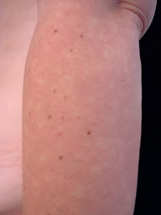
Fig. 106.17 Angiospastic macules (Bier spots).
of the donor site (donor dominance). No histologic abnormalities have been reported. No treatment is required or effective. Of note, nevus anemicus can co-exist with vascular malformations and is seen in up to half of patients with neurofibromatosis type 1.