血管痙攣性斑(Bier 斑)(ANGIOSPASTIC MACULES (BIER SPOTS))
這些病灶最早由 Bier 於 1898 年描述,代表一種血管斑駁的紋路,由白色斑狀區域被紅色、偶爾為藍色發紺的背景所環繞而構成。Bier 斑最常發生於年輕成人的上肢與下肢(Fig. 106.17),但也可能發生於軀幹。它們在膚色較淺的病人身上更為明顯。血管痙攣性斑可由靜脈鬱血所誘發,並可藉由將肢體置於下垂位置或在肢體周圍綁上止血帶而引出。這些斑點會隨肢體抬高或止血帶解除而消失。與貧血性痣一樣,病灶的邊界會在加壓(壓片檢查 diascopy)時變得無法辨識。此血管紋路被認為源自一種由血管收縮所構成的良性生理反應。Bier 斑也曾與懷孕、淋巴水腫、冷凝球蛋白血症、微血管畸形-動靜脈畸形症候群(capillary malformation-arteriovenous malformation syndrome,特別是因 EPHB4 突變所致的第 2 型)、結節性硬化症複合體,以及主動脈發育不全相關。BASCULE 症候群的特徵為 Bier 貧血性斑(Bier anemic spots)、(肢端)發紺,以及類似蕁麻疹的疹子,並與姿勢性不耐(orthostatic intolerance)相關。
更多圖片可於我們的電子書中取得(存取碼請見封面內頁)。
- Weir NU, Snowden JA, Greaves M, Davies-Jones GAB.
Clin. 1990;8:347–354.2. Gibbs MB, English JC, Zirwas MJ. Livedo reticularis: an
- Fischer TZ, Waxman SG. Familial pain syndromes from
Livedo reticularis associated with hereditary protein C deficiency and recurrent thromboembolism. Br J Dermatol. 1995;132:283–285.21. Donnet A, Khalil R, Terrier G, et al. Cerebral infarction, update. J Am Acad Dermatol. 2005;52:1009–1019.3. Bashir S, Chew A. Cutaneous reactions to cold and heat.
In: Griffiths C, Barker J, Bleiker T, et al. , editors. Textbook of Dermatology. 8th ed. Blackwell Science; 2017. p. 125. 6.4. Kelly RI, Wee E, Balta S, Williams RA. Lymphocytic throm- mutations of the NaV1. 7 sodium channel. Ann N Y Acad Sci. 2010;1184:196–207.39. Burns TM, Te Morsche RH, Jansen JB, Drenth JP. Genetic heterogeneity and exclusion of a modifying locus at 2q in a family with autosomal dominant primary erythermalgia. Br J Dermatol. 2005;153:174–177.40. Davis MDP, O’Fallon WM, Rogers III RS, Rooke TW.
livedo reticularis, and familial deficiency in antithrombin-III. Stroke. 1992;23:611–612.22. Freeman EF, Devon E, Lipoff JB, et al. The spectrum of bophilic arteritis and cutaneous polyarteritis nodosa: clinicopathologic comparison with blinded histologic assessment. J Am Acad Dermatol. 2020;83:501–508.5. Lee JS, Kossard S, McGrath MA. Lymphocytic thrombo-
COVID-19 associated dermatologic manifestations: an international registry of 716 patients from 31 countries. J Am Acad Dermatol. 2020;83:1118–1129.23. Spiers EM, Sanders DY, Omura EF. Clinical and histo- philic arteritis: a newly described medium-sized vessel arteritis of the skin. Arch Dermatol. 2008;144:1175–1182.6. Francés C, Piette JC. The mystery of Sneddon syndrome:
Natural history of erythromelalgia. Arch Dermatol. 2000;136:330–336.41. Iqbal J, Bhat MI, Charoo BA, et al. Experience with oral mexiletine in primary erythromelalgia in children. Ann Saudi Med. 2009;29:316–318.42. Natkunarajah J, Atherton D, Elmslie F, et al. Treatment logic features of primary oxalosis. J Am Acad Dermatol. 1990;22:952–956.24. Sladden MJ, Nicolaou N, Johnston GA, Hutchinson PE.
relationship with antiphospholipid syndrome and systemic lupus erythematosus. J Autoimmun. 2000;15:139–143.7. Francés C, Papo T, Wechsler B, et al. Sneddon syndrome with carbamazepine and gabapentin of a patient with primary erythermalgia (erythromelalgia) identified to have a mutation in the SCN9A gene, encoding a voltage-gated sodium channel. Clin Exp Dermatol. 2009;34:e640–e642.43. Cohen JS. Erythromelalgia: new theories and new
Livedo reticularis induced by amantadine. Br J Dermatol. 2003;149:655–680.25. Wilkin JK. Why is flushing limited to a mostly facial with or without antiphospholipid antibodies: a comparative study in 46 patients. Medicine (Baltimore). 1999;78:209–219.8. Devillers AC, de Waard-van der Spek FB, Oranje AP. Cutis cutaneous distribution? J Am Acad Dermatol. 1988;19:309–313.26. Freeman R, Waldorf HA, Dover JS. Autonomic neuroder- marmorata telangiectatica congenita: clinical features in 35 cases. Arch Dermatol. 1999;135:34–38.9. Gerritsen MJP, Steijlen PM, Brunner HG, Rieu P. Cutis therapies. J Am Acad Dermatol. 2000;43:841–847.44. Bean WB. Vascular Spiders and Related Lesions of the Skin.
matology (Part II): disorders of sweating and flushing. Semin Neurol. 1992;12:394–407.27. Wilkin JK. The red face: flushing disorders. Clin Dermatol.
marmorata telangiectatica congenita: report of 18 cases. Br J Dermatol. 2000;142:366–369.10. Speight EL, Lawrence CM. Reticulate purpura,
Oxford: Blackwell Scientific; 1958.45. Rothe MJ, Grant-Kels JM. Nomenclature of the primary
1993;11:211–223.28. Wilkin JK. Flushing reactions. Recent Adv Dermatol.
telangiectasias. Int J Dermatol. 1992;31:320.46. McGrae Jr JD, Winkelmann RK. Generalized essential
1983;6:157–187.29. Clement D, Ramage J, Srirajaskanthan R. Update on cryoglobulinaemia and livedo reticularis. Br J Dermatol. 1993;129:319–323.11. Filo V, Brezová D, Hlavcák P, Filová A. Livedo reticularis telangiectasia: report of a clinical and histochemical study of 13 patients with acquired cutaneous lesions. J Am Med Assoc. 1963;185:909–913.47. Salama S, Rosenthal DJ. Cutaneous collagenous vascu- pathophysiology, treatment, and complications of carcinoid syndrome. J Oncol. 2020;2020;8341426.30. Thompson GH, Hahn G, Rang M. Erythromelalgia. Clin as a presenting symptom of polycythemia vera. Clin Exp Dermatol. 1999;24:428.12. Bauzá A, España A, Idoate M. Cutaneous polyarteritis lopathy with generalized telangiectasia: an immunohistochemical and ultrastructural study. J Cutan Pathol. 2000;27:40–48.48. Davis TL, Mandal RV, Bevona C, et al. Collagenous
Orthop Relat Res. 1979;144:249–254.31. Kvernebo K. Erythromelalgia: a condition caused by nodosa. Br J Dermatol. 2002;146:694–699.13. Vakili S, Zampella JG, Kwatra SG, et al. Lymphocytic microvascular arteriovenous shunting. Vasa. 1998(Suppl. 51):1–40.32. van Genderen PJ, Lucas IS, van Strik R, et al. Erythromelalgia thrombophilic arteritis: a review. J Clin Rheumatol. 2019;25:147–152.14. Anavekar NS, Kelly R. Heterozygous prothrombin vasculopathy: a report of three cases. J Cutan Pathol. 2008;35:967–970.49. Wilken JK. Unilateral dermatomal superficial telangiec- in essential thrombocythemia is characterized by platelet activation and endothelial cell damage but not by thrombin generation. Thromb Haemost. 1996;76:333–338.33. Mork C, Asker CL, Salerud EG, Kvernebo K. Microvascular gene mutation associated with livedoid vasculopathy. Australas J Dermatol. 2007;48:120–123.15. Zhou Q, Yang D, Ombrello AK, et al. Early-onset stroke tasia. Arch Dermatol. 1984;120:579–580.50. Uhlin SR, McCarty Jr KS. Unilateral nevoid telangiec- tatic syndrome: the role of estrogen and progesterone receptors. Arch Dermatol. 1983;119:226–228.51. Hunt SJ, Santa Cruz DJ. Acquired benign and ‘borderline’
arteriovenous shunting is a probable pathogenetic mechanism in erythromelalgia. J Invest Dermatol. 2000;114:643–646.34. Berlin AL, Pehr K. Coexistence of erythromelalgia and vasculopathy associated with mutations in ADA2. N Engl J Med. 2014;370:911–920.16. Navon Elkan P, Pierce SB, Segel R, et al. Mutant adenosine deaminase 2 in a polyarteritis nodosa vasculopathy. N Engl J Med. 2014;370:921–931.17. Nigwekar SU, Kroshinsky D, Nazarian RM, et al.
vascular lesions. Dermatol Clin. 1992;10:97–115.52. Marriott PJ, Munro DD, Ryan T. Angioma serpiginosum:
and Raynaud’s phenomenon. J Am Acad Dermatol. 2004;50:456–460.35. Yang Y, Wang Y, Li S, et al. Mutations in SCN9A, encoding
Calciphylaxis: risk factors, diagnosis, and treatment. Am J Kidney Dis. 2015;66:133–146.18. Rosman HS, Davis TP, Reddy D, Goldstein S. Cholesterol familial incidence. Br J Dermatol. 1975;93:701–706.53. Abrahamian LM, Rothe MJ, Grant-Kels JM. Primary telan- giectasia of childhood. Int J Dermatol. 1992;31:307–313.54. Smith LL, Conerly SL. Ataxia-telangiectasia or Louis-Bar a sodium channel alpha subunit, in patients with primary erythermalgia. J Med Genet. 2004;41:171–174.36. Choi JS, Dib-Hajj SD, Waxman SG. Inherited embolization: clinical findings and implications. J Am Coll Cardiol. 1990;15:1296–1299.19. Gibson GE, Su WPD, Pittelkow MR. Antiphospholipid syndrome. J Am Acad Dermatol. 1985;12:681–696.55. Bean WB, Walsh JR. Venous lakes. Arch Dermatol.
erythermalgia: limb pain from an S4 charge-neutral Na channelopathy. Neurology. 2006;67:1563–1567.37. Han C, Dib-Hajj SD, Lin Z, et al. Early- and late-onset syndrome and the skin. J Am Acad Dermatol. 1997;36:970–982.
1956;74:459–463.56. Alcalay J, Sandbank M. The ultrastructure of cutaneous venous lakes. Int J Dermatol. 1987;26:645–646.57. Mountcastle EA, Diestelmeier MR, Lupton GP. Nevus inherited erythromelalgia: genotype-phenotype correlation. Brain. 2009;132:1711–1722.
anemicus. J Am Acad Dermatol. 1986;14:628–632.
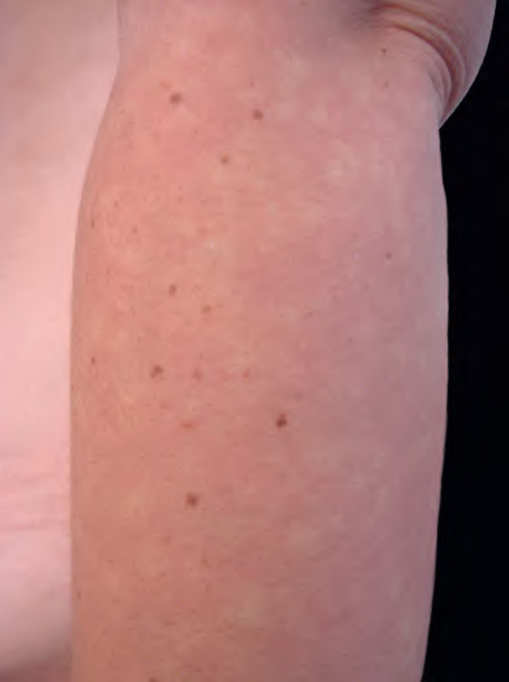
Fig. 106.17 血管痙攣性斑(Bier spots)。
圖 106-17:血管痙攣性斑(Bier spots)
供體部位的特性(供體優勢 donor dominance)。尚無組織學異常的報告。不需要治療,治療也無效。值得注意的是,貧血性痣可與血管畸形共存,並見於高達半數的第 1 型神經纖維瘤病(neurofibromatosis type 1)病人。