PATHOGENESIS
The pathogenetic mechanisms of the underlying diseases will be discussed in the respective chapters. The pathways involved in the de novo genesis of erythroderma or the generalization of pre-existing skin lesions are not well understood. The number of germinative keratinocytes as well as their mitotic rate is increased in erythrodermic skin, and the transit time of cells through the epidermis is shortened. Consequently, scales consist of material normally retained by the skin (nucleic acids, amino acids, soluble protein), and the daily loss of scales increases from 500–1000 mg to 20–30 g14. In acute erythroderma, the desquamated material usually has marginal metabolic significance, but in chronic erythroderma protein loss can be significant, leading to hypoalbuminemia and contributing to anemia of chronic disease. Given that the peak age of onset is during the 6th to 7th decade of life, it is possible that age-related immune senescence is a contributory factor in the development of idiopathic erythroderma. Likewise, the striking presentation of erythroderma in children with immunodeficiencies supports a role for immune dysregulation. Erythroderma in neonates often has a genetic basis and molecular diagnostics can play an important role in identifying causative mutations.
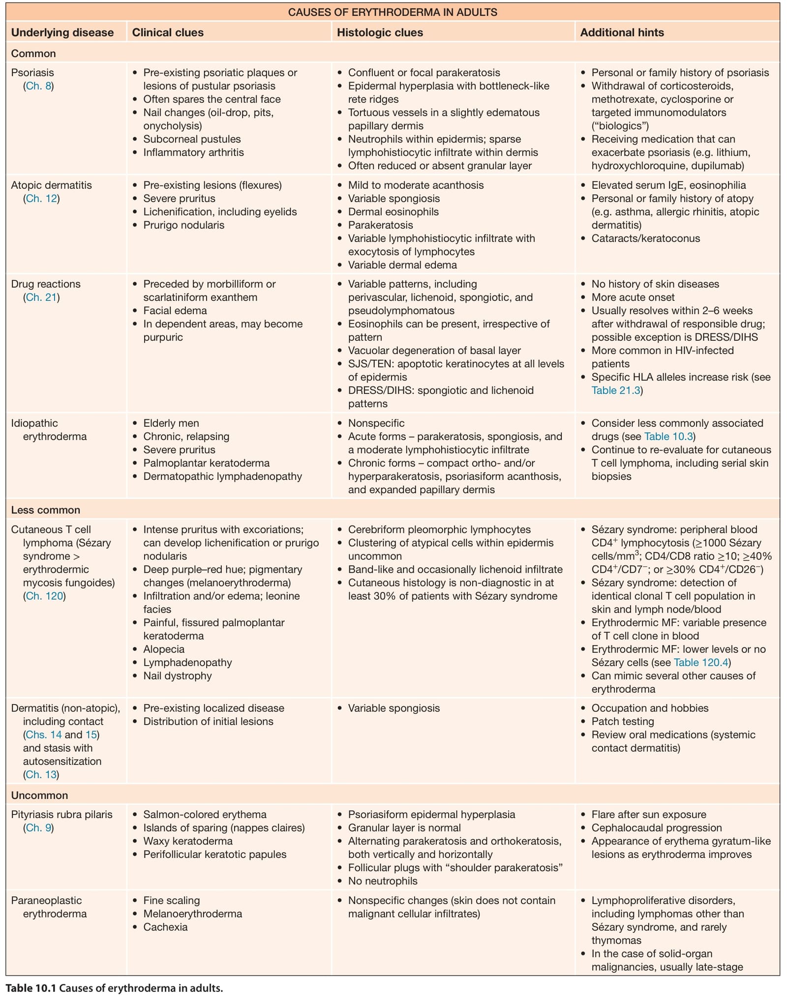
Table 10.1 Causes of erythroderma in adults.
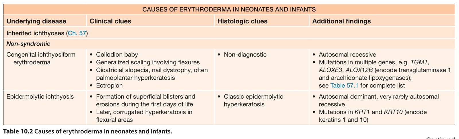
Table 10.2 Causes of erythroderma in neonates and infants.