CLINICAL FEATURES
Cutaneous Manifestations
Erythroderma is clinically defined by the presence of erythema and scaling involving more than 80% of the skin surface. Based upon its clinical course, erythroderma can be classified into primary or secondary types. In the primary form, the erythema (often initially on the trunk) extends within a few days or weeks to involve the entire skin surface, accompanied by the development of scaling (Fig. 10.1). The secondary form of erythroderma is defined as a generalization of a preceding localized skin disease – for example, psoriasis or atopic dermatitis. An acute onset most commonly occurs when the underlying cause is a drug reaction or dermatitis.
With the exception of very slowly progressing secondary erythroderma, erythema precedes the development of exfoliation by 2–6 days. The associated scaling varies extensively in size and color depending upon the stage of the erythroderma and nature of the underlying disease. During acute phases, scales are usually large and crusted, whereas in chronic states they tend to be smaller and drier. Occasionally, the cause of the erythroderma is suggested by the character of the scale – for example, fine in atopic dermatitis or dermatophytosis, bran-like in seborrheic dermatitis, crusted in pemphigus foliaceus, and exfoliative (i.e. peeling) in drug reactions. The color of the skin can also vary, from pink or red to reddish brown to deep red or purple.
Pruritus, the most frequent complaint, is observed in up to 90% of patients and it varies according to the underlying cause, but is most severe in patients with dermatitis or Sézary syndrome. Given the itch–scratch cycle, the skin may become thickened, and areas of lichenification are seen in one-third of cases. Patients, especially those with a
Obvious exfoliation of scale with underlying erythema.
chronic erythroderma, may develop dyspigmentation, with hyperpigmentation (40/90 patients, 45%) observed more frequently than hypoor depigmentation (18/90 patients, 20%) in one series. Palmoplantar keratoderma appears in ~30% of erythrodermic patients and is often an early sign in pityriasis rubra pilaris. Keratoderma with scale-crust can point to crusted scabies, whereas a painful and fissured keratoderma can occur in Sézary syndrome.
In cases of pre-existing dermatoses, nail changes may precede the erythroderma (e.g. pits in psoriasis or horizontal ridging in atopic dermatitis), whereas others develop subsequently. Nail changes are said to be present in ~40% of patients. Most often “shiny” nails are observed, but discoloration, brittleness, dullness, subungual hyperkeratosis, Beau’s lines, paronychia, and splinter hemorrhages can be seen. The nails may even be totally shed. Diffuse non-scarring alopecia of the scalp or other sites appears in 20% of patients with chronic erythroderma, including those with CTCL. Cicatricial alopecia can also develop in the setting of Sézary syndrome or erythrodermic MF.
Patients with erythroderma can develop multiple seborrheic keratoses, which may be pale in color compared with the background erythema. Colonization of the skin with Staphylococcus aureus is common and can lead to secondary cutaneous infections as well as bacteremia. Bilateral ectropion and purulent conjunctivitis can also develop as ocular complications (Fig. 10.2). Lastly, an exacerbation of erythroderma may follow UV irradiation or drug ingestion; this is not necessarily restricted to patients with photosensitive eczema or drug-induced erythroderma.
Systemic Manifestations
An awareness of potential systemic complications of erythroderma is essential. Pedal or pretibial edema is observed in ~50% of patients (182/380), and results from hypoalbuminemia and a shift of fluid into extracellular spaces. In patients with CTCL-associated or druginduced erythroderma, generalized anasarca can develop. Because of the markedly increased blood flow through the skin and increased fluid loss by transpiration, tachycardia affects 40% of patients, and there is a risk of high-output cardiac failure, particularly in the elderly. Furthermore, increased skin perfusion leads to thermoregulatory disturbances. Although hyperthermia is more frequently observed than hypothermia, most patients describe chilly, rigor-like sensations. The chronic
A There is diffuse involvement of the trunk but a clue to the diagnosis is the individual lesions on the arm. B Significant exfoliation of the lateral trunk and arms. A, Courtesy Luis Requena, MD; B, Courtesy Lorenzo Cerroni, MD.
and excessive loss of heat can lead to compensatory hypermetabolism with subsequent development of cachexia. Anemia, reflecting both iron deficiency and chronic disease, may also be observed in patients with chronic erythroderma.
The most common extracutaneous manifestation of erythroderma is peripheral lymphadenopathy, which is found in approximately half of the patients. Even in the absence of an underlying lymphoproliferative disorder, lymphadenopathy may be prominent and, if so, histologic and molecular examination of a lymph node is recommended (in particular, immunophenotypic studies and T cell receptor gene analysis). A core or excisional lymph node biopsy is preferable to a fine needle aspirate as the former provides sufficient tissue for accurate assessment of nodal architecture and cellular morphology. The major differential diagnosis is between lymphomatous involvement and reactive dermatopathic lymphadenopathy. Prior to histopathologic examination of an enlarged lymph node, a CT or PET-CT scan can identify the most morphologically abnormal and/or fluorodeoxyglucose (FDG)-avid node.
Hepatomegaly occurs in 20% of the cases (113/578), with a slight predominance in erythroderma due to drug-induced hypersensitivity. Splenomegaly is rarely seen and occurs most often in association with systemic lymphoma.
Specific Findings of the Underlying Disease
In addition to the general features previously discussed, the clinical presentation may have features suggestive of the underlying etiology (see Tables 10.1 and 10.2).
Psoriasis
Psoriasis is the most common underlying disorder in adults with erythroderma. Invariably, psoriatic erythroderma is preceded by typical psoriatic lesions (see Ch. 8). Its onset is most often due to withdrawal of potent topical or oral corticosteroids, cyclosporine or methotrexate; occasionally, widespread flares follow a systemic infection, phototoxicity, an irritant contact dermatitis to topical medications such as tar, or withdrawal of targeted immunomodulators (“biologics”). After generalization of the erythema, the typical features of psoriasis are often lost (Fig. 10.3), and generalized pustular psoriasis with disseminated sterile subcorneal pustules may develop (see Fig. 1.6). Due to a slower turnover rate, nail changes, such as oil-drop changes, onycholysis or nail pits, may still be visible and provide clues to the diagnosis of psoriatic erythroderma. Treatment of the erythroderma may result in the subsequent reappearance of characteristic psoriatic plaques.
Atopic dermatitis
Although occurring at any age, erythroderma develops most frequently in patients with a history of moderate to severe atopic dermatitis (Fig. 10.4). As a result, well-established pre-existing lesions can be found, especially when the erythroderma is of recent onset. The pruritus is intense, and secondary excoriations or prurigo-like lesions are frequently observed. Lichenification is often prominent and atrophy of the skin due to topical corticosteroids may be seen. Increased serum IgE and eosinophilia may accompany other signs and symptoms of atopy.
Drug reactions
The number of drugs implicated as causes of erythroderma is staggering (Table 10.3; see also Ch. 21). Whereas erythroderma resulting from topical medications usually begins as an irritant or allergic contact dermatitis, erythroderma due to systemic drugs often begins with the acute onset of a morbilliform or scarlatiniform exanthem. There are occasional exceptions such as the chronic eczematous eruptions associated with calcium channel blockers. In areas of greatest hydrostatic pressure (distal lower extremities), the eruption may become secondarily purpuric. While drug-induced erythrodermas have the shortest duration, usually resolving 2–6 weeks after withdrawal of the responsible drug, rarely erythroderma persists despite withdrawal of the putative drug and such cases may then be designated as idiopathic. Occasionally, there is a pseudolymphomatous presentation which can be associated with a transient peripheral blood lymphocytosis.
A minority of patients with DRESS (drug reaction with eosinophilia and systemic symptoms) syndrome, also referred to as drug-induced hypersensitivity syndrome (DIHS), become erythrodermic. In addition to fever, these patients can develop peripheral blood eosinophilia, hepatitis, and peripheral lymphadenopathy (see Table 21.9).
Idiopathic erythroderma
In ~25% of erythrodermic patients, no underlying disease is detectable. This group consists primarily of elderly men with a chronic pruritic erythroderma in association with dermatopathic lymphadenopathy and extensive palmoplantar keratoderma (Fig. 10.5); this constellation is often referred to as “red man syndrome” or “l’homme rouge” (not to be confused with the infusion reaction [“red man syndrome”] that accompanies rapid intra-venous infusions of vancomycin). When this group was compared with the entire group of erythrodermic patients, lymphadenopathy (68% vs 44%) and peripheral edema (54% vs 40%) were found to be more common than in other types of erythroderma, and hypothermia exceeded hyperthermia.
In one series of patients with idiopathic erythroderma, atopic dermatitis, drug-induced, and CTCL were the most common etiologies eventually identified. In a second series, in which detection of a clonal T cell population in the skin was a diagnostic criterion for CTCL, the latter was the most common underlying disease. However, it is important to note that in the absence of additional clinicopathologic features, the sole presence of a T cell clone in the skin is insufficient for the diagnosis of CTCL. Furthermore, peripheral blood T cell clones can be detected in elderly individuals and may reflect a diminished T cell receptor repertoire due to immune senescence rather than CTCL. Nonetheless, the detection of identical T cell clones in multiple biopsy specimens can serve as an important diagnostic clue for CTCL. Lastly, chronic actinic dermatitis should be considered in this group of patients.
A Widespread erythema with fine white scaling and excoriations. B Fine white scaling and obvious pruritus in addition to circumoral pallor. A, Courtesy Lorenzo Cerroni, MD; B, Courtesy Antonio Torrelo, MD.
Cutaneous T cell lymphoma (Sézary syndrome and
CTCL can mimic other causes of erythroderma and is frequently a significant diagnostic challenge. Erythroderma due to CTCL is subdivided into Sézary syndrome and erythrodermic MF (see Ch. 120). Sézary syndrome is defined by the triad of erythroderma, generalized lymphadenopathy, and peripheral blood lymphocytosis with morphologically abnormal Sézary cells; however, patients may have more limited skin involvement prior to the development of erythroderma. Additional clinical features include a painful and fissured keratoderma, diffuse alopecia, edema, and leonine facies. The skin is often infiltrated or hyperpigmented (melanoerythroderma) and severe pruritus is common (Fig. 10.6).
To distinguish between Sézary syndrome and erythrodermic MF, revised criteria were proposed by the International Society of Cutaneous Lymphomas and the Cutaneous Lymphoma Task Force of the EORTC for the diagnosis of Sézary syndrome, namely erythroderma and evidence of an identical T cell clone in the blood and skin plus one of the following: (1) ≥1000 Sézary cells/mm; (2) a CD4:CD8 ratio of ≥10:1; or (3) an increased percentage of CD4+ cells with an abnormal phenotype (≥40% CD4+/CD7− or ≥30% CD4+/CD26−) by peripheral blood flow cytometry (B in the TNMB classification; see Table 120.4). The latter cellular analysis provides a more objective measure of circulating tumor burden. Erythrodermic MF can also have an identical T cell clone in the skin and blood, but flow cytometry results are normal or demonstrate low-level blood involvement (B or B). In addition, diagnosis of erythrodermic MF can be challenging because the histopathologic findings are not diagnostic in at least one-third of patients.
Longitudinal evaluation, including repeated skin biopsies and PET-CT scans as well as lymph node biopsies and peripheral blood flow cytometry, is required to distinguish CTCL from idiopathic erythroderma. Identification of identical dominant T cell clones via more sensitive and specific high-throughput T cell receptor sequencing may
prove helpful. Rarely patients with other hematologic malignancies, in particular angioimmunoblastic T cell lymphoma, adult T cell leukemia/ lymphoma or T cell prolymphocytic leukemia, can present with erythroderma due to cutaneous infiltration of malignant T cells.
Pityriasis rubra pilaris
Erythrodermic pityriasis rubra pilaris (PRP) can be observed in children and adults (see Ch. 9). Usually, the lesions have a salmon to orange–red color. The degree of scaling varies, but it can be marked with large scales (Fig. 10.7). The combination of follicular keratotic papules on the knees, elbows and dorsal aspects of the fingers plus nappes claires (islands of uninvolved skin within the erythroderma) favors the diagnosis of PRP (Fig. 10.8). When the classic histologic features are present (see below), this is also helpful in distinguishing PRP from psoriasis. Occasionally, CTCL and the Wong type of dermatomyositis can have an appearance similar to PRP.
Papuloerythroderma of Ofuji
This represents a distinctive reaction pattern most often described in older men. Pruritic, monomorphic reddish brown papules often become confluent, with sparing of skin folds (“deck-chair” sign). In one review of
170 reported cases, a causative factor was not identified in the majority of patients. Of the remaining individuals, CTCL represented the most common underlying etiology and thus needs to be excluded (see Table 10.1 for additional etiologies).
Paraneoplastic erythroderma
Paraneoplastic erythroderma is most commonly associated with lymphoproliferative disorders, including systemic lymphomas and rarely thymomas. In patients with solid organ malignancies, erythroderma is rare and usually only appears during the late stages of the disease. Of note, in these patients the skin does not contain malignant cellular infiltrates. Fine scales and erythema can be accompanied by a brownish hue (i.e. melanoerythroderma). Additional signs of malignancy, such as cachexia or fatigue, may be seen.
Bullous dermatoses
Among the bullous dermatoses, pemphigus foliaceus is the one most likely, albeit rarely, to present as erythroderma; erythrodermic forms of paraneoplastic pemphigus and bullous pemphigoid have also been reported (see Table 10.1; see also Chs. 29 and 30). In pemphigus foliaceus, impetigo-like blisters and erosions are followed by collarettes of scale and scale-like crusts (Fig. 10.9). The erythroderma is usually preceded by localized lesions on the face and upper trunk.
Ichthyoses
Erythroderma due to one of the syndromic or non-syndromic inherited ichthyoses is usually present from birth or infancy (see Table 57.1). In neonates, congenital ichthyosiform erythroderma (CIE; previously referred to as non-bullous CIE), epidermolytic ichthyosis (previously referred to as bullous CIE), and Netherton syndrome should be considered (see Table 10.2). As a rule, CIE presents as a collodion baby (90% of the cases), and within a few days after birth, an erythroderma with fine white scaling appears. Epidermolytic ichthyosis initially presents as generalized erythema with superimposed superficial blisters and erosions. This disorder may be mistakenly diagnosed as staphylococcal scalded skin syndrome or as a form of epidermolysis bullosa. Later, children develop spiny, corrugated hyperkeratoses, particularly in flexural areas, and blisters and erosions become less prevalent. Netherton syndrome manifests as an ichthyosiform erythroderma in neonates. It is associated with trichorrhexis invaginata (“bamboo hair”), elevated serum levels of IgE, and an immune defect that can result in life-threatening infections, particularly within the first years of life. Later, either the ichthyosiform erythroderma persists or ichthyosis linearis circumflexa develops, which is characterized by garland-like scaly eruptions. Because there are very rare ichthyoses that can present with an ichthyosiform erythroderma as well as multiple genes associated with a specific ichthyosis (see Table 57.1), molecular diagnostics consisting of next generation sequencing of large gene panels or whole exome sequencing have become an important evaluation tool.
Staphylococcal scalded skin syndrome
Staphylococcal scalded skin syndrome (SSSS; see Ch. 74) is seen primarily in children (<5 years of age), and it is due to a circulating exfoliative toxin produced by S. aureus. Since desmoglein 1 is cleaved by exfoliative toxin A, pemphigus foliaceus and SSSS exhibit clinical similarities, with the latter often becoming erythrodermic. Common foci for the initial infection include the nares, nasopharynx, conjunctivae, and umbilicus. After a period of 1–2 days of a low-grade fever, large areas of tender erythema develop. The skin appears tense and subcorneal blisters appear, which subsequently desquamate. A very similar syndrome, the toxic shock syndrome, can be caused by staphylococcal or streptococcal exotoxins, but more commonly affects adults and acral exfoliation has a delayed appearance, usually 2 weeks later.
Omenn syndrome
Omenn syndrome represents an autosomal recessive form of severe combined immunodeficiency (SCID; see Table 60.16). It is characterized by failure to thrive, leukocytosis with prominent eosinophilia, hypogammaglobulinemia, and elevated serum IgE levels. Cutaneous findings, which appear in neonates, are generally the first sign of the syndrome and include an erythroderma with diffuse alopecia. Systemic manifestations include prominent lymphadenopathy, hepatosplenomegaly, and recurrent diarrhea. In the absence of hematopoietic stem cell or bone marrow transplantation, severe infections lead to early death. Additional forms of SCID can also cause exfoliative dermatitis in neonates (see Table 60.16).
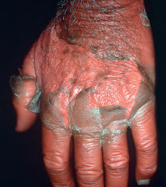
Fig. 10.1 Erythroderma with desquamation.
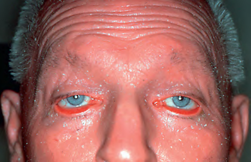
Fig. 10.2 Ectropion in the setting of erythroderma.
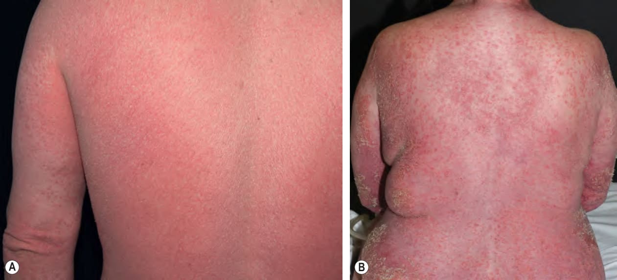
Fig. 10.3 Psoriatic erythroderma.
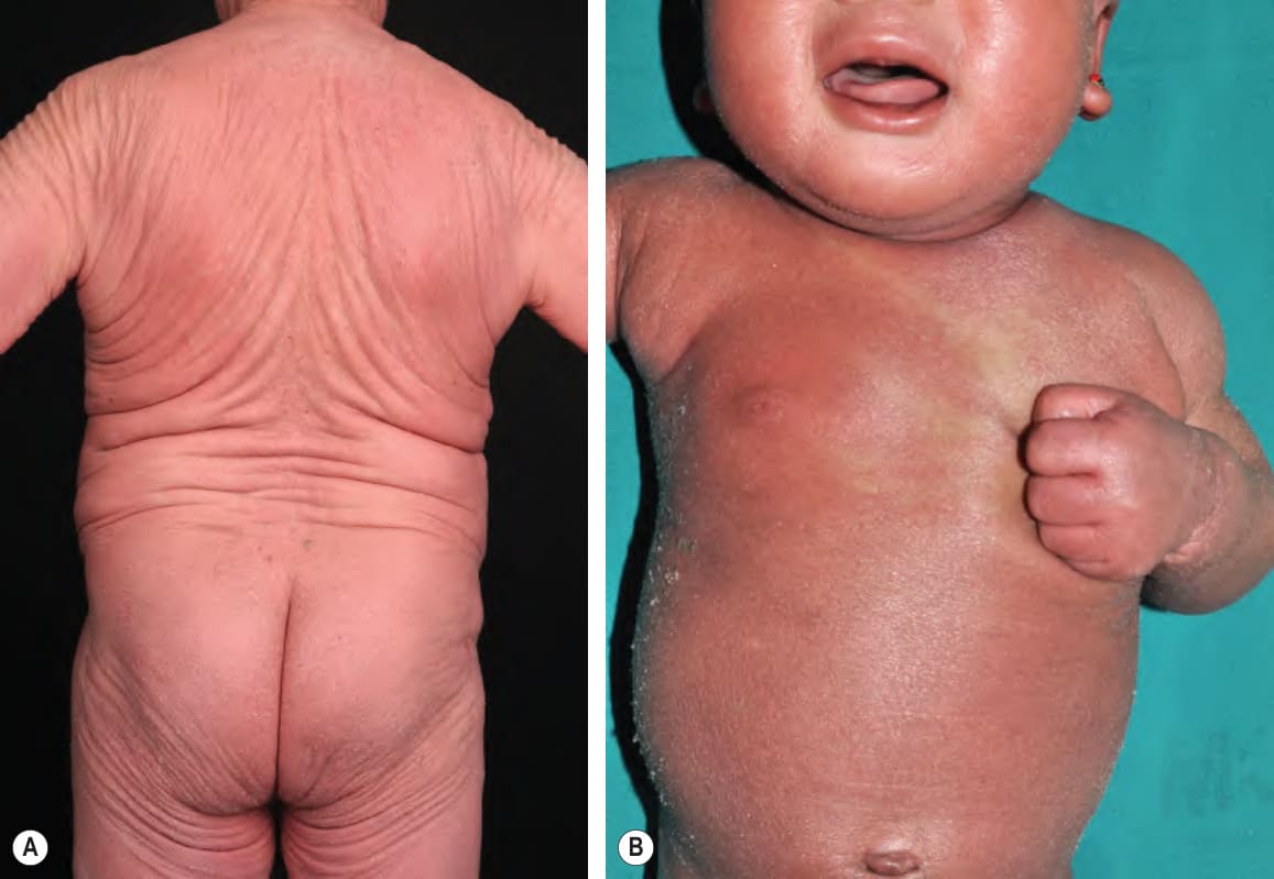
Fig. 10.4 Erythroderma due to atopic dermatitis.
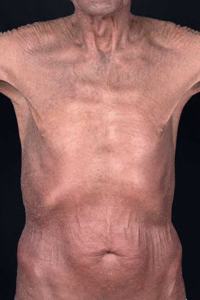
Fig. 10.5 Idiopathic erythroderma. This is the type of patient who requires longitudinal evaluation to exclude the development of cutaneous T cell lymphoma. Courtesy Lorenzo Cerroni, MD.
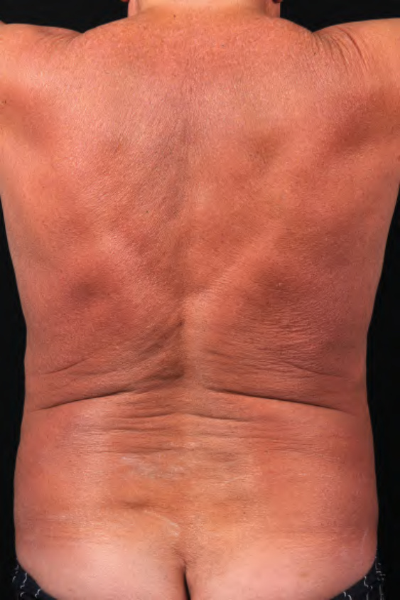
Fig. 10.6 Sézary syndrome. Diffuse erythroderma with a red–brown color. Note the evidence of scratching on the lower back. Courtesy Lorenzo Cerroni, MD.
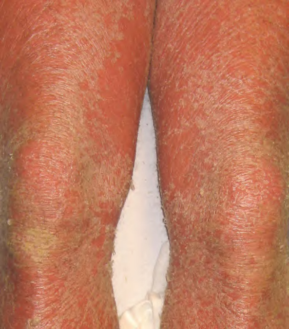
Fig. 10.7 Erythroderma due to pityriasis rubra pilaris. Large thin scales are seen as well as the characteristic orange–red color.
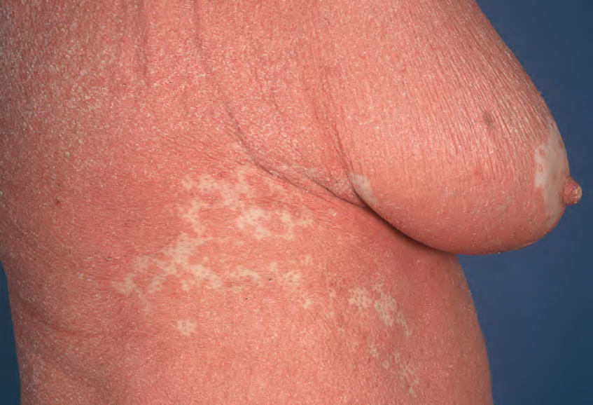
Fig. 10.8 Erythroderma secondary to pityriasis rubra pilaris. Islands of sparing are noted on the flank and breast. Note the salmon color.
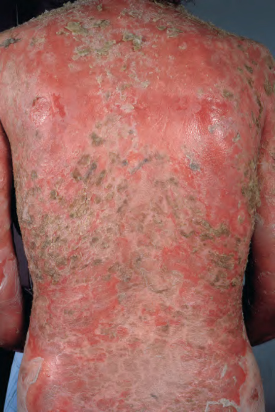
Fig. 10.9 Erythroderma due to pemphigus foliaceus. Generalized erythema with widespread scale-crusts and large areas of erosion.
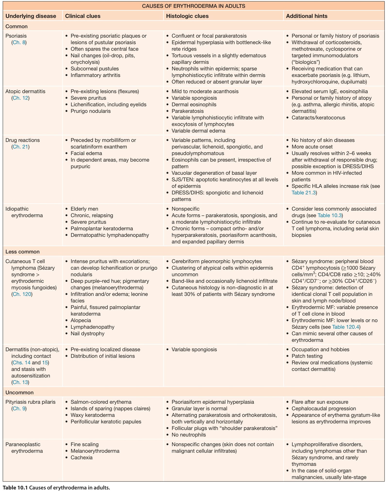
Table 10.1 Causes of erythroderma in adults.
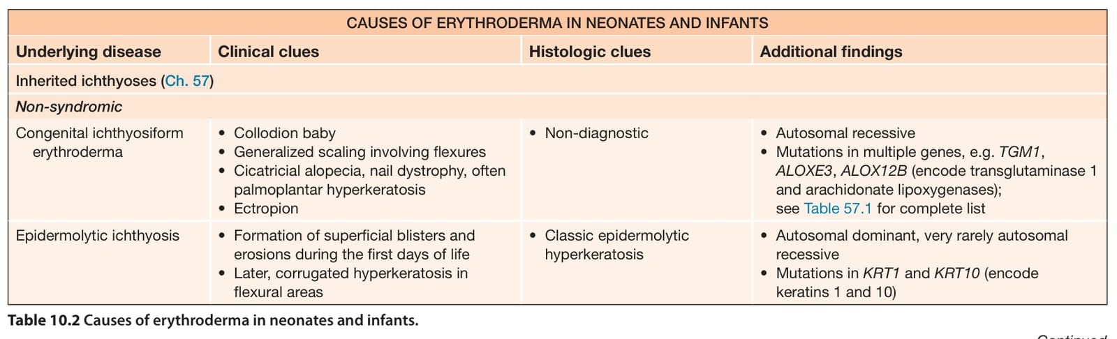
Table 10.2 Causes of erythroderma in neonates and infants.
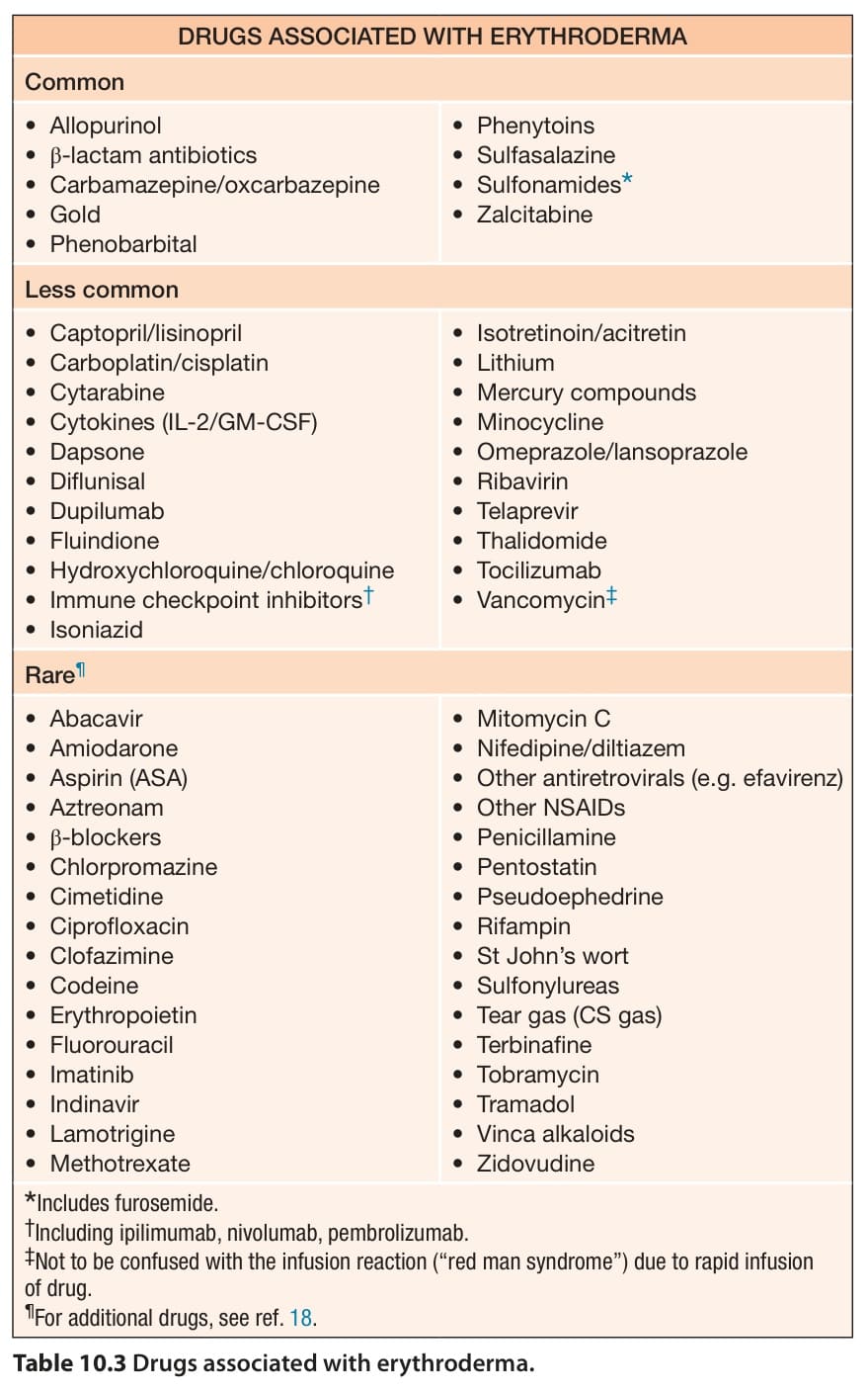
Table 10.3 Drugs associated with erythroderma.