DIFFERENTIAL DIAGNOSIS
Erythroderma is a clinical presentation for a variety of diseases, and identification of the underlying etiology represents one of the most complex challenges in dermatology (Figs. 10.10 and 10.11; see Tables 10.1 and 10.2). The evaluation starts with a complete medical history: up to 45% of the patients will have a prior history of a more localized skin disease, and ~20% of cases represent drug reactions. A comprehensive clinical examination may reveal additional clues to the under-lying disease. However, it is important to recognize that pre-morbid susceptibilities may modify the clinical morphology of an erythroderma, e.g. a drug reaction in an atopic individual may appear lichenified. Once the number of possible underlying diseases is reduced, further laboratory investigation can aid in establishing the final diagnosis. Hopefully in the future, specific diagnostic biomarkers will emerge that can assist in establishing a precise etiology. For erythroderma in neonates and young children, molecular diagnostics represent a critical laboratory investigation (see above).
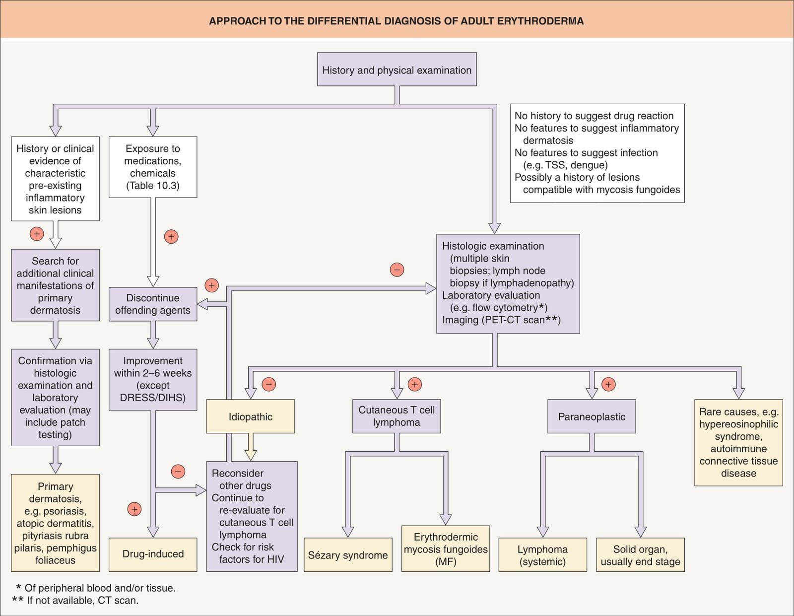
Fig. 10.10 Approach to the differential diagnosis of adult erythroderma. CT, computed tomography; DRESS/DIHS, drug reaction with eosinophilia and systemic symptoms/drug-induced hypersensitivity syndrome; PET, positron emission tomography; TSS, toxic shock syndrome.
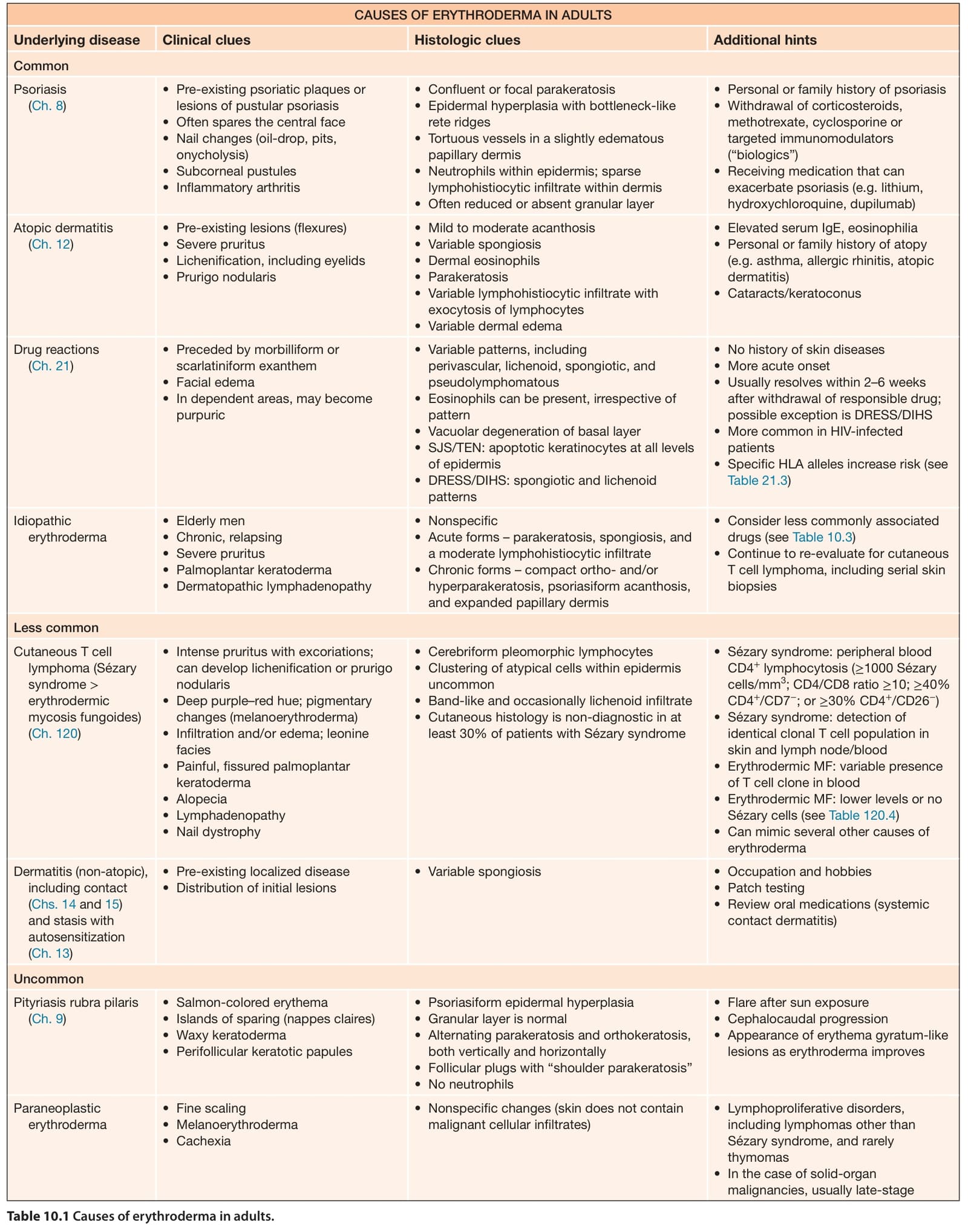
Table 10.1 Causes of erythroderma in adults.