Epidermoid Cyst
Synonyms: Epidermal inclusion cyst Infundibular cyst Epidermal cyst
Epidermoid cysts are the most common cutaneous cysts. They can occur anywhere on the skin, but are most common on the face and upper trunk. These lesions are well-demarcated, skin-colored to yellowish dermal nodules, and they may have a clinically visible punctum representing the follicle from which the cyst is derived (Fig. 110.2). Epidermoid cysts range from a few millimeters to several centimeters in diameter. Tiny superficial epidermoid cysts are known as milia (see below). These cysts derive from the follicular infundibulum, hence
Cysts 110
Mary Seabury Stone
the synonym infundibular cysts (Fig. 110.3). They may be primary, or they may arise from disrupted follicular structures or traumatically implanted epithelium (hence the synonym epidermal “inclusion” cyst).
As follicular disruption is important in the pathogenesis of many epidermoid cysts, multiple epidermoid cysts may occur in individuals with a history of significant acne vulgaris. Multiple cysts may also occur in the setting of Gardner syndrome (familial adenomatous polyposis) and in basal cell nevus syndrome. Patients receiving cyclosporine to prevent solid organ transplant rejection or BRAF inhibitors, both non-selective (e.g. sorafenib) and selective (e.g. vemurafenib), can develop multiple epidermoid cysts or milia, often on the face. Multiple scrotal cysts (Fig. 110.4) may lead to scrotal calcinosis via dystrophic calcification.
Non-inflamed epidermoid cysts are usually asymptomatic, but with pressure, cyst contents may be expressed that may have an objectionable odor. Rupture of the cyst wall can result in an intensely painful inflammatory reaction, and this is a common reason for presentation to a physician (Fig. 110.5). Development of a basal cell carcinoma (BCC) or squamous cell carcinoma (SCC) within an epidermoid cyst is a very rare event.
Pathology
Histologic examination shows a cystic cavity filled with laminated keratin lined by a stratified squamous epithelium that includes a granular layer (Fig. 110.6). A surrounding inflammatory response with both acute suppurative and chronic granulomatous inflammation as well as variable fibrosis may be seen as evidence of prior rupture. In individuals with Gardner syndrome, some cysts have, as a characteristic feature, columns of pilomatricoma-like shadow cells projecting into the cyst cavity.
Treatment
If treatment is desired, excision is curative. Removal may be accomplished by simple excision, or incision and expression of the cyst contents and wall through the surgical defect. If the entire cyst wall is not removed, the cyst may recur. Excision is best performed when the cyst is non-inflamed (“cold”). Inflamed epidermoid cysts may require incision and drainage, and occasionally, antibiotic therapy. Intralesional injections of triamcinolone may speed resolution of the inflammation.
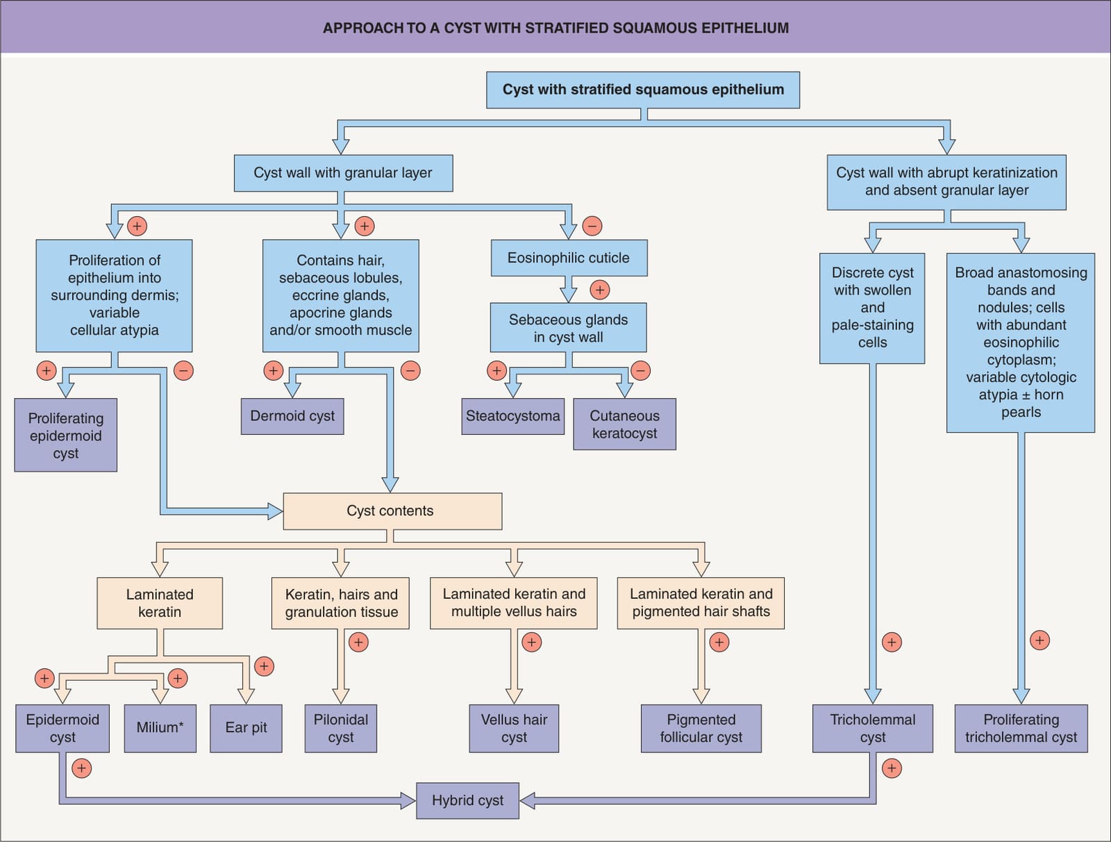
*Fig. 110.1 Approach to a cyst with stratified squamous epithelium. Diameter 1–2 mm.
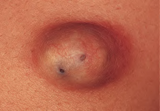
Fig. 110.2 Epidermoid cyst. Typical clinical appearance of an epidermoid cyst with a yellowish hue. Two pores are present in this example.
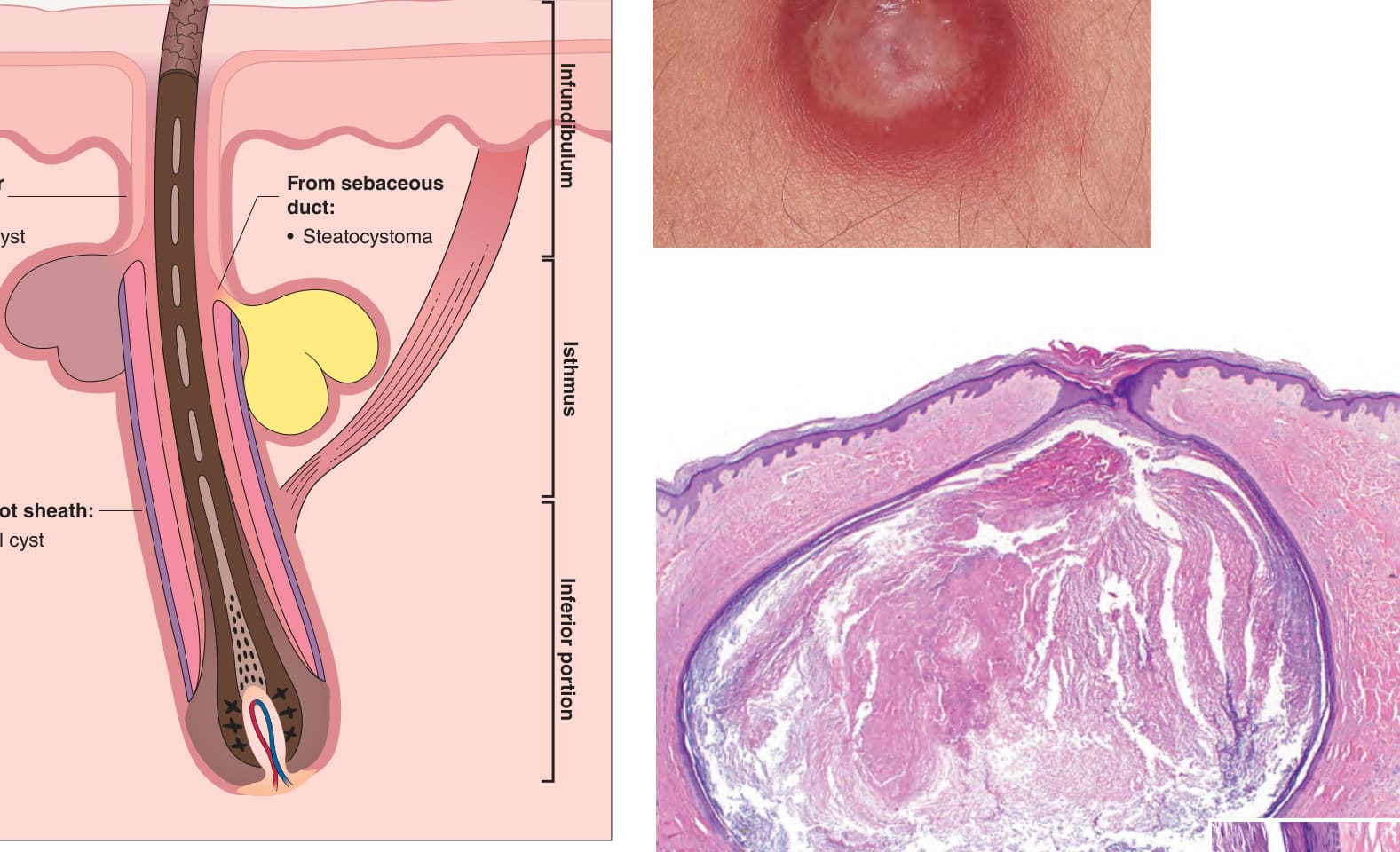
Fig. 110.3 Primary cysts of follicular origin. Anatomic origin of cysts derived from the pilosebaceous unit. Adapted from Requena L, Sanchez Yus E. Follicular hybrid cysts. An expanded spectrum. Am J Dermatopathol 1991;13:228–33.
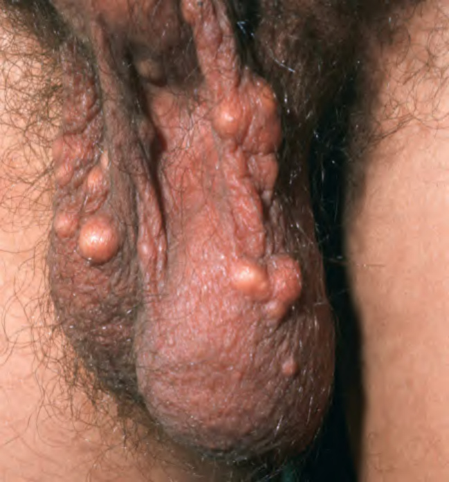
Fig. 110.4 Multiple epidermoid cysts of the scrotum.Courtesy Lorenzo Cerroni, MD.
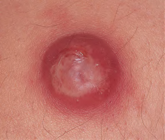
Fig. 110.5 Inflamed epidermoid cyst. Such painful inflammatory reactions to cyst rupture are a frequent cause for presentation to a physician.
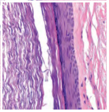
Fig. 110.6 Histopathology of an epidermoid cyst. Laminated keratin within an epithelial-lined cyst in the dermis. The cyst wall shows infundibular keratinization including a granular layer (inset).
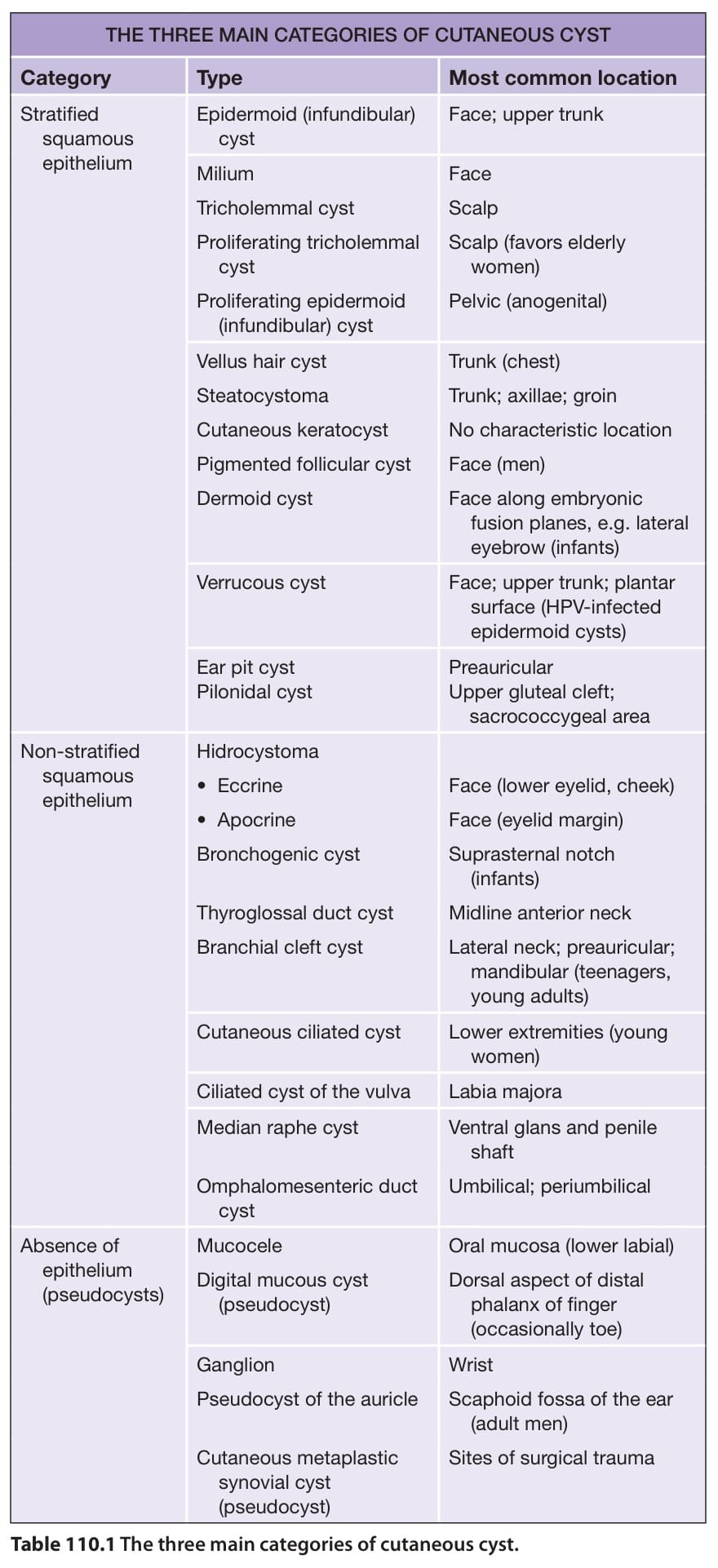
Table 110.1 The three main categories of cutaneous cyst.