Milium
Milia are small superficial cysts and are quite common, occurring in individuals of all ages. They may originate from the infundibulum of hair follicles or from eccrine ducts, and in the mouth from minor salivary gland ducts or from epithelium entrapped within embryologic fusion planes. Milia present as 1–2 mm, firm, white to yellow, subepidermal papules (Fig. 110.7).
Between 40% and 50% of infants will have milia, most commonly on the face. Most milia in newborns will resolve spontaneously during the first 4 weeks of life. Milia in newborns may also occur on the hard palate (Epstein pearls) or on the alveolar ridges (Bohn nodules). These also resolve spontaneously. Milia may occur as a primary phenomenon, especially on the face, or as secondary phenomena following blistering processes (e.g. porphyria cutanea tarda, epidermolysis bullosa acquisita) or superficial ulceration from trauma or cosmetic procedures. Milia
may also occur in areas of topical corticosteroid-induced atrophy, in follicular mycosis fungoides, and as a side effect of BRAF inhibitors (see above).
Milia en plaque is characterized by multiple milia within an erythematous edematous plaque, and the most common location is the postauricular area. Numerous milia in an infant may be a sign of oral–facial–digital syndrome type 1, an X-linked disorder that is lethal in males, in which milia are associated with facial and skull malformations, cleft lip and palate, a lobulated tongue, developmental delay, and polycystic kidneys; the associated alopecia follows the lines of Blaschko on the scalp. Milia are also seen in the setting of a number of other syndromes, including the BCC-associated syndromes basal cell nevus syndrome, Rombo syndrome, and Bazex–Dupré–Christol syndrome, as well as atrichia with papular lesions, Loeys–Dietz syndrome, basaloid follicular hamartoma syndrome, Basan syndrome, hypohidrotic ectodermal dysplasia, and a subset of Brooke–Spiegler syndrome (Rasmussen syndrome).
Pathology
Histologic features are those of a small epidermoid cyst with a stratified squamous epithelial lining that includes a granular layer. The cyst contents consist of laminated keratin.
Treatment
Milia may be removed by incising the epidermis over the milium with a needle, scalpel, or lancet and expressing the milium. The latter can be aided by the use of a comedo extractor. Laser ablation and electrodesiccation are also reported options. For multiple facial milia, topical retinoid therapy may be helpful in reducing the number of new milia and aiding in the ease of removal.
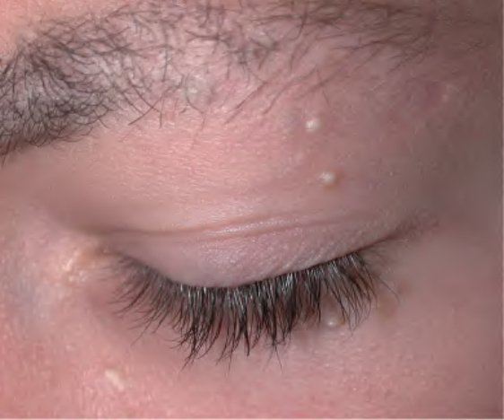
Fig. 110.7 Milia. Tiny (1–2 mm), white, dome-shaped papules on the face.