Tricholemmal Cyst
Synonyms: Trichilemmal cyst Pilar cyst Wen Isthmus–catagen cyst
Tricholemmal cysts are clinically indistinguishable from epidermoid cysts, but they are fourfold to fivefold less common. Ninety percent of tricholemmal cysts are located on the scalp (Fig. 110.8A). They may be solitary, but frequently are multiple, and they may be inherited as an autosomal dominant trait due to a germline variant of PLCD1, which encodes phospholipase C delta 1a. Interestingly, the somatic mutations in the cysts were found to be on the same chromosome as the germline variant5a.
Pathology
The walls of tricholemmal cysts show keratinization analogous to that of the outer root sheath of the hair follicle at the isthmus and the sac surrounding catagen and telogen hairs (hence the synonym isthmus– catagen cyst). Tricholemmal cysts are lined by stratified squamous epithelial cells that become swollen and pale close to the cystic cavity and show abrupt keratinization without an intervening granular layer (Fig. 110.9). The cyst contents consist of homogeneous, eosinophilic, compact material that often contains foci of calcification. A suppurative
and/or granulomatous response may surround the cyst if prior wall rupture has occurred. Occasionally, if the outermost epithelial portion of the cyst wall separates from the remainder of the cyst, the histologic features can resemble a hidrocystoma.
Differential diagnosis
In addition to epidermoid cysts, ruptured pilar cysts of the scalp can overlap with alopecic and aseptic nodules of the scalp.
Treatment
Treatment is by excision. Tricholemmal cysts typically “deliver” themselves through an incision without rupture more easily than do epidermoid cysts, and, therefore, the distinction between a tricholemmal cyst and an epidermoid cyst can often be correctly made at the time of excision.
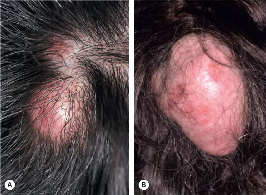
Fig. 110.8 Tricholemmal cyst versus proliferating tricholemmal cyst.A A tricholemmal cyst is a well circumscribed, mobile, mid dermal nodule with a smooth surface. B A proliferating tricholemmal cyst presenting as a large skin-colored to light-pink nodule on the scalp. Both types of cyst favor the scalp. B, Courtesy Luis Requena, MD.
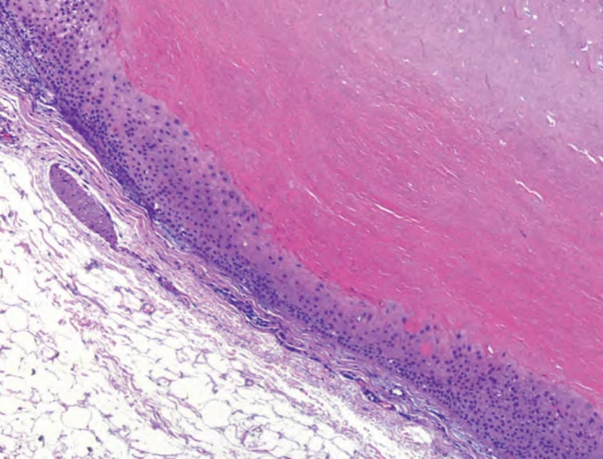
Fig. 110.9 Histopathology of a tricholemmal cyst. The cyst wall shows swollen keratinocytes with abrupt keratinization without formation of a granular layer. Compact, eosinophilic keratin fills the cyst. Courtesy Luis Requena, MD.