Proliferating Tricholemmal Cyst
Synonyms: Proliferating trichilemmal cyst Proliferating tricholemmal tumor Proliferating pilar tumor Proliferating follicular cystic neoplasm
A proliferating tricholemmal cyst classically occurs as a slow-growing nodule on the scalp (see Fig. 110.8B). Ninety percent occur on the scalp and ~85% occur in women, with a median age of 63 years. Proliferating tricholemmal cysts vary in size from a few millimeters to up to 25 cm in diameter. These tumors generally behave in a benign fashion, although on occasion, aggressive local growth with recurrences and metastases has been observed. Extremely rarely, spindle cell carcinomas may develop within a proliferating tricholemmal cyst. Distant metastases are rare.
Pathology
The histologic features of a proliferating tricholemmal cyst consist of broad anastomosing bands and nodules of squamous epithelium (Fig. 110.10). The epithelium consists of a proliferation of cells with abundant eosinophilic cytoplasm; the cells undergo abrupt keratinization and form dense homogeneous keratin that fills cystic spaces. There may be areas of epidermoid keratinization with formation of horn pearls as well as areas of foreign body giant cell reaction. One-quarter of cases show an epidermal connection. Cytologic atypia can vary markedly. Most tumors show well-circumscribed, pushing borders surrounded by compressed collagen. The lack of infiltrative growth into the surrounding stroma and abrupt tricholemmal keratinization helps to differentiate these cysts from SCC. Areas of marked atypia and infiltrative borders are features associated with a potential for aggressive behavior.
Treatment
Treatment is by complete surgical excision.
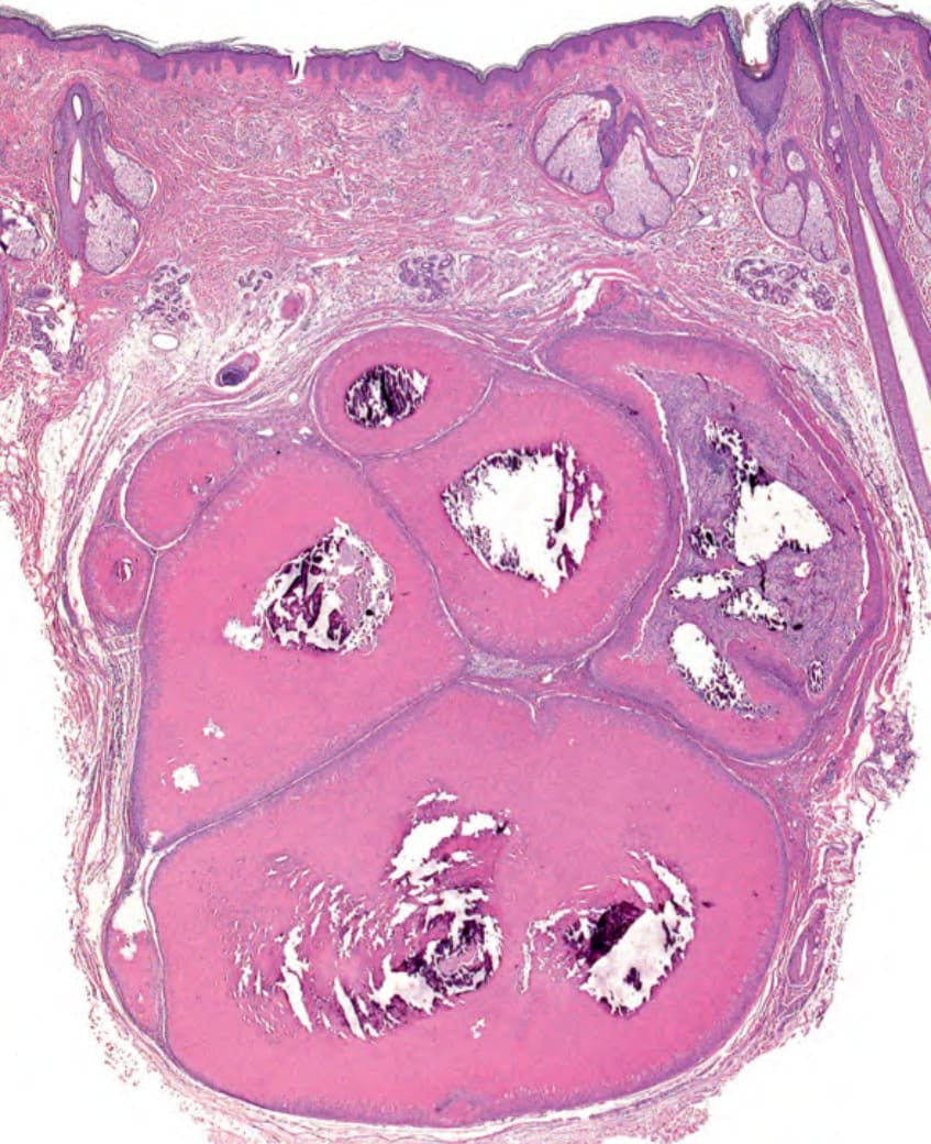
Fig. 110.10 Histopathology of a proliferating tricholemmal cyst.