Vellus Hair Cysts
Vellus hair cysts, described by Esterly, Fretzin, and Pinkus in 1977, most commonly present as numerous, small, dome-shaped papules, ranging from skin-colored to darkly pigmented (Fig. 110.11). These cysts are most commonly located on the trunk, and they may be inherited in an autosomal dominant pattern. Multiple lesions are known as “eruptive” vellus hair cysts; occasionally, solitary cysts are seen. Although some lesions may resolve via transepidermal elimination of cyst products, most lesions persist indefinitely. Vellus hair cysts may become inflamed, but in general, they are asymptomatic except for cosmetic concerns.
Eruptive vellus hair cysts may be seen in conjunction with steatocystoma multiplex, and both types of cysts have been reported in the setting of pachyonychia congenita, most commonly PC-17 (see Ch. 58).
Pathology
Histologically, one sees a small cystic structure lined by stratified squamous epithelium with epidermoid (infundibular) keratinization. The cysts contain loose laminated keratin and numerous vellus hair shafts (Fig. 110.12). A follicle may be found entering the lower portion of the cyst.
Visualization of the vellus hair shafts via microscopic examination of expressed cyst contents allows for a bedside diagnosis.
Treatment
Vellus hair cysts may be treated by a number of modalities, including incision and drainage, needle evacuation, puncture followed by forcepsassisted extraction, topical retinoic or lactic acid, and laser ablation.
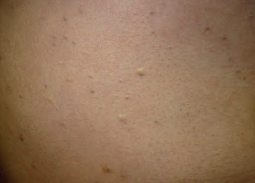
Fig. 110.11 Eruptive vellus hair cysts. Admixture of pigmented, skin-colored, and yellow–white papules of varying sizes on the trunk.
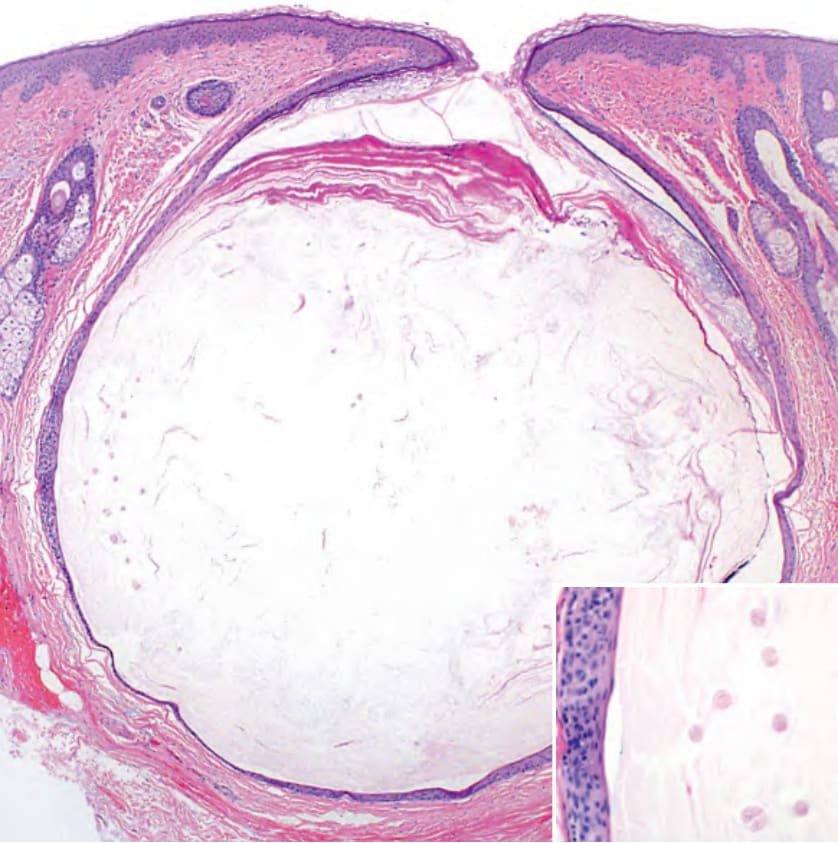
Fig. 110.12 Histopathology of a vellus hair cyst. A cyst shows epidermoid keratinization of its wall as well as laminated keratin and several vellus hair shafts within its cavity (inset).