Ear Pit
Synonyms: Preauricular cyst Congenital auricular fistula Preauricular fistula
Ear pits are congenital defects, and they may present as a true cystic nodule or an invagination in the preauricular area (Fig. 110.14). During development, the ear is formed by the fusion of six tubercles: three each from the first two branchial arches. Preauricular cysts reflect defective embryologic fusion with epithelial entrapment. These defects are relatively common, occurring in ~0.5%–1% of the normal population, and they may be transmitted in an autosomal dominant fashion. Ear pits are usually unilateral and right-sided. Infection with tenderness and purulent drainage may prompt presentation to a physician.
Although ear pits are usually not associated with other significant abnormalities, they can occur in conjunction with deafness or with deafness plus renal anomalies in the branchio-otic syndrome and branchio-oto-renal syndrome, respectively. Ear pits are also seen in a number of other congenital syndromes, including Treacher Collins, Goldenhar, and cat-eye syndromes.
Pathology
Histologically, preauricular pits or cysts are lined by stratified squamous epithelium with a granular layer.
Treatment
If removal is desired, simple excision is curative. Although most ear pits are incidental findings, in a newborn, a physical examination to exclude one of the associated syndromes and an evaluation for hearing loss is indicated. Infected lesions may require antibiotic therapy.
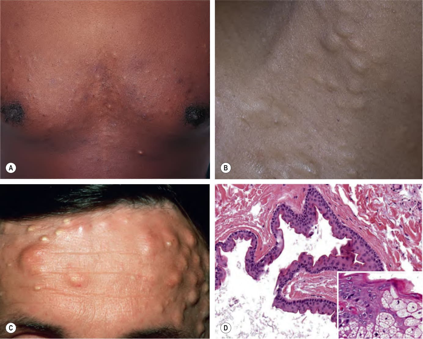
Fig. 110.13 Steatocystoma multiplex.A, B Numerous cystic papules on the trunk and multiple cystic nodules on the neck. C Larger cystic nodules on the forehead. D Histopathology of steatocystoma. The cyst wall is characterized by an undulated, thin stratified epithelium lacking a stratum granulosum and lined by an eosinophilic cuticle. A search may be required to identify small sebaceous lobules in or immediately adjacent to the cyst wall (inset). C, Courtesy Luis Requena, MD; D, Courtesy Lorenzo Cerroni, MD.
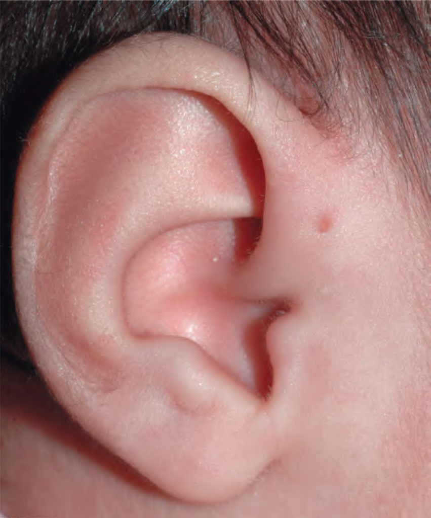
Fig. 110.14 Ear pit.Courtesy Julie V. Schaffer, MD.