CYSTS LINED WITH NON-STRATIFIED SQUAMOUS EPITHELIUM
Hidrocystoma
Synonyms: Cystadenoma Sudoriferous cyst Moll gland cyst
Hidrocystomas typically present as translucent, skin-colored to bluish cysts on the face, although they may occur in other sites. Hidrocystomas are traditionally divided into apocrine and eccrine hidrocystomas based upon histologic features, and as solitary (Smith type) or multiple (Robinson type). Hidrocystomas may be associated with specific syndromes of ectodermal dysplasia, including Schöpf–Schulz–Passarge syndrome.
Apocrine hidrocystomas are usually solitary (Fig. 110.15), whereas eccrine hidrocystomas may be solitary or multiple and are occasionally quite numerous (Fig. 110.16). Eccrine hidrocystomas can enlarge with heat exposure or during the summer and regress with cooler temperatures. In general, eccrine hidrocystomas are thought to develop from cystic dilation of eccrine ducts due to retention of eccrine secretions, while apocrine hidrocystomas are thought to represent adenomas of apocrine gland coils. Apocrine hidrocystomas are sometimes referred to as cystadenomas, although it has been recommended that this term be reserved for lesions with true papillomatous projections histologically. Lesions along the lower eyelid margin are also known as Moll gland cysts.
Pathology
Histologically, apocrine hidrocystomas are unilocular to multi-locular dermal cysts lined by two layers of epithelial cells that show luminal bulbous protrusions (“snouting”) and decapitation secretion (Fig. 110.17). Similarly lined papillary projections may extend into the cyst lumen. Histologic features of eccrine hidrocystomas are those of a uniloculate cyst containing clear fluid, lined by two layers of cuboidal to flattened epithelium (Fig. 110.18). Hidrocystomas that appear by light microscopy to be eccrine include a subset of lesions that stain positively for milk fat globulin 1, as in lesions of apocrine origin.
Treatment
Hidrocystomas may be removed by simple excision, including via Gradle scissors, or by electrodesiccation. Multiple eccrine hidrocystomas
may also be treated with daily application of topical 1% atropine in aqueous solution, although lesions reappear within days of discontinuing therapy. Flattening after botulinum toxin A injections has been reported.
Bronchogenic Cyst
Cutaneous bronchogenic cysts are most commonly found in the suprasternal notch, and rarely they appear on the anterior neck or chin (see
Fig. 64.2). A fistulous tract may connect to the epidermis. Rarely, they present as a pedunculated growth. Bronchogenic cysts are solitary, and they are typically noted at birth. Malignant transformation is very rare. They represent respiratory epithelium sequestered during embryologic development of the tracheobronchial tree.
Pathology
Bronchogenic cysts are lined by pseudostratified, ciliated, columnar epithelium with interspersed goblet cells. The cyst wall often contains smooth muscle and mucous glands and rarely cartilage.
Treatment
Treatment is by excision.
Thyroglossal Duct Cyst
Thyroglossal duct cysts present as deep midline cystic nodules on the anterior neck in children or young adults (see Fig. 64.2). During development, the thyroid gland descends from the floor of the pharynx to the anterior neck. The tract it forms is known as the thyroglossal duct. Thyroglossal duct cysts arise from remnants of the thyroglossal duct. A tract connecting these cysts to the hyoid bone is frequently present,
resulting in characteristic movement of the cyst with swallowing. Rarely, thyroid carcinoma may originate in a thyroglossal duct cyst.
Pathology
Histologically, thyroglossal duct cysts may be lined with cuboidal, columnar, or stratified squamous epithelium, and it may contain some ciliated columnar cells. The characteristic histologic feature is the presence of thyroid follicles, characterized by low cuboidal cells surrounding homogeneous pink material, in the cyst wall (Fig. 110.19).
Treatment
Treatment is surgical, with excision of the cyst and any residual tract.
Branchial Cleft Cyst
Synonyms: Lymphoepithelial cyst Lateral cervical cyst
Branchial cleft cysts occur in the preauricular area, mandibular region, or along the anterior border of the sternocleidomastoid muscle (see Fig. 64.2). The origin of these cysts is controversial. There are two major theories regarding their origin:
●they arise from branchial cleft remnants
●they represent cystic alteration of embryologic epithelium or tonsillar epithelium within cervical lymph nodes. Branchial cleft cysts most commonly manifest during the second or third decade of life. Infection of these cysts is a frequent cause of presentation to a physician.
Pathology
Histologically, these cysts are lined by stratified squamous epithelium or by pseudostratified ciliated columnar epithelium, and they are surrounded by lymphoid tissue.
Treatment
Treatment is by excision of the cyst and its associated tract after delineation of the extent of the lesion by CT or MRI.
Cutaneous Ciliated Cyst and Ciliated Cyst of the Vulva
Synonyms: Cutaneous müllerian cyst Cutaneous ciliated cystadenoma Paramesonephric mucinous cyst of the vulva
Cutaneous ciliated cysts are uncommon cysts that typically occur on the lower extremities of young women, although a few cases have been reported in men. They are usually a few centimeters in diameter and,
on rupture, drain clear to amber fluid. The histogenesis of these cysts is controversial. Most authors have suggested a müllerian duct origin, hence the term cutaneous müllerian cyst. However, the few occurrences in men and the rare reports of ciliated cysts on the scalp have led to the alternative hypothesis that some cases represent ciliated metaplasia of eccrine glands.
Ciliated cysts of the vulva are müllerian heterotopias, and they are located most commonly on the labia majora. They usually measure between 1 and 3 cm in diameter.
Pathology
Cutaneous ciliated cysts may be unilocular or multiloculated. The cyst wall is composed of simple cuboidal to columnar ciliated epithelium that may have papillary projections into the cyst lumen (Fig. 110.20).
Differential diagnosis
Bartholin gland cysts and abscesses occur on either side of the vaginal opening and are more common than ciliated cysts of the vulva.
Treatment
Excision is curative.
Median Raphe Cyst
Median raphe cysts are solitary and usually only a few millimeters in diameter, although they may extend over several centimeters linearly. They occur in young men on the ventral aspect of the penis, most commonly on or near the glans. These cysts are thought to develop from aberrant urethral epithelium, but do not connect to the urethra.
Pathology
Histologically, median raphe cysts are lined by stratified columnar epithelium (one to four cell layers thick) without a connection to the overlying epithelium. Occasionally, mucin-containing cells are seen in the lining. Very rarely, a ciliated lining has been observed. Positive immunohistochemical staining of the cyst wall epithelium for cytokeratin 7 and GATA3 supports the urothelial origin of these cysts30a.
Treatment
Excision is curative.
Omphalomesenteric Duct Cyst
Synonyms: Vitelline cyst Omphalomesenteric duct remnant
Omphalomesenteric duct cysts represent a developmental defect in the closure of the omphalomesenteric duct. The omphalomesenteric duct is the fetal connection between the midgut and the yolk sac. It is usually
obliterated and loses its intestinal attachment by 6 weeks of gestation. Remnants of this duct may occur anywhere along its course between the intestines and the umbilicus. The spectrum of defects resulting from this faulty closure includes Meckel diverticulum, umbilical–enteric fistulae, umbilical sinuses, and omphalomesenteric duct cysts (internal or external); the latter can present as an umbilical polyp (Fig. 110.21A).
Pathology
These lesions are characterized histologically by ectopic gastrointestinal mucosa (Fig. 110.21B) and must be distinguished from umbilical metastases of gastrointestinal adenocarcinomas.
Differential diagnosis
In postmenarcheal female patients, the possibility of cutaneous endometriosis should be considered.
Treatment
Management of omphalomesenteric duct cysts includes appropriate radiologic studies to exclude communication to the gastrointestinal tract prior to surgical excision.
Urachal Cyst
The urachus is a tube connecting the fetal bladder to the umbilicus and it normally closes during development, resulting in a fibrous tract. A persistent urachus presents during infancy with urine leakage from the umbilicus. A urachal cyst results from an incomplete urachal duct remnant. Urachal cysts are rare, and they usually present as a painful umbilical mass due to secondary infection. The diagnosis can be established by ultrasound in the majority of patients.
Pathology
These cysts are characterized by a urothelial lining of cuboidal or columnar cells.
Treatment
Excision is recommended to prevent secondary infections as well as late development of adenocarcinoma.
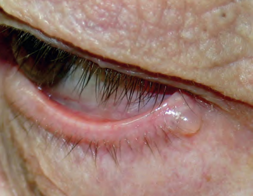
Fig. 110.15 Apocrine hidrocystoma. A single, clear fluid-filled cyst on the lateral lower eyelid margin.
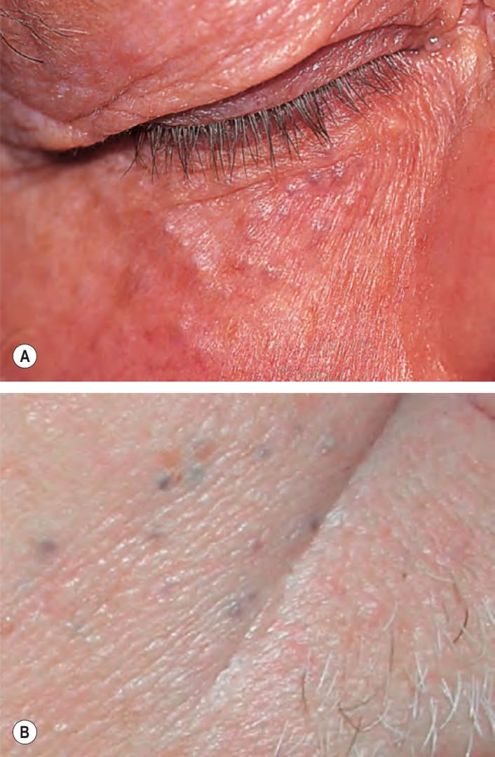
Fig. 110.16 Eccrine hidrocystomas. Numerous, tiny, translucent or bluish papules on the lower eyelid (A) or the cheek (B).
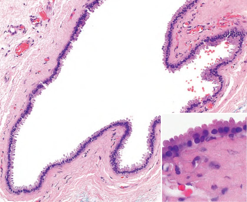
Fig. 110.17 Histopathology of an apocrine hidrocystoma. A unilocular cyst whose wall shows typical apocrine decapitation secretion (inset).
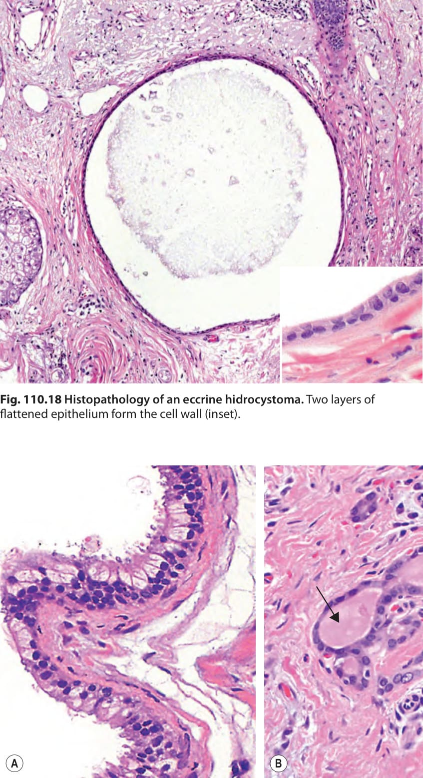
Fig. 110.18 Histopathology of an eccrine hidrocystoma. Two layers of flattened epithelium form the cell wall (inset).
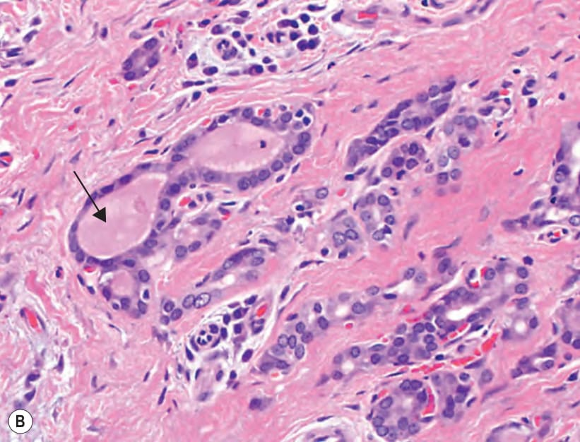
Fig. 110.19 Histopathology of a thyroglossal duct cyst.A The cyst is lined by ciliated columnar epithelium; some of the cells contain clear cytoplasm. B Thyroid follicles are present in the tissue surrounding the cyst; note the homogeneous colloid within the lumina (arrow).
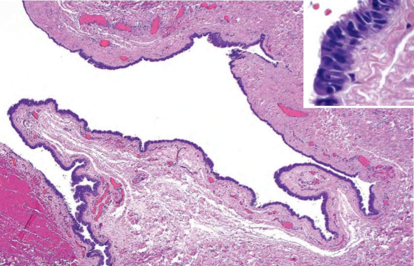
Fig. 110.20 Histopathology of a ciliated cyst of the vulva. The cyst wall is composed of columnar ciliated epithelium (inset).
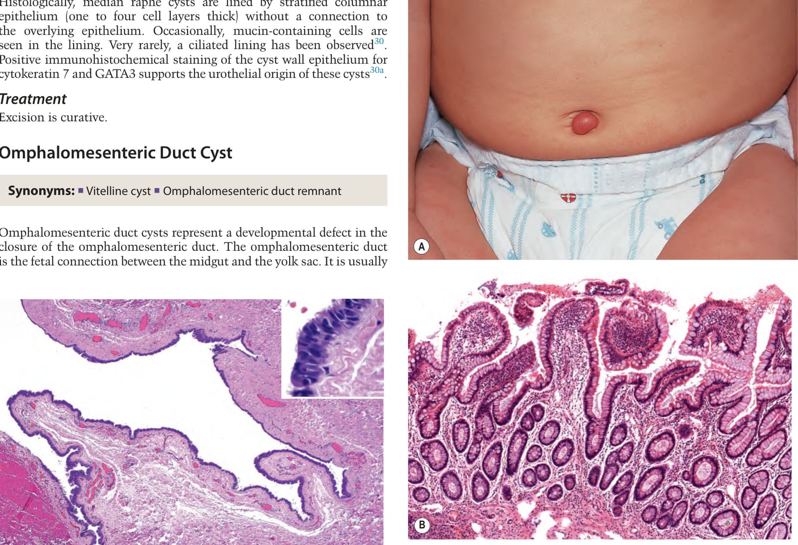
Fig. 110.21 Omphalomesenteric duct cyst.A An umbilical pink papule in an infant. The clinical differential diagnosis includes an umbilical (pyogenic) granuloma and urachal cyst/remnant. B Histologically, ectopic gastrointestinal epithelium is seen; note the villi and goblet cells. B, Courtesy Luis Requena, MD.