CYSTS WITHOUT AN EPITHELIAL LINING
Mucocele
Synonyms: Mucous cyst of oral mucosa Ranula (when located on the floor of the mouth)
Mucoceles most frequently develop on the lower labial mucosa, but they also occur on the floor of the mouth, buccal mucosa, and tongue. They appear as dome-shaped, mucosa-colored to bluish, translucent papules or nodules that range in size from a few millimeters to over a centimeter (Fig. 110.22; see Fig. 72.22). Mucoceles arise as a result of disruption of the ducts of minor salivary glands. This disruption leads to an accumulation of mucinous material, a reactive inflammatory response, and the development of surrounding granulation and fibrous tissue. Multiple mucoceles can develop in patients with GVHD.
A variant of mucocele, superficial mucocele, presents as a clear tense vesicle that is a few millimeters in diameter. Superficial mucoceles are most commonly found on the retromolar pad, posterior buccal mucosa, and soft palate. These lesions are short-lived, asymptomatic, and recurrent. They may be confused clinically with an immunobullous or viral process.
Pathology
Biopsy specimens of mucoceles show one or several spaces within the connective tissue filled with mucinous material, but without an epithelial lining. The spaces are surrounded by chronic inflammation and mucin-containing macrophages, as well as granulation and fibrous tissue. A salivary duct may be seen at the periphery of these findings. Adjacent minor salivary glands may show chronic inflammation and fibrosis. The mucinous material is sialomucin, and the latter contains both neutral and acid mucopolysaccharides, which stain with PAS (diastase-resistant) and with Alcian blue or colloidal iron, respectively.
Histologically, a superficial mucocele shows a subepithelial vesicle filled with mucin and a surrounding sparse to moderate mixed inflammatory infiltrate. Salivary gland ducts are seen opening into the vesicle or immediately adjacent to the vesicle.
Treatment
Mucoceles may resolve spontaneously. If they do not, treatment options include excision, marsupialization, electrodesiccation, intralesional corticosteroid injection, or cryosurgery.
Digital Mucous Cyst (Pseudocyst)
Synonyms: Digital myxoid cyst Cutaneous myxoid cyst
Digital mucous cysts (pseudocysts) most commonly occur on the dorsal surface of the distal phalanx of the finger (Fig. 110.23). Toe lesions are less commonly observed. A characteristic longitudinal depression in the nail plate may be seen distal to the cyst. These cysts are skin-colored to bluish and drain clear gelatinous material when punctured. The etiology of digital mucous cysts is controversial, with some authors stating that they are degenerative in origin while others believe they extend from the distal interphalangeal joint space. A pedicle connecting the cyst to the adjacent joint space can usually be demonstrated.
Pathology
Histologically, a digital mucous cyst resembles focal mucinosis, often with clefts in the dermis; an epithelial lining is absent (Fig. 110.24). The clefts and the surrounding loose connective tissue contain abundant acid mucopolysaccharides, which can be highlighted by Alcian blue or colloidal iron stains.
A A skin-colored, compressible, subcutaneous nodule is present on the wrist (arrow), the most common location. B A cystic space of acral skin is surrounded by fibrous tissue. A, Courtesy Jean L. Bolognia, MD; B, Courtesy Lorenzo Cerroni, MD.
Treatment
Resolution may be seen after intralesional injection of corticosteroids, injection of sclerosing agents, or repeated puncture and drainage. Surgical excision may lead to even higher success rates.
Ganglion
Synonyms: Ganglion cyst Synovial cyst
Ganglia are soft cystic masses up to 4 cm in diameter that most commonly occur on the dorsal aspect of the wrist; they may also be found on the volar wrist or fingers, the dorsal aspect of the feet, or the knees (Fig. 110.25A). Ganglia rarely develop on the lateral elbow or anterior shoulder. They occur more commonly in women and may cause discomfort with activity, impairment of mobility, or cosmetic concerns. In the rare disorder cystic ganglionosis, multiple lesions appear during childhood or adolescence.
Ganglia are frequently attached to a tendon sheath or the joint capsule, but usually do not communicate with the joint space. The mucin present within a ganglion is thought to be produced by local fibroblasts.
Pathology
Myxoid change is seen within the connective tissue that ultimately forms cystic spaces. These spaces coalesce into a dominant cystic space lined by variably thick fibrous tissue (Fig. 110.25B), sometimes with a synovial lining.
Erythematous firm nodule on the ear.
Treatment
Early lesions may respond to several weeks of compression therapy. Other options include aspiration plus intralesional corticosteroid injection or excision. Recurrences are common, even with excisional therapy.
Pseudocyst of the Auricle
Synonyms: Endochondral pseudocyst Cystic chondromalacia Intracartilaginous cyst
Pseudocyst of the auricle usually arises in the scaphoid fossa of the ear in middle-aged men. Lesions are usually unilateral. They present as a painless swelling (Fig. 110.26), and they tend to arise over the course of a few weeks. The etiology of pseudocyst of the auricle is unknown, but chronic trauma as well as a developmental defect have been suggested.
Pathology
Biopsy specimens show a cavity within the auricular cartilage (without an epithelial lining) that contains clear fluid. Fibrous tissue and granulation tissue may be found in the cavity as well. The cartilage lining the cavity may show degenerative changes. No inflammation is seen within the cartilage, a feature that distinguishes a pseudocyst from relapsing polychondritis, which may be in the clinical differential diagnosis.
Treatment
Treatment options include aspiration, with or without intralesional injection of corticosteroids, as well as incision and drainage with destruction of the cavity. Each of these modalities should be followed by pressure dressings.
Cutaneous Metaplastic Synovial Cyst (Pseudocyst)
Cutaneous metaplastic synovial cysts (pseudocysts) typically present as a solitary tender subcutaneous nodule, although multiple lesions have been reported. They occur primarily in areas of prior trauma, particularly prior surgical trauma. Frequently, the preoperative diagnosis is that of a suture granuloma.
Pathology
A cystic cavity is seen within the dermis that is not lined by epithelium. The cavity may communicate with the overlying epidermis via fistulas. Variably cellular villous structures mimicking hyperplastic synovium protrude into the cavity. These villi are covered with a fibrinous exudate. The base of the villi tends to merge with surrounding scar tissue.
Treatment
Excision is curative.
Additional figures available in our eBook (see inside front cover for access code).
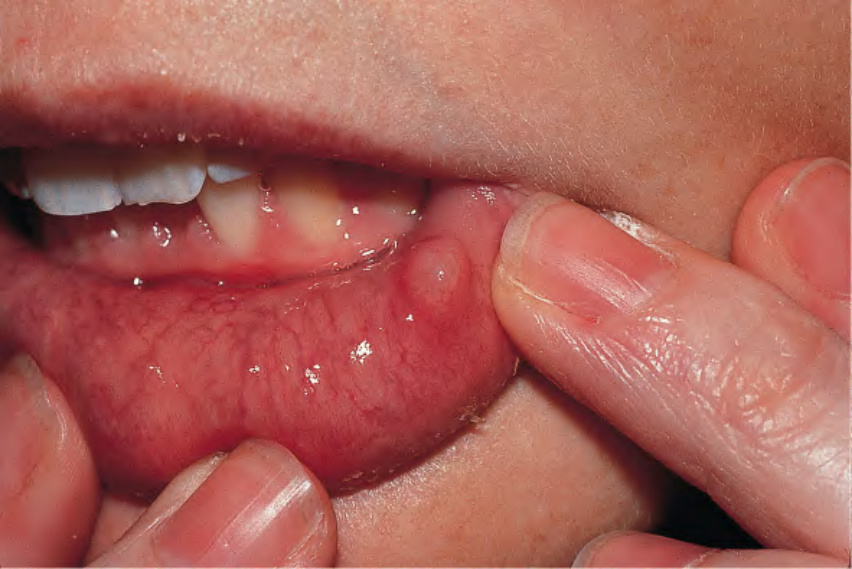
Fig. 110.22 Mucocele. A bluish to mucosa-colored translucent papule on the lower mucosal lip.
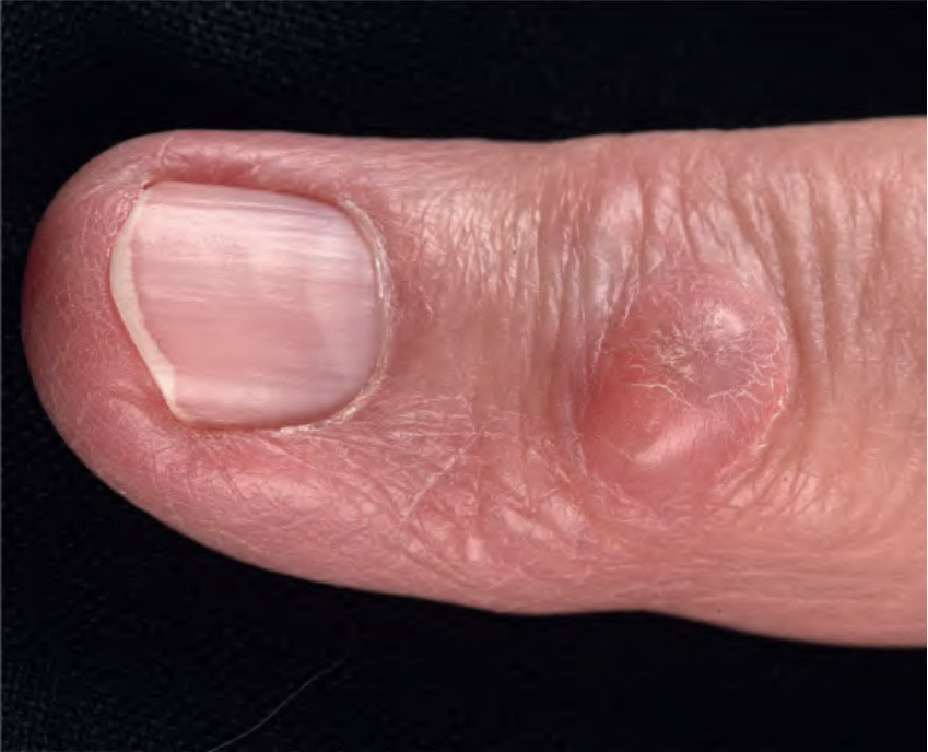
Fig. 110.23 Digital mucous cyst. Translucent cystic papule overlying the distal interphalangeal joint. Courtesy Lorenzo Cerroni, MD.
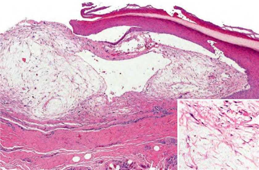
Fig. 110.24 Histopathology of a digital mucous cyst. A subepidermal cystic cavity is surrounded by mucin-rich dermis (inset). Note the prominent stratum lucidum of acral skin. Courtesy Lorenzo Cerroni, MD.
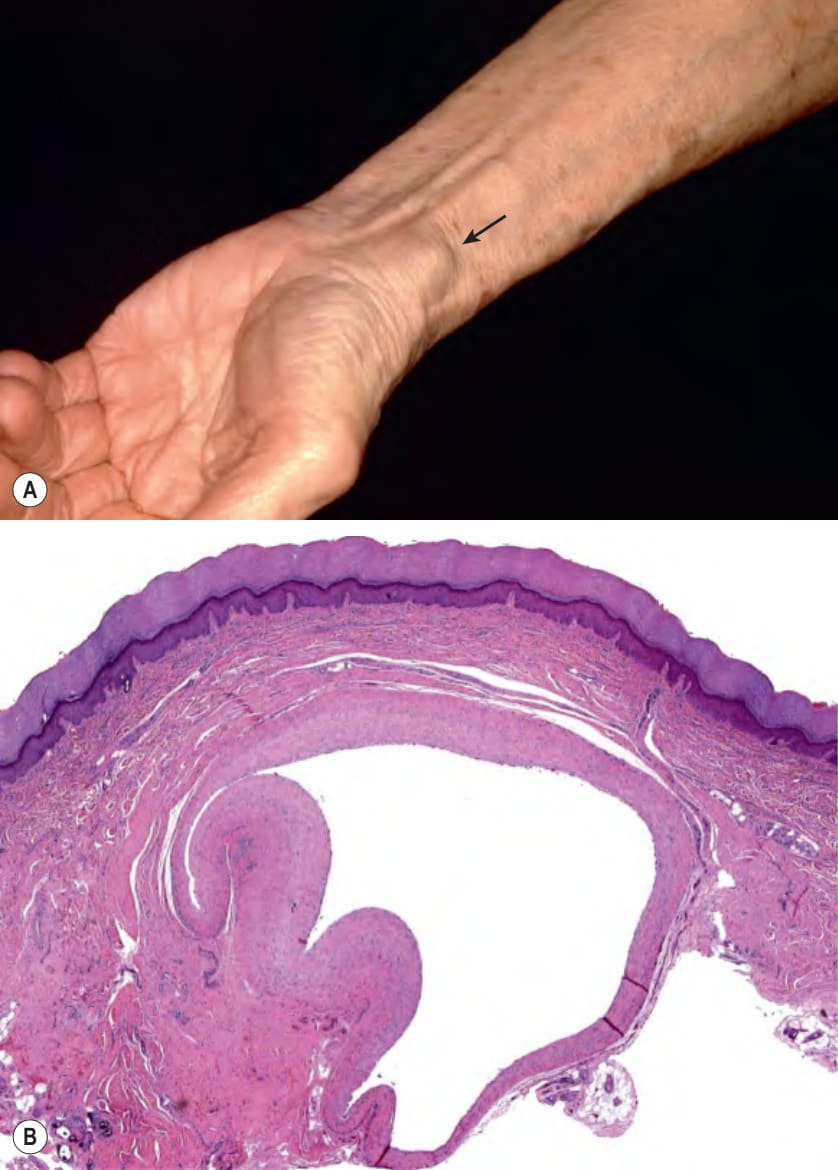
Fig. 110.25 Ganglion (cyst) – clinical presentation and histologic features.
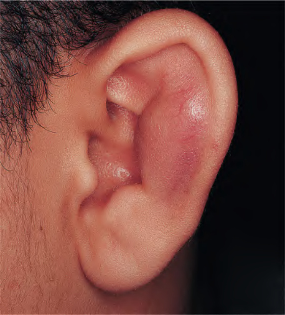
Fig. 110.26 Pseudocyst of the auricle.