Introduction
Timothy H. McCalmont and Laura B. Pincus Adnexal Neoplasms
Chapter Contents
Neoplasms and Proliferations with Follicular Differentiation … 1948
Neoplasms and Proliferations with Sebaceous Differentiation . . 1960
Neoplasms and Proliferations with Apocrine (or Eccrine)
Key features
Terminology and classification of adnexal tumors has varied considerably according to different authorities
Adnexal neoplasms can differentiate toward the folliculosebaceous- apocrine unit or the eccrine apparatus
As a reflection of joint ontogeny, tumors often exhibit combinations of follicular, sebaceous, and apocrine differentiation
Many adnexal tumors historically classified as eccrine are probably apocrine
The nosology of adnexal neoplasms has been confused for decades, and a key problem has been the lack of a logical classification scheme. Postulated classifications and inferences regarding lineage have often been contradictory, a reflection of the fact that broad conclusions regarding lineage and classification were formerly based on enzyme histochemical attributes of dubious specificity.
The crucial principle to keep in mind when considering adnexal neoplasms is that the development of the eccrine apparatus is distinct from the folliculosebaceous-apocrine unit. Eccrine glands develop directly from embryonic epidermal anlage during early months of fetal development. Follicles arise directly from the epidermis concurrently, but their development differs in that mesenchymal cells, precursors of the follicular papilla, descend jointly into the dermis with the developing follicle (see Ch. 2 and Fig. 2.4). Subsequently, sebaceous and apocrine glands and their ducts elaborate as secondary structures from bulges on the side of the developing follicle.
These ontogenetic relationships reflect relationships observed repeatedly in clinical disease. As one would expect from ontogeny, follicular, sebaceous, and apocrine differentiation occur conjointly, and combinations of eccrine and folliculosebaceous differentiation probably do not exist.
Both the differentiation of the tumor (Fig. 111.1) and the topography of adnexal structures offer insights into logical classification. There is striking variation in anatomic distribution among adnexal neoplasms, and some of these differences hold implications regarding lineage. Based on topography, it is nonsensical to pigeonhole spiradenoma, a tumor that commonly occurs within apocrine skin and rarely, if ever, develops within acral skin, as an eccrine tumor. In contrast, an acral predilection suggests that poroma is commonly of eccrine lineage.
Lastly, microscopy and morphology play a role in the assessment of lineage. For some lines of differentiation, the meaning ascribed to specific attributes is indisputable. Cells with coarsely vacuolated cytoplasm and scalloped nuclei signify sebaceous differentiation. Follicular differentiation is established by the presence of bulbar basaloid cells accompanied by mesenchymal cells resembling the follicular papilla. Follicular lineage is also connoted by attributes such as matrical or outer sheath differentiation. In contrast, there are no specific attributes that permit recognition of eccrine or apocrine differentiation, although decapitation configuration at the luminal border can be considered suggestive of apocrine differentiation.
More recently, molecular pathology has been employed given the various alterations, including gene fusions, that have been observed in benign and malignant adnexal tumors.
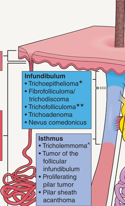
Fig. 111.1 The folliculosebaceousapocrine unit and eccrine sweat gland with examples of adnexal tumors that differentiate toward the hair follicle, sebaceous gland, apocrine gland, and eccrine gland.